
Recommended
More Related Content
What's hot
What's hot (20)
Similar to Phylum Platyhelminths.pptx
Similar to Phylum Platyhelminths.pptx (20)
More from Dr. Muhammad Moosa
More from Dr. Muhammad Moosa (15)
Recently uploaded
Recently uploaded (20)
Phylum Platyhelminths.pptx
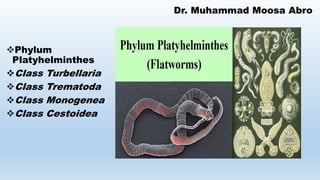
- 1. Dr. Muhammad Moosa Abro Phylum Platyhelminthes Class Turbellaria Class Trematoda Class Monogenea Class Cestoidea
- 2. PHYLUM PLATYHELMINTHES The phylum Platyhelminthes (Gr. platys, flat 1 helmins, worm) contains over 34,000 animal species. Flatworms range in adult size from 1 mm or less to 25 m (Taeniarhynchus saginatus; in length. Their mesodermally derived tissues include a loose tissue called parenchyma (Gr. parenck, anything poured in beside) that fills spaces between other more specialized tissues, organs, and the body wall. Depending on the species, parenchyma may provide skeletal support, nutrient storage, motility, reserves of regenerative cells, transport of materials, structural interactions with other tissues, modifiable tissue for morphogenesis, oxygen storage, and perhaps other functions yet to be determined. Dr. Muhammad Moosa Abro
- 3. This is the first phylum covered that has an organ-system level of organization—a significant evolutionary advancement over the tissue level of organization. The phylum is divided into four classes : (1) the Turbellaria consist of mostly free-living flatworms, whereas the (2) Monogenea, (3) Trematoda, and (4) Cestoidea contain solely parasitic species. Turbellaria is a paraphyletic group. PHYLUM PLATYHELMINTHES Dr. Muhammad Moosa Abro
- 4. Some general characteristics of the phylum Platyhelminthes include: 1. Usually flattened dorsoventrally, triploblastic, acoelomate, bilaterally symmetrical 2. Unsegmented worms (members of the class Cestoidea are strobilated) 3. Incomplete gut usually present (gut absent in Cestoidea) 4. Somewhat cephalized, with an anterior cerebral ganglion and usually longitudinal nerve cords 5. Protonephridia as excretory/osmoregulatory structures 6. Most forms monoecious; complex reproductive systems 7. Nervous system consists of a pair of anterior ganglia with longitudinal nerve cords connected by transverse nerves and located in the mesenchyme PHYLUM PLATYHELMINTHES Dr. Muhammad Moosa Abro
- 6. Class Turbellaria Members of the class Turbellaria (L. turbellae, a commotion 1 aria, like) are mostly free-living bottom dwellers in freshwater and marine environments, where they crawl on stones, sand, or vegetation. Turbellarians are named for the turbulence that their beating cilia create in the water. Over 3,000 species have been described. PHYLUM PLATYHELMINTHES Dr. Muhammad Moosa Abro
- 7. Class Turbellaria Turbellarians are predators and scavengers The few terrestrial turbellarians known live in the humid tropics and subtropics. Although most turbellarians are less than 1 cm long, the terrestrial, tropical ones may reach 60 cm in length. Coloration is mostly in shades of black, brown, and gray, although some groups display brightly colored patterns. PHYLUM PLATYHELMINTHES Dr. Muhammad Moosa Abro
- 8. Body Wall As in the Cnidaria, the ectodermal derivatives include an epidermis that is in direct contact with the environment (figure 10.3). Some epidermal cells are ciliated, and others contain microvilli. A basement membrane of connective tissue separates the epidermis from mesodermally derived tissues. PHYLUM PLATYHELMINTHES Dr. Muhammad Moosa Abro
- 9. An outer layer of circular muscle and an inner layer of longitudinal muscle lie beneath the basement membrane. Other muscles are located dorsoventrally and obliquely between the dorsal and ventral surfaces. Between the longitudinal muscles and the gastrodermis are the loosely organized parenchymal cells. PHYLUM PLATYHELMINTHES Dr. Muhammad Moosa Abro Body Wall
- 10. The innermost tissue layer is the endodermally derived gastrodermis. It consists of a single layer of cells that comprise the digestive cavity. The gastrodermis secretes enzymes that aid in digestion, and it absorbs the end products of digestion. On the ventral surface of the body wall are several types of glandular cells of epidermal origin. Rhabdites are rodlike cells that swell and form a protective mucous sheath around the body, possibly in response to attempted predation or desiccation. Adhesive glands open to the epithelial surface and produce a chemical that attaches part of the turbellarian to a substrate. Releaser glands secrete a chemical that dissolves the attachment as needed. PHYLUM PLATYHELMINTHES Dr. Muhammad Moosa Abro
- 11. Locomotion Turbellarians were one of the earliest groups of bilaterally symmetrical animals to appear. Bilateral symmetry is usually characteristic of animals with an active lifestyle—those that move from one locale to another. Turbellarians are primarily bottom dwellers that glide over the substrate. They move using both cilia and muscular undulations, with the muscular undulations being more important in their movement. Just beneath the epithelium are layers of muscle cells. The outer layer runs in a circular direction and the inner layer in a longitudinal direction. Muscles also run vertically and obliquely, making agile bending and twisting movements possible. PHYLUM PLATYHELMINTHES Dr. Muhammad Moosa Abro
- 12. (The dorsoventral muscles are essential for maintaining the flatness of flatworms. This flattened shape provides adequate surface area for diffusion of respiratory gases and metabolic wastes across body surfaces.) As they move, turbellarians lay down a sheet of mucus that aids in adhesion and helps the cilia gain some traction. The densely ciliated ventral surface and the flattened bodies of turbellarians enhance the effectiveness of this locomotion. All of these movements thus result from two mechanisms: (1) The gliding is both muscular and ciliary. (2) The rapid movements pass from the head backwards, propelling the animal forward, and are wholly muscular. Locomotion PHYLUM PLATYHELMINTHES Dr. Muhammad Moosa Abro
- 13. Digestion and Nutrition The digestive tract of turbellarians is incomplete—it has a mouth opening but lacks an anus. This blind cavity varies from a simple, unbranched chamber (figure 10.4a) to a highly branched system of digestive tubes (figure 10.4c and d). Other turbellarians have digestive tracts that are lobed (figure 10.4b). Highly branched digestive systems result in more gastrodermis closer to the sites of digestion and absorption, reducing the distance nutrients must diffuse. This aspect of digestive tract structure is especially important in some of the larger turbellarians and partially compensates for the absence of a circulatory system. PHYLUM PLATYHELMINTHES Dr. Muhammad Moosa Abro
- 14. The turbellarian pharynx functions as an ingestive organ. It varies in structure from a simple, ciliated tube to a complex organ developed from the folding of muscle layers. In the latter, the free end of the tube lies in a pharyngeal sheath and can project out of the mouth when feeding (figure 10.5). Most turbellarians, such as the common planarian, are carnivores and feed on small, live invertebrates or scavenge on larger, dead animals; some are herbivores and feed on algae that they scrape from rocks. PHYLUM PLATYHELMINTHES Dr. Muhammad Moosa Abro Digestion and Nutrition
- 15. Sensory cells (chemoreceptors) on their heads help them detect food from a considerable distance. Food digestion is partially extracellular. Pharyngeal glands secrete enzymes that help break down food into smaller units that can be taken into the pharynx. In the digestive cavity, phagocytic cells engulf small units of food, and digestion is completed in intracellular vesicles. PHYLUM PLATYHELMINTHES Dr. Muhammad Moosa Abro Digestion and Nutrition
- 16. The turbellarians do not have respiratory organs; thus, respiratory gases (CO2 and O2) are exchanged by diffusion through the body wall. Most metabolic wastes (e.g., ammonia) are also removed by diffusion through the body wall. In marine environments, invertebrates are often in osmotic equilibrium with their environment. In freshwater, invertebrates are hypertonic to their aquatic environment and thus must regulate the osmotic concentration (water and ions) of their body tissues. The evolution of osmoregulatory structures in the form of protonephridia enabled turbellarians to invade freshwater, PHYLUM PLATYHELMINTHES Dr. Muhammad Moosa Abro Exchanges with the Environment
- 17. Protonephridia (Gr. protos, first 1 nephros, kidney) (sing., protonephridium) are networks of fine tubules that run the length of the turbellarian, along each of its sides (figure 10.6a). Numerous, fine side branches of the tubules originate in the parenchyma as tiny enlargements called flame cells (figure 10.6b). Flame cells (so named because, in the living organism, they resemble a candle flame) have numerous cilia that project into the lumen of the tubule. Slitlike fenestrations (openings) perforate the tubule wall surrounding the flame cell. The beating of the cilia drives fluid down the tubule, creating a negative pressure in the tubule. As a result, fluid from the surrounding tissue is sucked through the fenestrations into the tubule. The tubules eventually merge and open to the outside of the body wall through a minute opening called a nephridiopore. PHYLUM PLATYHELMINTHES Dr. Muhammad Moosa Abro
- 18. Nervous and Sensory Functions Turbellarians have subepidermal nervous tissues. In some cases, nerves are netlike and fibers coalesce to form cerebral ganglia (figure 10.7a). The nervous tissues of most other turbellarians, such as the planarian Dugesia, consists of a subepidermal nerve net and several pairs of long nerve cords (figure 10.7b). Lateral branches called commissures (points of union) connect the nerve cords. Nerve cords and their commissures give a ladderlike appearance to the turbellarian nervous organization. PHYLUM PLATYHELMINTHES Dr. Muhammad Moosa Abro
- 19. Neurons are organized into sensory (going to the primitive brain), motor (going away from the primitive chemicals. Auricles (sensory lobes) may project from the side of the head (figure 10.7b). Chemoreceptors that aid in food location are especially dense in these auricles. Most turbellarians have two simple eyespots called ocelli (sing., ocellus). These ocelli orient the animal to the direction of light. (Most turbellarians are negatively phototactic and move away from light.) Each ocellus consists of a cuplike depression lined with black pigment. Photoreceptor nerve endings in the cup are part of the neurons that leave the eye and connect with a cerebral ganglion. PHYLUM PLATYHELMINTHES Dr. Muhammad Moosa Abro
- 20. Reproduction and Development Many turbellarians reproduce asexually by transverse fission. Fission usually begins as a constriction behind the pharynx ( figure 10.8). The two (or more) animals that result from fission are called zooids (Gr., zoon, living being or animal), and they regenerate missing parts after separating from each other. Sometimes, the zooids remain attached until they have attained a fairly complete degree of development, at which time they detach as independent individuals. PHYLUM PLATYHELMINTHES Dr. Muhammad Moosa Abro
- 21. Turbellarians are monoecious, and reproductive systems arise from the mesodermal tissues in the parenchyma. Numerous paired testes lie along each side of the worm and are the sites of sperm production. Sperm ducts (vas deferens) lead to a seminal vesicle (a sperm storage organ) and a protrusible penis (figure 10.9). The penis projects into a genital chamber. The female system has one to many pairs of ovaries. Oviducts lead from the ovaries to the genital chamber, which opens to the outside through the genital pore. Even though turbellarians are monoecious, reciprocal sperm exchange between two animals is usually the rule. PHYLUM PLATYHELMINTHES Dr. Muhammad Moosa Abro
- 22. This cross-fertilization ensures greater genetic diversity than does self-fertilization. During cross-fertilization, the penis of each individual is inserted into the copulatory sac of the partner. After copulation, sperm move from the copulatory sac to the genital chamber and then through the oviducts to the ovaries, where fertilization occurs. Yolk may either be directly incorporated into the egg during egg formation or yolk cells may be laid around the zygote as it passes down the female reproductive tract past the vitellaria (yolk glands). PHYLUM PLATYHELMINTHES Dr. Muhammad Moosa Abro
- 23. • Eggs are laid with or without a gel-like mass. • A hard capsule called a cocoon (L., coccum, eggshell) encloses many turbellarian eggs. • These cocoons attach to the substrate by a stalk and contain several embryos per capsule. Two kinds of capsules are laid. Summer capsules hatch in two to three weeks, and immature animals emerge. • Autumn capsules have thick walls that can resist freezing and drying, and they hatch after overwintering. • Development of most turbellarians is direct—a gradual series of changes transforms embryos into adults. • A few turbel- larians have a free-swimming stage called a Müller’s larva. • It has ciliated extensions for feeding and locomotion. • The larva eventually settles to the substrate and develops into a young turbellarian. PHYLUM PLATYHELMINTHES Dr. Muhammad Moosa Abro
- 24. Class Trematoda The approximately 10,000 species of parasitic flatworms in the class Trematoda (trem0ah- to9dah) (Gr. trematodes, perforated form) are collectively called flukes, which describes their wide, flat shape. Almost all adult flukes are parasites of vertebrates, whereas immature stages may be found in vertebrates or invertebrates, or encysted on plants. Many species are of great economic and medical importance. Most flukes are flat and oval to elongate, and range from less than 1 mm to 6 cm in length (figure 10.10). PHYLUM PLATYHELMINTHES Dr. Muhammad Moosa Abro
- 25. They feed on host cells and cell fragments. The digestive tract includes a mouth and a muscular, pumping pharynx. Posterior to the pharynx, the digestive tract divides into two blind-ending, variously branched pouches called cecae (sing., cecum). Some flukes supplement their feeding by absorbing nutrients across their body walls. PHYLUM PLATYHELMINTHES Dr. Muhammad Moosa Abro Class Trematoda
- 26. Body wall Body-wall structure is similar for all flukes and represents an evolutionary adaptation to the parasitic way of life. The epidermis consists of an outer layer called the tegument (figure 10.11), which forms a syncytium (a continuous layer of fused cells). The outer zone of the tegument consists of an organic layer of proteins and carbohydrates called the glycocalyx. The glycocalyx aids in the transport of nutrients, wastes, and gases across the body wall, and protects the fluke against enzymes and the host’s immune system. Also found in this zone are microvilli that facilitate nutrient exchange. PHYLUM PLATYHELMINTHES Dr. Muhammad Moosa Abro
- 27. Cytoplasmic bodies that contain the nuclei and most of the organelles lie below the basement membrane. Slender cell processes called cytoplasmic bridges connect the cytoplasmic bodies with the outer zone of the tegument. PHYLUM PLATYHELMINTHES Dr. Muhammad Moosa Abro Body wall
- 28. Subclass There are two subclasses of trematodes. The subclass Aspidogastrea is a small group of flukes that are endoparasites of molluscs, and in some cases a second host may be a fish or turtle. The subclass Digenea contains the vast majority of flukes and will be covered in the following discussion. • . PHYLUM PLATYHELMINTHES Dr. Muhammad Moosa Abro
- 29. Subclass Digenea The flukes that comprise the subclass Digenea (Gr. di, two 1 genea, birth) include many medically important species. In this subclass, at least two different forms, an adult and one or more larval stages, Because digenetic flukes require at least two different hosts to complete their life cycles, these animals possess the most complex life cycles in the entire animal kingdom. As adults, they are all endoparasites in the bloodstreams, digestive tracts, ducts of the digestive organs, or other visceral organs in a wide variety of vertebrates that serve as definitive, or final, hosts. PHYLUM PLATYHELMINTHES Dr. Muhammad Moosa Abro
- 30. The one or more intermediate hosts (the hosts that harbor immature stages) may harbor several different larval stages. The adhesive organs are two large suckers. The anterior sucker is the oral sucker and surrounds the mouth. The other sucker, the acetabulum, is located below the oral sucker on the middle portion of the body (see figure 10.10). PHYLUM PLATYHELMINTHES Dr. Muhammad Moosa Abro Subclass Digenea
- 31. Lifecycle • The eggs of digenetic trematodes are oval and usually have a lidlike hatch called an operculum (figure 10.12a). • When an egg reaches freshwater, the operculum opens, and a ciliated larva called a miracidium (pl., miracidia) swims out (figure 10.12b). • The miracidium swims until it finds a suitable first intermediate host (a snail) to which it is chemically attracted. • The miracidium penetrates the snail, loses its cilia, and develops into a sporocyst (figure 10.12c). • (Alternately, the miracidium may remain in the egg and hatch after a snail eats it.) Sporocysts are baglike and contain embryonic cells that develop into either daughter sporocysts or rediae (sing., redia) (figure 10.12d). PHYLUM PLATYHELMINTHES Dr. Muhammad Moosa Abro
- 32. Lifecycle At this point in the life cycle, asexual reproduction first occurs. From a single miracidium, hundreds of daughter sporocysts, and in turn, hundreds of rediae, can form by asexual reproduction Embryonic cells in each daughter sporocyst or redia produce hundreds of the next larval stage, called cercariae (sing., cercaria) (figure 10.12e). (This phenomenon of producing many cercariae is called polyembryony. It greatly enhances the chances that one or two of these cercaria will further the life cycle.) PHYLUM PLATYHELMINTHES Dr. Muhammad Moosa Abro
- 33. A cercaria has a digestive tract, suckers, and a tail. Cercariae leave the snail and swim freely until they encounter a second intermediate or final host, which may be a vertebrate, invertebrate, or plant. The cercaria penetrates this host and encysts as a metacercaria (pl., metacercariae) (figure 10.12f). When the definitive host eats the second intermediate host, the metacercaria excysts and develops into an adult (figure 10.12g). PHYLUM PLATYHELMINTHES Dr. Muhammad Moosa Abro Lifecycle
- 34. Some Important Trematode Parasites of Humans The Chinese liver fluke Clonorchis sinensis is a common parasite of humans in Asia, where more than 30 million people are infected. The adult lives in the bile ducts of the liver, where it feeds on epithelial tissue and blood (figure 10.13a). The adults release embryonated eggs into the common bile duct. The eggs make their way to the intestine and are eliminated with feces ( figure 10.13b). The miracidia are released when a snail ingests the eggs. PHYLUM PLATYHELMINTHES Dr. Muhammad Moosa Abro
- 35. Following the sporocyst and redial stages, cercariae emerge into the water. If a cercaria contacts a fish (the second intermediate host), it penetrates the epidermis of the fish, loses its tail, and encysts. The metacercaria develops into an adult in a human who eats raw or poorly cooked fish, a delicacy in Asian countries and gaining in popularity in the Western world (e.g., sushi, sashimi, ceviche). PHYLUM PLATYHELMINTHES Dr. Muhammad Moosa Abro Some Important Trematode Parasites of Humans
- 36. Fasciola hepatica is called the sheep liver fluke (see figure 10.12a– g) because it is common in sheep- raising areas and uses sheep or humans as its definitive host. The adults live in the bile duct of the liver. Eggs pass via the common bile duct to the intestine, from which they are eliminated. Eggs deposited in freshwater hatch, and the miracidia must locate the proper species of snail. PHYLUM PLATYHELMINTHES Dr. Muhammad Moosa Abro Some Important Trematode Parasites of Humans
- 37. If a snail is found, miracidia penetrate the snail’s soft tissue and develop into sporocysts that develop into rediae and give rise to cercariae. After the cercariae emerge from the snail, they encyst on aquatic vegetation. Sheep or other animals become infected when they graze on the aquatic vegetation. Humans may become infected with Fasciola hepatica by eating a freshwater plant called watercress that contains the encysted metacercaria. PHYLUM PLATYHELMINTHES Dr. Muhammad Moosa Abro Some Important Trematode Parasites of Humans
- 38. Schistosomes are blood flukes with vast medical significance. The impact these flukes have had on history is second only to that of Plasmodium (see figure 8.15). They infect more than 200 million people throughout the world. Infections are most common in Africa (Schistosoma haematobium and S. mansoni), South and Central America (S. mansoni), and Southeast Asia (S. japonicum). The adult dioecious worms live in the human bloodstream (figure 10.14a). The male fluke is shorter and thicker than the female, and the sides of the male body curve under to form a canal along the ventral surface (schistosoma means “split body”). The female fluke is long and slender and is carried in the canal of the male (figure 10.14b). Copulation is continuous, and the female produces thousands of eggs over her lifetime. PHYLUM PLATYHELMINTHES Dr. Muhammad Moosa Abro Some Important Trematode Parasites of Humans
- 39. Each egg contains a spine that mechanically aids it in moving through host tissue until it is eliminated in either the feces or urine (figure 10.14c). Unlike other flukes, schistosome eggs lack an operculum. The miracidium escapes through a slit that develops in the egg when the egg reaches freshwater (figure 10.14d). The miracidium seeks, via chemotaxis, a snail (figure 10.14e). The miracidium penetrates it, and develops into a sporocyst, then daughter sporocysts, and finally forktailed cercariae (figure 10.14f). There is no redial generation. The cercariae leave the snail and penetrate the skin of a human (figure 10.14g). Anterior glands that secrete digestive enzymes aid in penetration. Once in a human, the cercariae lose their tails and develop into adults in the intestinal veins, skipping the metacercaria stage. PHYLUM PLATYHELMINTHES Dr. Muhammad Moosa Abro Some Important Trematode Parasites of Humans
- 40. PHYLUM PLATYHELMINTHES Dr. Muhammad Moosa Abro
- 42. Class Monogenea Monogenetic flukes are so named because they have only one generation in their life cycle; that is, one adult develop from one egg. Monogeneans are mostly external parasites (ectoparasites) of freshwater and marine fishes, where they attach to the gill filaments and feed on epithelial cells, mucus, or blood. A large, posterior attachment organ called an opisthaptor facilitates attachment (figure 10.15). Adult monogeneans produce and release eggs that have one or more sticky threads that attach the eggs to the fish gill. PHYLUM PLATYHELMINTHES Dr. Muhammad Moosa Abro
- 43. Eventually, a ciliated larva called an oncomiracidium hatches from the egg and swims to another host fish, where it attaches by its opisthaptor and develops into an adult. Although monogeneans have been traditionally aligned with the trematodes, some structural and chemical evidence suggests that they are more closely related to tapeworms than to trematodes. PHYLUM PLATYHELMINTHES Dr. Muhammad Moosa Abro Class Monogenea
- 44. Class Cestoidea The most highly specialized class of flatworms are members of the class Cestoidea (ses-toid9e-ah) (Gr. kestos, girdle 1 eidos, form), commonly called either tapeworms or cestodes. All of the approximately 3,500 species are endoparasites that usually reside in the vertebrate digestive system. Because they lack pigment as adults, their color is often white with shades of yellow or gray. Adult tapeworms range from 1 mm to 25 m in length. Two unique adaptations to a parasitic lifestyle characterize tapeworms: (1) Tapeworms lack a mouth and digestive tract in all of their life-cycle stages; they absorb nutrients directly across their body wall. (2) Most adult tapeworms consist of a long series of repeating units called proglottids. PHYLUM PLATYHELMINTHES Dr. Muhammad Moosa Abro
- 45. Each proglottid contains one or two complete sets of reproductive structures. As with most endoparasites, adult tapeworms live in a very stable environment. The vertebrate intestinal tract has very few environmental variations that would require the development of great anatomical or physiological complexity in any single tapeworm body system. The physiology of the tapeworm’s host maintains the tapeworm’s homeostasis (internal constancy). In adapting to such a specialized environment, tapeworms have lost some of the structures believed to have been present in ancestral flatworms. Tapeworms are, therefore, a good example of evolution not always resulting in greater complexity. There are two subclasses of tapeworms. The subclass Cestodaria contains about 15 species of fish parasites. The subclass Eucestoda contains medically important tapeworms. PHYLUM PLATYHELMINTHES Dr. Muhammad Moosa Abro Class Cestoidea
- 46. Subclass Eucestoda Almost all of the cestodes belong to the subclass Eucestoda and are called true tapeworms. They represent the ultimate degree of specialization of any parasitic animal. The body is divided into three regions (figure 10.16a). At one end is a holdfast structure called the scolex that contains circular or leaflike suckers and sometimes a rostellum of hooks (figure 10.16b). With the scolex, the tapeworm firmly anchors itself to the intestinal wall of its definitive vertebrate host. No mouth is present. Posteriorly, the scolex narrows to form the neck. Transverse constrictions in the neck give rise to the third body region, PHYLUM PLATYHELMINTHES Dr. Muhammad Moosa Abro
- 47. the strobila (Gr. strobilus, a linear series) (pl., strobilae). The strobila consists of a series of linearly arranged proglottids, which function primarily as reproductive units. As a tapeworm grows, new proglottids are added in the neck region, and older proglottids are gradually pushed posteriorly. As they move posteriorly, proglottids mature and begin producing eggs. Thus, anterior proglottids are said to be immature, those in the midregion of the strobila are mature, and those at the posterior end that have accumulated eggs are gravid (L., gravida, heavy, loaded, pregnant). The outer body wall of tapeworms consists of a tegument similar in structure to that of trematodes (see figure 10.11). It plays a vital role in nutrient absorption because tapeworms have no digestive system. The tegument even absorbs some of the host’s own enzymes to facilitate digestion. With the exception of the reproductive systems, the body systems of tapeworms are reduced in structural complexity. PHYLUM PLATYHELMINTHES Dr. Muhammad Moosa Abro Subclass Eucestoda
- 48. The nervous system consists of only a pair of lateral nerve cords that arise from a nerve mass in the scolex and extend the length of the strobila. A protonephridial system also runs the length of the tapeworm (see figure 10.6). Tapeworms are monoecious, and most of their physiology is devoted to producing large numbers of eggs. Each proglottid contains one or two complete sets of male and female reproductive organs (figure 10.16a). Numerous testes are scattered throughout the proglottid and deliver sperm via a duct system to a copulatory organ called a cirrus. PHYLUM PLATYHELMINTHES Dr. Muhammad Moosa Abro Subclass Eucestoda
- 49. The cirrus opens through a genital pore, which is an opening shared with the female system. The male system of a proglottid matures before the female system, so that copulation usually occurs with another mature proglottid of the same tapeworm or with another tapeworm in the same host. As previously mentioned, the avoidance of self-fertilization leads to hybrid vigor. PHYLUM PLATYHELMINTHES Dr. Muhammad Moosa Abro Subclass Eucestoda
- 50. PHYLUM PLATYHELMINTHES Dr. Muhammad Moosa Abro
- 51. Some Important Tapeworm Parasites of Humans One medically important tapeworm of humans is the beef tapeworm Taeniarhynchus saginatus (figure 10.17). Adults live in the small intestine and may reach lengths of 25 m. About 80,000 eggs per proglottid are released as proglottids break free of the adult worm. As an egg develops, it forms a six-hooked (hexacanth) larva called the oncosphere. As cattle (the intermediate host) graze in pastures contaminated with human feces, they ingest oncospheres (or proglottids). Digestive enzymes of the cattle free the oncospheres, and the larvae use their hooks to bore through the intestinal wall into the bloodstream. PHYLUM PLATYHELMINTHES Dr. Muhammad Moosa Abro
- 52. use their hooks to bore through the intestinal wall into the bloodstream. The bloodstream carries the larvae to skeletal muscles, where they encyst and form a fluid-filled bladder called a cysticercus (pl., cysticerci) or bladder worm. When a human eats infected meat (termed “measly beef”) that is raw or improperly cooked, the cysticercus is released from the meat, the scolex attaches to the human intestinal wall, and the tapeworm matures. A closely related tapeworm, Taenia solium (the pork tapeworm), has a life cycle similar to that of Taeniarhynchus saginatus, except that the intermediate host is the pig. PHYLUM PLATYHELMINTHES Dr. Muhammad Moosa Abro Some Important Tapeworm Parasites of Humans
- 53. The strobila has been reported as being 10 m long, but 2 to 3 m is more common. The pathology is more serious in the human than in the pig. Gravid proglottids frequently release oncospheres before the proglottids have had a chance to leave the small intestine of the human host. When these larvae hatch, they move through the intestinal wall, enter the bloodstream, and are distributed throughout the body, where they eventually encyst in human tissue as cysticerci. The disease that results is called cysticercosis and can be fatal if the cysticerci encyst in the brain. PHYLUM PLATYHELMINTHES Dr. Muhammad Moosa Abro Some Important Tapeworm Parasites of Humans
- 54. The broad fish tapeworm Diphyllobothrium latum is relatively common in the northern parts of North America, in the Great Lakes area of the United States, and throughout northern Europe. This tapeworm has a scolex with two longitudinal grooves (bothria; sing., bothrium) that act as holdfast structures(figure 10.18). The adult worm may attain a length of 10 m and shed up to a million eggs a day. Many proglottids release eggs through uterine pores. When eggs are deposited in freshwater, they hatch, and ciliated larvae called coracidia (sing., coracidium) emerge. PHYLUM PLATYHELMINTHES Dr. Muhammad Moosa Abro Some Important Tapeworm Parasites of Humans
- 55. These coracidia swim about until small crustaceans called copepods ingest them. The larvae shed their ciliated coats in the copepods and develop into procercoid larvae. When fish eat the copepods, the procercoids burrow into the muscle of the fish and become plerocercoid larvae. Larger fishes that eat smaller fishes become similarly infected with plerocercoids. When humans (or other carnivores) eat infected, raw, or poorly cooked fishes, the plerocercoids attach to the small intestine and grow into adult worms. PHYLUM PLATYHELMINTHES Dr. Muhammad Moosa Abro Some Important Tapeworm Parasites of Humans
- 56. References 56 1. Miller, A.S. and Harley, J.B. ; 1999 , 2002., 2007, 2009, 2012 & 2016 Zoology, 4th , 5th, 6th, 7th, 8th , 9th& 10th Edition (International), Singapore : McGraw Hill. 2. Hickman, C.P., Roberts, L.C/, AND Larson, A., 2018. INTEGRATED PRINCIPLES OF ZOOLOGY, 15th Edition (International), Singapore: McGRAW-Hill