PET CT Scan Price_House of Diagnostics
•
0 likes•3 views

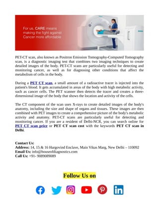
PET-CT scan, also known as Positron Emission Tomography-Computed Tomography scan, is a diagnostic imaging test that combines two imaging techniques to create detailed images of the body.
Report
Share
Report
Share
Download to read offline

Recommended
Difference between PET scan.ppt
A computed tomography (CT) scan is a particular imaging method. A CT scan delivers a detailed 3-D image of the inside of your body. This scan is often prescribed to check for abnormalities in the brain, spine, neck, chest, or abdomen. By looking at CT scan images, your doctor can consider hard tissues, such as your bones, and soft tissues, such as your muscles and organs. This indicates that a CT scan will show everything from a bone fracture to a tumour...
visit our website to get treatment from best oncologist in Delhi NCR : https://cancerconsultindia.com/
Positron Emission Tomography
PET scans use small amounts of radioactive tracers injected into the body to produce images showing how organs and tissues are functioning. A PET scan works by detecting gamma rays emitted by the tracers, allowing visualization of processes like blood flow, metabolic activity, and biochemical processes. PET scans are used to diagnose and manage conditions like cancer, heart disease, and neurological disorders.
Positron emission tomography
Positron emission tomography (PET) is an imaging technique that uses radiolabeled tracers to produce images showing their distribution in the body. During a PET scan, a tracer containing a radioactive isotope is injected and decays, emitting positrons. The positrons interact with electrons, producing pairs of gamma rays detected by the PET scanner to reconstruct images. PET scans are used to study brain function, detect and characterize cancers, and examine heart disease. Advantages include showing tissue function, but disadvantages include expense and limited availability.
P.e.t
Positron emission tomography (PET) is a nuclear imaging technique that produces 3D images of functional processes in the body. A radioactive tracer isotope is injected and accumulates in tissues of interest, undergoing positron emission decay which produces gamma photons detected by the PET scanner to create images. PET scans are used to detect cancer, evaluate brain abnormalities, and examine heart function by tracking radioisotope-labeled molecules processed by the body.
PET.docx
PET scans use radioactive tracers to detect metabolic activity in tissues and organs. The tracer is introduced and detected by the PET scanner, which produces images showing tracer distribution. Main applications include detecting cancer spread or recurrence, assessing heart damage after attack, and discovering brain disorders like Alzheimer's. However, PET scans involve radiation exposure and require nearby radioactive isotope production facilities.
medical imaging anatomy.pptx
Medical imaging refers to techniques that create visual representations of the body's interior, including X-rays, computed tomography (CT), magnetic resonance imaging (MRI), ultrasound, nuclear medicine, and positron emission tomography (PET). X-rays produce images of bones and dense tissues while CT scans create detailed 3D images; MRI uses magnets and radio waves to image soft tissues; ultrasound uses sound waves to view organs; nuclear medicine and PET involve radioactive tracers; and medical imaging plays a key role in diagnosis and treatment but risks like radiation exposure must be considered.
Ct urinary system in radiology
CT scans use X-rays and computer calculations to produce detailed cross-sectional images of the body's internal structures. A CT scan of the urinary system can detect tumors, masses, stones, or cysts. The test is performed in a radiology department where the patient lies on a table that slides into a CT scanner. Dye may be injected into the patient's vein to enhance image quality. CT scans can distinguish between solid and liquid tissues and provide valuable information for examining kidney tumors and other abnormalities of the urinary tract. Risks of CT scanning are generally low but may include minor reactions to dye in rare cases more severe complications.
Pet CT Scan for Cancer Diagnosis - Onco Life Cancer Centre
Find the best doctor of PET CT Scan in Pune at Onco Life Cancer Centre, Satara.
For more info, please visit: https://www.oncolifehospitals.com/services/pet-ct-scan/
Project 1
The document summarizes various medical imaging machines including CT scans, PET scans, MRI scans, DSR scans, and sonograms. CT scans use X-rays and computers to produce cross-sectional images of the body. PET scans use radioactive tracers to detect cancer and other diseases. MRI scans use magnetic fields to produce detailed images of organs and soft tissues without radiation. DSR scans produce real-time 3D images while sonograms use ultrasound to image fetuses and internal organs in real-time without radiation.
Project 1 (Skeletal System Multi Media) Mc Sheet
Medical imaging machines such as CT scans, PET scans, MRIs, DSR scans, and sonograms are described. CT scans use X-rays and computers to produce cross-sectional images of the body. PET scans use radioactive tracers to detect cancer and brain disorders. MRIs use magnetic fields to produce detailed images without radiation. DSR scans create 3D images in real time. Sonograms use ultrasound to safely image fetuses and internal organs.
Project 1 (skeletal system multi media) mc sheet
The document summarizes various medical imaging machines including CT scans, PET scans, MRI scans, DSR scans, and sonograms. CT scans use X-rays and computers to produce cross-sectional images of the body. PET scans use radioactive tracers to detect cancer and other diseases. MRI scans use magnetic fields to produce detailed images of organs and soft tissues without radiation. DSR scans produce real-time 3D images while sonograms use ultrasound to image fetuses and internal organs in real-time without radiation.
Best CT Scan Centre of Delhi | Ganesh Diagnostic & Imaging Centre Pvt Ltd
A CT Scan typically combines various rotating X-Rays along with hi-end computerised processing to initiate a more detailed ‘picture’ of the inner structures of a body – including bones, tissues, and organs. A CT Scan is generally done on a patient's spine, heart, head, chest, abdomen, face, and knee. During a CT Scan, the body is made to pass by a tunnel-like machine that rotates through a 360-degree arc as it takes pictures in rapid succession. These images are eventually fed into a computer to produce an ‘all-around’ 2D snap of any part of the body. While the process of CT scan starts the contrast (substance) is taken by mouth or through an injection to see all the organs more clearly. Top CT scan centre of Delhi, India for getting the best laboratory services at an affordable price visit Ganesh diagnostic centre.
RADIOLOGY OF LUNG CANCER.pptx
RADIOLOGY OF LUNG CANCER
HOW DOES LUNG CANCER APPER IN XRAY
RADIOLOGICAL FINDINGS OF LUNG CANCER ON XRAY
Pet scanning.pptx
What is pet scan, it's principle, components of pet, pet working , cases of pet , pet clinical applications PET/CT, Disadvantages and accuracy.#PETSCAN
Anuja.ppt
This document provides information about PET scans and endoscopies. It explains that a PET scan uses radioactive tracers and detects gamma ray pairs to produce 3D images showing functional body processes. PET scans are often used alongside other scans like MRI and CT scans to aid in diagnosis and evaluate treatment effectiveness. An endoscopy uses a thin, flexible tube with a camera to examine the interior of organs. It is used to investigate conditions like cancer, ulcers and bleeding. Both procedures provide internal views of the body but PET scans show function while endoscopies allow visual examination and sample collection. Potential risks of endoscopy include tearing or puncturing organs.
Skeletal System Multi Media
Medical imaging machines such as CT scans, PET scans, MRIs, DSR scans, sonograms, and x-rays are used to examine the inside of the body. CT scans combine x-rays and computers to produce cross-sectional images of organs and tissues. PET scans use radioactive tracers and scanning technology to find cancer inside the body. MRIs use magnetic fields to create detailed images of organs and soft tissues without radiation. DSR scans produce 3D images in real-time. Sonograms use ultrasound to safely examine fetuses and moving internal structures.
Positron emission tomography
Positron emission tomography (PET) is a nuclear medicine imaging technique that uses radiotracers introduced into the body to produce 3D images of functional processes. PET detects gamma ray pairs from the radiotracer to construct images of tracer concentration throughout the body. The most common PET scan uses the glucose analogue FDG tracer to image tissue metabolic activity and detect possible cancer metastasis.
Automatic Threshold based Liver Lesion Segmentation in Abdominal 2D-CT Images
Liver lesion segmentation using single threshold in 2D abdominal CT images proves insufficient. The variations in gray level between liver and liver lesion, presence of similar gray levels in adjoining liver regions and type of lesion may vary from person to person. Thus, with threshold based segmentation, choice of appropriate thresholds for each case becomes a crucial task. An automatic threshold based liver lesion segmentation method for 2D abdominal CT pre contrast and post contrast image is proposed in this paper. The two thresholds, Lower Threshold and Higher Threshold are determined from statistical moments and texture measures. In pre contrast images, gray level difference in liver and liver lesion is very feeble as compared to post contrast images, which makes segmentation of lesion difficult. Proposed method is able to determine the accurate lesion boundaries in pre-contrast images also. It is able to segment lesions of various types and sizes in both pre contrast and post contrast images and also improves radiological analysis and diagnosis. Algorithm is tested on various cases and four peculiar cases are discussed in detail to evaluate the performance of algorithm.
virtopsy.pptx
This document discusses Virtopsy, which is a non-invasive autopsy procedure that uses medical imaging techniques like CT scans and MRI instead of scalpels. It allows examiners to view the internal and external structures of the body in 3D without dissecting the corpse. The document outlines the various technologies used in Virtopsy like 3D surface scanning, MSCT, MRI, and digital photography. It notes the benefits of Virtopsy are that it avoids infecting wounds or mutilating the body, allows remote sharing of findings, and is less time-consuming. However, it also has limitations like an insufficient database for comparison and difficulty determining some pathological conditions or distinguishing ante- vs. post-mortem wounds.
Positron Emission Tomography
A brief introduction about the Neuro-cognitive technique Positron Emission Tomography widely used in neurolinguistics and for medical purposes like tumor detection etc.
Science Project Skelatal Scan
The document provides an overview and comparison of different medical scanning technologies, including CAT scans, MRI, DSR, PET scans, and sonograms. It explains what each scan is, how it works, its advantages and disadvantages, and common medical uses. The CAT scan uses X-rays to create cross-sectional images of the body, while MRI uses magnets and radio waves for soft tissue imaging without radiation. DSR is a prototype high-speed 3D CT scanner. PET scans use radioactive tracers to examine organ function, and sonograms use ultrasound to image areas like the womb during pregnancy in a non-invasive manner.
CT-scan
The document discusses computed tomography (CT) scans, including what they are, how they work, how to prepare for one, what happens during the test, risks, and differences from standard X-rays. A CT scan uses X-rays and a computer to create detailed images of the inside of the body by obtaining multiple cross-sectional slices. It is a painless and noninvasive test that takes 10-30 minutes and allows doctors to diagnose various medical conditions.
Nuclear Medicine - PET/CT
PET/CT is a medical imaging technique that combines a positron emission tomography (PET) scanner and an x-ray computed tomography (CT) scanner into a single gantry system. This allows it to obtain both functional metabolic information from PET and anatomic information from CT in a single imaging session. The PET data provides physiological functional imaging while the CT data provides accurate structural information. By combining the PET and CT images, diagnostic accuracy and localization of lesions is improved for conditions like cancer, infections, and inflammation. The PET/CT scan involves intravenous injection of FDG, a CT scan, a PET scan, and generation of thousands of fused PET/CT images which are reconstructed, reformatted and analyzed.
Pet scan shows cancer
Pet scan is used for doctors in various ways. It helps the doctors to identify the disease, view the overall results of the treatment and prepare for surgery. It is mostly connected with brain conditions, heart disease, and cancer. The doctors use the PET scan identify cancer, see whether it has been spread, check the level of treatment, how the treatment is working and find out whether it can arrive even after treatment. The doctor uses PET scan results for evaluating brain conditions such as Parkinson’s disease, Alzheimer’s disease, seizures, stroke, and tumors. Go through this website for best PET scan tests.
https://www.andersondiagnostics.com/
BetaSense - Pitch Deck - Centech Propulsion 2019
BetaSense is developing a non-invasive biomarker imaging detector called BetaSense that simplifies dynamic positron emission tomography (PET) scans. Dynamic PET scans currently require an invasive blood sampling step but BetaSense replaces this with a non-invasive detector placed on the wrist. This could enable dynamic PET scans in more clinics by making the process easier. BetaSense has validated their prototype detector, filed a provisional patent, and is partnering with clinical research centers and strategic clients.
The International Journal of Engineering and Science (The IJES)
This document summarizes a research paper on developing a computer-aided diagnosis system for early detection of liver cancer from CT chest images. The proposed system involves extracting features from segmented liver regions of CT images using techniques like noise removal, segmentation, and morphological operations. Features are then extracted and can be classified using Hidden Markov Models to identify liver cancer cells at an early stage and improve diagnosis. The authors suggest future work to refine cancer cell classification and reduce time complexity for improved diagnosis confidence.
The International Journal of Engineering and Science (The IJES)
The International Journal of Engineering & Science is aimed at providing a platform for researchers, engineers, scientists, or educators to publish their original research results, to exchange new ideas, to disseminate information in innovative designs, engineering experiences and technological skills. It is also the Journal's objective to promote engineering and technology education. All papers submitted to the Journal will be blind peer-reviewed. Only original articles will be published.
The papers for publication in The International Journal of Engineering& Science are selected through rigorous peer reviews to ensure originality, timeliness, relevance, and readability.
Science Project Skelatal Scan
The document summarizes and compares different medical scanning technologies, including CT scans, MRI scans, DSR scans, PET scans, and sonograms. CT scans use X-rays to create slice images of the body, while MRI scans use magnets and radio waves without radiation. DSR is a prototype scanner that can create high-resolution 3D images of moving organs. PET scans use radioactive tracers to measure organ function, and sonograms use sound waves to safely image pregnancy and other areas.
NAVIGATING THE HORIZONS OF TIME LAPSE EMBRYO MONITORING.pdf
Time-lapse embryo monitoring is an advanced imaging technique used in IVF to continuously observe embryo development. It captures high-resolution images at regular intervals, allowing embryologists to select the most viable embryos for transfer based on detailed growth patterns. This technology enhances embryo selection, potentially increasing pregnancy success rates.
Nano-gold for Cancer Therapy chemistry investigatory project
chemistry investigatory project
The development of nanogold-based cancer therapy could revolutionize oncology by providing a more targeted, less invasive treatment option. This project contributes to the growing body of research aimed at harnessing nanotechnology for medical applications, paving the way for future clinical trials and potential commercial applications.
Cancer remains one of the leading causes of death worldwide, prompting the need for innovative treatment methods. Nanotechnology offers promising new approaches, including the use of gold nanoparticles (nanogold) for targeted cancer therapy. Nanogold particles possess unique physical and chemical properties that make them suitable for drug delivery, imaging, and photothermal therapy.
More Related Content
Similar to PET CT Scan Price_House of Diagnostics
Project 1
The document summarizes various medical imaging machines including CT scans, PET scans, MRI scans, DSR scans, and sonograms. CT scans use X-rays and computers to produce cross-sectional images of the body. PET scans use radioactive tracers to detect cancer and other diseases. MRI scans use magnetic fields to produce detailed images of organs and soft tissues without radiation. DSR scans produce real-time 3D images while sonograms use ultrasound to image fetuses and internal organs in real-time without radiation.
Project 1 (Skeletal System Multi Media) Mc Sheet
Medical imaging machines such as CT scans, PET scans, MRIs, DSR scans, and sonograms are described. CT scans use X-rays and computers to produce cross-sectional images of the body. PET scans use radioactive tracers to detect cancer and brain disorders. MRIs use magnetic fields to produce detailed images without radiation. DSR scans create 3D images in real time. Sonograms use ultrasound to safely image fetuses and internal organs.
Project 1 (skeletal system multi media) mc sheet
The document summarizes various medical imaging machines including CT scans, PET scans, MRI scans, DSR scans, and sonograms. CT scans use X-rays and computers to produce cross-sectional images of the body. PET scans use radioactive tracers to detect cancer and other diseases. MRI scans use magnetic fields to produce detailed images of organs and soft tissues without radiation. DSR scans produce real-time 3D images while sonograms use ultrasound to image fetuses and internal organs in real-time without radiation.
Best CT Scan Centre of Delhi | Ganesh Diagnostic & Imaging Centre Pvt Ltd
A CT Scan typically combines various rotating X-Rays along with hi-end computerised processing to initiate a more detailed ‘picture’ of the inner structures of a body – including bones, tissues, and organs. A CT Scan is generally done on a patient's spine, heart, head, chest, abdomen, face, and knee. During a CT Scan, the body is made to pass by a tunnel-like machine that rotates through a 360-degree arc as it takes pictures in rapid succession. These images are eventually fed into a computer to produce an ‘all-around’ 2D snap of any part of the body. While the process of CT scan starts the contrast (substance) is taken by mouth or through an injection to see all the organs more clearly. Top CT scan centre of Delhi, India for getting the best laboratory services at an affordable price visit Ganesh diagnostic centre.
RADIOLOGY OF LUNG CANCER.pptx
RADIOLOGY OF LUNG CANCER
HOW DOES LUNG CANCER APPER IN XRAY
RADIOLOGICAL FINDINGS OF LUNG CANCER ON XRAY
Pet scanning.pptx
What is pet scan, it's principle, components of pet, pet working , cases of pet , pet clinical applications PET/CT, Disadvantages and accuracy.#PETSCAN
Anuja.ppt
This document provides information about PET scans and endoscopies. It explains that a PET scan uses radioactive tracers and detects gamma ray pairs to produce 3D images showing functional body processes. PET scans are often used alongside other scans like MRI and CT scans to aid in diagnosis and evaluate treatment effectiveness. An endoscopy uses a thin, flexible tube with a camera to examine the interior of organs. It is used to investigate conditions like cancer, ulcers and bleeding. Both procedures provide internal views of the body but PET scans show function while endoscopies allow visual examination and sample collection. Potential risks of endoscopy include tearing or puncturing organs.
Skeletal System Multi Media
Medical imaging machines such as CT scans, PET scans, MRIs, DSR scans, sonograms, and x-rays are used to examine the inside of the body. CT scans combine x-rays and computers to produce cross-sectional images of organs and tissues. PET scans use radioactive tracers and scanning technology to find cancer inside the body. MRIs use magnetic fields to create detailed images of organs and soft tissues without radiation. DSR scans produce 3D images in real-time. Sonograms use ultrasound to safely examine fetuses and moving internal structures.
Positron emission tomography
Positron emission tomography (PET) is a nuclear medicine imaging technique that uses radiotracers introduced into the body to produce 3D images of functional processes. PET detects gamma ray pairs from the radiotracer to construct images of tracer concentration throughout the body. The most common PET scan uses the glucose analogue FDG tracer to image tissue metabolic activity and detect possible cancer metastasis.
Automatic Threshold based Liver Lesion Segmentation in Abdominal 2D-CT Images
Liver lesion segmentation using single threshold in 2D abdominal CT images proves insufficient. The variations in gray level between liver and liver lesion, presence of similar gray levels in adjoining liver regions and type of lesion may vary from person to person. Thus, with threshold based segmentation, choice of appropriate thresholds for each case becomes a crucial task. An automatic threshold based liver lesion segmentation method for 2D abdominal CT pre contrast and post contrast image is proposed in this paper. The two thresholds, Lower Threshold and Higher Threshold are determined from statistical moments and texture measures. In pre contrast images, gray level difference in liver and liver lesion is very feeble as compared to post contrast images, which makes segmentation of lesion difficult. Proposed method is able to determine the accurate lesion boundaries in pre-contrast images also. It is able to segment lesions of various types and sizes in both pre contrast and post contrast images and also improves radiological analysis and diagnosis. Algorithm is tested on various cases and four peculiar cases are discussed in detail to evaluate the performance of algorithm.
virtopsy.pptx
This document discusses Virtopsy, which is a non-invasive autopsy procedure that uses medical imaging techniques like CT scans and MRI instead of scalpels. It allows examiners to view the internal and external structures of the body in 3D without dissecting the corpse. The document outlines the various technologies used in Virtopsy like 3D surface scanning, MSCT, MRI, and digital photography. It notes the benefits of Virtopsy are that it avoids infecting wounds or mutilating the body, allows remote sharing of findings, and is less time-consuming. However, it also has limitations like an insufficient database for comparison and difficulty determining some pathological conditions or distinguishing ante- vs. post-mortem wounds.
Positron Emission Tomography
A brief introduction about the Neuro-cognitive technique Positron Emission Tomography widely used in neurolinguistics and for medical purposes like tumor detection etc.
Science Project Skelatal Scan
The document provides an overview and comparison of different medical scanning technologies, including CAT scans, MRI, DSR, PET scans, and sonograms. It explains what each scan is, how it works, its advantages and disadvantages, and common medical uses. The CAT scan uses X-rays to create cross-sectional images of the body, while MRI uses magnets and radio waves for soft tissue imaging without radiation. DSR is a prototype high-speed 3D CT scanner. PET scans use radioactive tracers to examine organ function, and sonograms use ultrasound to image areas like the womb during pregnancy in a non-invasive manner.
CT-scan
The document discusses computed tomography (CT) scans, including what they are, how they work, how to prepare for one, what happens during the test, risks, and differences from standard X-rays. A CT scan uses X-rays and a computer to create detailed images of the inside of the body by obtaining multiple cross-sectional slices. It is a painless and noninvasive test that takes 10-30 minutes and allows doctors to diagnose various medical conditions.
Nuclear Medicine - PET/CT
PET/CT is a medical imaging technique that combines a positron emission tomography (PET) scanner and an x-ray computed tomography (CT) scanner into a single gantry system. This allows it to obtain both functional metabolic information from PET and anatomic information from CT in a single imaging session. The PET data provides physiological functional imaging while the CT data provides accurate structural information. By combining the PET and CT images, diagnostic accuracy and localization of lesions is improved for conditions like cancer, infections, and inflammation. The PET/CT scan involves intravenous injection of FDG, a CT scan, a PET scan, and generation of thousands of fused PET/CT images which are reconstructed, reformatted and analyzed.
Pet scan shows cancer
Pet scan is used for doctors in various ways. It helps the doctors to identify the disease, view the overall results of the treatment and prepare for surgery. It is mostly connected with brain conditions, heart disease, and cancer. The doctors use the PET scan identify cancer, see whether it has been spread, check the level of treatment, how the treatment is working and find out whether it can arrive even after treatment. The doctor uses PET scan results for evaluating brain conditions such as Parkinson’s disease, Alzheimer’s disease, seizures, stroke, and tumors. Go through this website for best PET scan tests.
https://www.andersondiagnostics.com/
BetaSense - Pitch Deck - Centech Propulsion 2019
BetaSense is developing a non-invasive biomarker imaging detector called BetaSense that simplifies dynamic positron emission tomography (PET) scans. Dynamic PET scans currently require an invasive blood sampling step but BetaSense replaces this with a non-invasive detector placed on the wrist. This could enable dynamic PET scans in more clinics by making the process easier. BetaSense has validated their prototype detector, filed a provisional patent, and is partnering with clinical research centers and strategic clients.
The International Journal of Engineering and Science (The IJES)
This document summarizes a research paper on developing a computer-aided diagnosis system for early detection of liver cancer from CT chest images. The proposed system involves extracting features from segmented liver regions of CT images using techniques like noise removal, segmentation, and morphological operations. Features are then extracted and can be classified using Hidden Markov Models to identify liver cancer cells at an early stage and improve diagnosis. The authors suggest future work to refine cancer cell classification and reduce time complexity for improved diagnosis confidence.
The International Journal of Engineering and Science (The IJES)
The International Journal of Engineering & Science is aimed at providing a platform for researchers, engineers, scientists, or educators to publish their original research results, to exchange new ideas, to disseminate information in innovative designs, engineering experiences and technological skills. It is also the Journal's objective to promote engineering and technology education. All papers submitted to the Journal will be blind peer-reviewed. Only original articles will be published.
The papers for publication in The International Journal of Engineering& Science are selected through rigorous peer reviews to ensure originality, timeliness, relevance, and readability.
Science Project Skelatal Scan
The document summarizes and compares different medical scanning technologies, including CT scans, MRI scans, DSR scans, PET scans, and sonograms. CT scans use X-rays to create slice images of the body, while MRI scans use magnets and radio waves without radiation. DSR is a prototype scanner that can create high-resolution 3D images of moving organs. PET scans use radioactive tracers to measure organ function, and sonograms use sound waves to safely image pregnancy and other areas.
Similar to PET CT Scan Price_House of Diagnostics (20)
Best CT Scan Centre of Delhi | Ganesh Diagnostic & Imaging Centre Pvt Ltd
Best CT Scan Centre of Delhi | Ganesh Diagnostic & Imaging Centre Pvt Ltd
Automatic Threshold based Liver Lesion Segmentation in Abdominal 2D-CT Images
Automatic Threshold based Liver Lesion Segmentation in Abdominal 2D-CT Images
The International Journal of Engineering and Science (The IJES)
The International Journal of Engineering and Science (The IJES)
The International Journal of Engineering and Science (The IJES)
The International Journal of Engineering and Science (The IJES)
Recently uploaded
NAVIGATING THE HORIZONS OF TIME LAPSE EMBRYO MONITORING.pdf
Time-lapse embryo monitoring is an advanced imaging technique used in IVF to continuously observe embryo development. It captures high-resolution images at regular intervals, allowing embryologists to select the most viable embryos for transfer based on detailed growth patterns. This technology enhances embryo selection, potentially increasing pregnancy success rates.
Nano-gold for Cancer Therapy chemistry investigatory project
chemistry investigatory project
The development of nanogold-based cancer therapy could revolutionize oncology by providing a more targeted, less invasive treatment option. This project contributes to the growing body of research aimed at harnessing nanotechnology for medical applications, paving the way for future clinical trials and potential commercial applications.
Cancer remains one of the leading causes of death worldwide, prompting the need for innovative treatment methods. Nanotechnology offers promising new approaches, including the use of gold nanoparticles (nanogold) for targeted cancer therapy. Nanogold particles possess unique physical and chemical properties that make them suitable for drug delivery, imaging, and photothermal therapy.
Test bank for karp s cell and molecular biology 9th edition by gerald karp.pdf
Test bank for karp s cell and molecular biology 9th edition by gerald karp.pdf
Test bank for karp s cell and molecular biology 9th edition by gerald karp.pdf
Test bank for karp s cell and molecular biology 9th edition by gerald karp.pdf
DECLARATION OF HELSINKI - History and principles
This SlideShare presentation provides a comprehensive overview of the Declaration of Helsinki, a foundational document outlining ethical guidelines for conducting medical research involving human subjects.
acne vulgaris -Mpharm (2nd semester) Cosmetics and cosmeceuticals
cosmetics and cosmeceuticals
Muskan
mpharm
2nd semester
Guru Gobind Singh college of pharmacy, yamunanagar, haryana
Osvaldo Bernardo Muchanga-GASTROINTESTINAL INFECTIONS AND GASTRITIS-2024.pdf
Osvaldo Bernardo Muchanga-GASTROINTESTINAL INFECTIONS AND GASTRITIS-2024.pdfOsvaldo Bernardo Muchanga
GASTROINTESTINAL INFECTIONS AND GASTRITIS
Osvaldo Bernardo Muchanga
Gastrointestinal Infections
GASTROINTESTINAL INFECTIONS result from the ingestion of pathogens that cause infections at the level of this tract, generally being transmitted by food, water and hands contaminated by microorganisms such as E. coli, Salmonella, Shigella, Vibrio cholerae, Campylobacter, Staphylococcus, Rotavirus among others that are generally contained in feces, thus configuring a FECAL-ORAL type of transmission.
Among the factors that lead to the occurrence of gastrointestinal infections are the hygienic and sanitary deficiencies that characterize our markets and other places where raw or cooked food is sold, poor environmental sanitation in communities, deficiencies in water treatment (or in the process of its plumbing), risky hygienic-sanitary habits (not washing hands after major and/or minor needs), among others.
These are generally consequences (signs and symptoms) resulting from gastrointestinal infections: diarrhea, vomiting, fever and malaise, among others.
The treatment consists of replacing lost liquids and electrolytes (drinking drinking water and other recommended liquids, including consumption of juicy fruits such as papayas, apples, pears, among others that contain water in their composition).
To prevent this, it is necessary to promote health education, improve the hygienic-sanitary conditions of markets and communities in general as a way of promoting, preserving and prolonging PUBLIC HEALTH.
Gastritis and Gastric Health
Gastric Health is one of the most relevant concerns in human health, with gastrointestinal infections being among the main illnesses that affect humans.
Among gastric problems, we have GASTRITIS AND GASTRIC ULCERS as the main public health problems. Gastritis and gastric ulcers normally result from inflammation and corrosion of the walls of the stomach (gastric mucosa) and are generally associated (caused) by the bacterium Helicobacter pylor, which, according to the literature, this bacterium settles on these walls (of the stomach) and starts to release urease that ends up altering the normal pH of the stomach (acid), which leads to inflammation and corrosion of the mucous membranes and consequent gastritis or ulcers, respectively.
In addition to bacterial infections, gastritis and gastric ulcers are associated with several factors, with emphasis on prolonged fasting, chemical substances including drugs, alcohol, foods with strong seasonings including chilli, which ends up causing inflammation of the stomach walls and/or corrosion. of the same, resulting in the appearance of wounds and consequent gastritis or ulcers, respectively.
Among patients with gastritis and/or ulcers, one of the dilemmas is associated with the foods to consume in order to minimize the sensation of pain and discomfort. Demystifying Fallopian Tube Blockage- Grading the Differences and Implication...
Fallopian tube blockage may cause female infertility. For treatment, herbal medicine Fuyan Pill can be a solution.
The Nervous and Chemical Regulation of Respiration
These lecture slides, by Dr Sidra Arshad, offer a simplified look into the mechanisms involved in the regulation of respiration:
Learning objectives:
1. Describe the organisation of respiratory center
2. Describe the nervous control of inspiration and respiratory rhythm
3. Describe the functions of the dorsal and respiratory groups of neurons
4. Describe the influences of the Pneumotaxic and Apneustic centers
5. Explain the role of Hering-Breur inflation reflex in regulation of inspiration
6. Explain the role of central chemoreceptors in regulation of respiration
7. Explain the role of peripheral chemoreceptors in regulation of respiration
8. Explain the regulation of respiration during exercise
9. Integrate the respiratory regulatory mechanisms
10. Describe the Cheyne-Stokes breathing
Study Resources:
1. Chapter 42, Guyton and Hall Textbook of Medical Physiology, 14th edition
2. Chapter 36, Ganong’s Review of Medical Physiology, 26th edition
3. Chapter 13, Human Physiology by Lauralee Sherwood, 9th edition
How to Control Your Asthma Tips by gokuldas hospital.
Respiratory issues like asthma are the most sensitive issue that is affecting millions worldwide. It hampers the daily activities leaving the body tired and breathless.
The key to a good grip on asthma is proper knowledge and management strategies. Understanding the patient-specific symptoms and carving out an effective treatment likewise is the best way to keep asthma under control.
How to choose the best dermatologists in Indore.
The skin is the largest organ and its health plays a vital role among the other sense organs. The skin concerns like acne breakout, psoriasis, or anything similar along the lines, finding a qualified and experienced dermatologist becomes paramount.
Alzheimer’s Disease Case Conference: Gearing Up for the Expanding Role of Neu...
Alzheimer’s Disease Case Conference: Gearing Up for the Expanding Role of Neu...PVI, PeerView Institute for Medical Education
Co-Chairs, Val J. Lowe, MD, and Cyrus A. Raji, MD, PhD, prepared useful Practice Aids pertaining to Alzheimer’s disease for this CME/AAPA activity titled “Alzheimer’s Disease Case Conference: Gearing Up for the Expanding Role of Neuroradiology in Diagnosis and Treatment.” For the full presentation, downloadable Practice Aids, and complete CME/AAPA information, and to apply for credit, please visit us at https://bit.ly/3PvVY25. CME/AAPA credit will be available until June 28, 2025.Hemodialysis: Chapter 5, Dialyzers Overview - Dr.Gawad
- Video recording of this lecture in English language: https://youtu.be/Pt1nA32sdHQ
- Video recording of this lecture in Arabic language: https://youtu.be/uFdc9F0rlP0
- Link to download the book free: https://nephrotube.blogspot.com/p/nephrotube-nephrology-books.html
- Link to NephroTube website: www.NephroTube.com
- Link to NephroTube social media accounts: https://nephrotube.blogspot.com/p/join-nephrotube-on-social-media.html
Breast cancer: Post menopausal endocrine therapy
Breast cancer in postmenopausal women with hormone receptor-positive (HR+) status is a common and complex condition that necessitates a multifaceted approach to management. HR+ breast cancer means that the cancer cells grow in response to hormones such as estrogen and progesterone. This subtype is prevalent among postmenopausal women and typically exhibits a more indolent course compared to other forms of breast cancer, which allows for a variety of treatment options.
Diagnosis and Staging
The diagnosis of HR+ breast cancer begins with clinical evaluation, imaging, and biopsy. Imaging modalities such as mammography, ultrasound, and MRI help in assessing the extent of the disease. Histopathological examination and immunohistochemical staining of the biopsy sample confirm the diagnosis and hormone receptor status by identifying the presence of estrogen receptors (ER) and progesterone receptors (PR) on the tumor cells.
Staging involves determining the size of the tumor (T), the involvement of regional lymph nodes (N), and the presence of distant metastasis (M). The American Joint Committee on Cancer (AJCC) staging system is commonly used. Accurate staging is critical as it guides treatment decisions.
Treatment Options
Endocrine Therapy
Endocrine therapy is the cornerstone of treatment for HR+ breast cancer in postmenopausal women. The primary goal is to reduce the levels of estrogen or block its effects on cancer cells. Commonly used agents include:
Selective Estrogen Receptor Modulators (SERMs): Tamoxifen is a SERM that binds to estrogen receptors, blocking estrogen from stimulating breast cancer cells. It is effective but may have side effects such as increased risk of endometrial cancer and thromboembolic events.
Aromatase Inhibitors (AIs): These drugs, including anastrozole, letrozole, and exemestane, lower estrogen levels by inhibiting the aromatase enzyme, which converts androgens to estrogen in peripheral tissues. AIs are generally preferred in postmenopausal women due to their efficacy and safety profile compared to tamoxifen.
Selective Estrogen Receptor Downregulators (SERDs): Fulvestrant is a SERD that degrades estrogen receptors and is used in cases where resistance to other endocrine therapies develops.
Combination Therapies
Combining endocrine therapy with other treatments enhances efficacy. Examples include:
Endocrine Therapy with CDK4/6 Inhibitors: Palbociclib, ribociclib, and abemaciclib are CDK4/6 inhibitors that, when combined with endocrine therapy, significantly improve progression-free survival in advanced HR+ breast cancer.
Endocrine Therapy with mTOR Inhibitors: Everolimus, an mTOR inhibitor, can be added to endocrine therapy for patients who have developed resistance to aromatase inhibitors.
Chemotherapy
Chemotherapy is generally reserved for patients with high-risk features, such as large tumor size, high-grade histology, or extensive lymph node involvement. Regimens often include anthracyclines and taxanes.
Cervical Disc Arthroplasty ORSI 2024.pptx
Indication and installation of a mobile cervical disc prosthesis. Benefits of the PRODISC C VIVO mobile disc prosthesis (Centinel Spine)
Acute Gout Care & Urate Lowering Therapy .pdf
In this document , the management of acute gout attacks and a description of urate lowering therapy is mentioned.
vonoprazan A novel drug for GERD presentation
Vonoprazan, a new potassium acid channel blocker used in the conditions of GERD and gastric ulcer, duodenal ulcer ....
Pharmacology of 5-hydroxytryptamine and Antagonist
5-hydroxytryptamine or 5-HT or Serotonin is a neurotransmitter that serves a range of roles in the human body. It is sometimes referred to as the happy chemical since it promotes overall well-being and happiness.
It is mostly found in the brain, intestines, and blood platelets.
5-HT is utilised to transport messages between nerve cells, is known to be involved in smooth muscle contraction, and adds to overall well-being and pleasure, among other benefits. 5-HT regulates the body's sleep-wake cycles and internal clock by acting as a precursor to melatonin.
It is hypothesised to regulate hunger, emotions, motor, cognitive, and autonomic processes.
STUDIES IN SUPPORT OF SPECIAL POPULATIONS: GERIATRICS E7
Unit 4: MRA 103T Regulatory affairs
This guideline is directed principally toward new Molecular Entities that are
likely to have significant use in the elderly, either because the disease intended
to be treated is characteristically a disease of aging ( e.g., Alzheimer's disease) or
because the population to be treated is known to include substantial numbers of
geriatric patients (e.g., hypertension).
Recently uploaded (20)
NAVIGATING THE HORIZONS OF TIME LAPSE EMBRYO MONITORING.pdf
NAVIGATING THE HORIZONS OF TIME LAPSE EMBRYO MONITORING.pdf
Nano-gold for Cancer Therapy chemistry investigatory project
Nano-gold for Cancer Therapy chemistry investigatory project
Test bank for karp s cell and molecular biology 9th edition by gerald karp.pdf
Test bank for karp s cell and molecular biology 9th edition by gerald karp.pdf
acne vulgaris -Mpharm (2nd semester) Cosmetics and cosmeceuticals
acne vulgaris -Mpharm (2nd semester) Cosmetics and cosmeceuticals
Osvaldo Bernardo Muchanga-GASTROINTESTINAL INFECTIONS AND GASTRITIS-2024.pdf
Osvaldo Bernardo Muchanga-GASTROINTESTINAL INFECTIONS AND GASTRITIS-2024.pdf
Demystifying Fallopian Tube Blockage- Grading the Differences and Implication...
Demystifying Fallopian Tube Blockage- Grading the Differences and Implication...
The Nervous and Chemical Regulation of Respiration
The Nervous and Chemical Regulation of Respiration
How to Control Your Asthma Tips by gokuldas hospital.
How to Control Your Asthma Tips by gokuldas hospital.
Alzheimer’s Disease Case Conference: Gearing Up for the Expanding Role of Neu...
Alzheimer’s Disease Case Conference: Gearing Up for the Expanding Role of Neu...
Hemodialysis: Chapter 5, Dialyzers Overview - Dr.Gawad
Hemodialysis: Chapter 5, Dialyzers Overview - Dr.Gawad
Pharmacology of 5-hydroxytryptamine and Antagonist
Pharmacology of 5-hydroxytryptamine and Antagonist
STUDIES IN SUPPORT OF SPECIAL POPULATIONS: GERIATRICS E7
STUDIES IN SUPPORT OF SPECIAL POPULATIONS: GERIATRICS E7
PET CT Scan Price_House of Diagnostics
- 1. PET-CT scan, also known as Positron Emission Tomography-Computed Tomography scan, is a diagnostic imaging test that combines two imaging techniques to create detailed images of the body. PET-CT scans are particularly useful for detecting and monitoring cancer, as well as for diagnosing other conditions that affect the metabolism of cells in the body. During a PET CT scan, a small amount of a radioactive tracer is injected into the patient's blood. It gets accumulated in areas of the body with high metabolic activity, such as cancer cells. The PET scanner then detects the tracer and creates a three- dimensional image of the body that shows the location and activity of the cells. The CT component of the scan uses X-rays to create detailed images of the body's anatomy, including the size and shape of organs and tissues. These images are then combined with PET images to create a comprehensive picture of the body's metabolic activity and anatomy. PET-CT scans are particularly useful for detecting and monitoring cancer. If you are a resident of Delhi-NCR, you can search online for PET CT scan price or PET CT scan cost with the keywords PET CT scan in Delhi. Contact Us: Address: 14, 15 & 16 Hargovind Enclave, Main Vikas Marg, New Delhi – 110092 Email Us: info@houseofdiagnostics.com Call Us: +91- 9089089089 Follow Us on