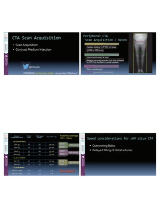
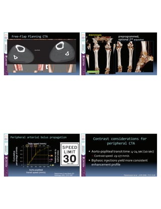
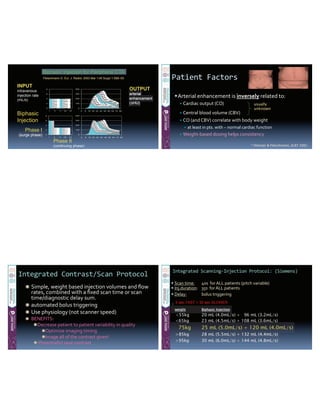
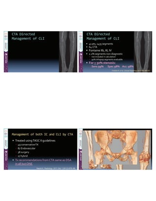
@CTterrific
DSA
DSA
• 
• 
• 
• 
@CTterrific@
• 
• 
Optional Scanning Range 2
above the kneesabove the knees toestoes
Always pre-programmed, but only initiated
by RT if no contrast in pedal vessels
Scanning Range 1
celiac artery (~T12)celiac artery ( T toes
(105 – 130 cm)
Recons:
Thin, overlapped
FOV = ggggggrererererereatatatatataterererererer trtrtrtrtrtrocococococochahahahahahantntntntntntererererererssssss
~35~35~35~35~35 mmmmmmmmm/////sssss
slslsllslowowowowow
slslsllslowowowowowslslslowowoowol
fafafastststfafafastststtf t
fafafastststt
Anatomic coverage:
105 – 130cm
fafafafafafaafastststststsststfafafafaaststststff t
vevevev ryryryryyyyyyyyyyyyyyyyyy
~~~~85858585 mm/mm/mm/mm/ssss
~~~656565 mm/mm/mm/sss
FLASH Modes
BBBBBLLLLLLAAAAAAZZZZZZIIIINNNNNNGGGGG
 
 
preprogrammed,
optional 2nd acquisition
Arteriomegaly
1st acquisition
0
0.2
0.4
0.6
0.8
1
0 30 60 90 120 150 180
p
0
0.2
0.4
0.6
0.8
1
Cumulative
percentageoflimbs
Table speed (mm/s)
Relativeriskto
outrunbolus
Aorto-popliteal
transit speed (mm/s)
0 30 60 90 120 150 180
0
< .01
~.33
> .50
Fleischmann D and Rubin GD.
Radiology 2005, 1076-1082


Fleischmann et al. JVIR 2006, 17(1) 3-26.
0
100
200
300
400
0 8 16 24 32 40 48 56 64 72 80
0
2
4
6
8
1 9 17 25 33
INPUT
intravenous
injection rate
(mL/s)
OUTPUT
arterial
enhancement
(ΔHU)
Phase I
(surge phase)
Phase II
(continuing phase)
Biphasic Injection for Peripheral CTA
0
100
200
300
400
0 8 16 24 32 40 48 56 64 72 80
0
2
4
6
8
1 9 17 25 33
Biphasic
Injection
Fleischmann D. Eur. J. Radiol. 2003 Mar 1;45 Suppl 1:S88–93.


1) Hittmair & Fleischmann, JCAT 2001
usually
unknown


weight Biphasic Injection
<55kg 20 mL (4.0mL/s) + 96 mL (3.2mL/s)
<65kg 23 mL (4.5mL/s) + 108 mL (3.6mL/s)
75kg 25 mL (5.0mL/s) + 120 mL (4.0mL/s)
>85kg 28 mL (5.5mL/s) + 132 mL (4.4mL/s)
>95kg 30 mL (6.0mL/s) + 144 mL (4.8mL/s)
5 sec FAST + 30 sec SLOWER


weight Biphasic Injection
<55 kg 20 mL (4.0mL/s) + 96 mL (3.2mL/s)
555-95 kg 25 mL (5.0mL/s) + 120 mL (4.0mL/s)
>95 kg 30 mL (6.0mL/s) + 144 mL (4.8mL/s)


Gurm HS, et al. J Am Coll Cardiol 2011; 58:907-14
 
 
** Low concentration CM (300 mgI-/mL)
Courtesy:D.Fleischmann,MD


Chapter 3: Contrast Medium Injection Technique. In: Schoepf and
Meinel, eds. Multidetector-Row CT of the Thorax (2016)


@CTterrific@


** Fleischmann D, Hallett RL, Rubin GR. JVIR 2006, 17: 3-26.
1 Meyer BC Eur Radiol (2010) 20:497-505
2 Ouwendijk R. Radiology (2006) 241, 603-608


Itoga NK et al. J Vasc Surg 2017, 835-43.
• 
• 
• 
• 
• 
Sommer Eur. Radiol (2010) 20: 2876-2881
@CTterrific@
Detection of >50%
Stenosis or Occlusion
By Anatomical Region
Performance
Met R et al. JAMA 2009;301:415-424


Napoli A. Radiology. 2011 Dec 1;261(3):976–86.


Schernthaner R, et al. AJR 2007; 189:1215-1222


Fotiadis N, et al. Clinical Radiology 2011; 66: 945-52


Napoli A. Radiology. 2011 Dec 1;261(3):976–86.
Willmann JK, et al. Radiology 2003; 229: 465-474.


1 Li X, et al. Eur J Radiol 2010; 98-103
2 Strotzer, Invest. Radiol. 2001:36(11)


1DeFreitas DJ, et al. J Vasc Surg 2013; 57(1): 5-55.
2Johnston WF, et al. J Vasc Surg 2012: 56(5) 1331-37.


@CTterrific@

Peripheral CTA

  • 1.
  • 2.
  • 3.
    @CTterrific@ •  •  Optional Scanning Range2 above the kneesabove the knees toestoes Always pre-programmed, but only initiated by RT if no contrast in pedal vessels Scanning Range 1 celiac artery (~T12)celiac artery ( T toes (105 – 130 cm) Recons: Thin, overlapped FOV = ggggggrererererereatatatatataterererererer trtrtrtrtrtrocococococochahahahahahantntntntntntererererererssssss ~35~35~35~35~35 mmmmmmmmm/////sssss slslsllslowowowowow slslsllslowowowowowslslslowowoowol fafafastststfafafastststtf t fafafastststt Anatomic coverage: 105 – 130cm fafafafafafaafastststststsststfafafafaaststststff t vevevev ryryryryyyyyyyyyyyyyyyyyy ~~~~85858585 mm/mm/mm/mm/ssss ~~~656565 mm/mm/mm/sss FLASH Modes BBBBBLLLLLLAAAAAAZZZZZZIIIINNNNNNGGGGG    
  • 4.
    preprogrammed, optional 2nd acquisition Arteriomegaly 1stacquisition 0 0.2 0.4 0.6 0.8 1 0 30 60 90 120 150 180 p 0 0.2 0.4 0.6 0.8 1 Cumulative percentageoflimbs Table speed (mm/s) Relativeriskto outrunbolus Aorto-popliteal transit speed (mm/s) 0 30 60 90 120 150 180 0 < .01 ~.33 > .50 Fleischmann D and Rubin GD. Radiology 2005, 1076-1082       Fleischmann et al. JVIR 2006, 17(1) 3-26.
  • 5.
    0 100 200 300 400 0 8 1624 32 40 48 56 64 72 80 0 2 4 6 8 1 9 17 25 33 INPUT intravenous injection rate (mL/s) OUTPUT arterial enhancement (ΔHU) Phase I (surge phase) Phase II (continuing phase) Biphasic Injection for Peripheral CTA 0 100 200 300 400 0 8 16 24 32 40 48 56 64 72 80 0 2 4 6 8 1 9 17 25 33 Biphasic Injection Fleischmann D. Eur. J. Radiol. 2003 Mar 1;45 Suppl 1:S88–93.             1) Hittmair & Fleischmann, JCAT 2001 usually unknown       weight Biphasic Injection <55kg 20 mL (4.0mL/s) + 96 mL (3.2mL/s) <65kg 23 mL (4.5mL/s) + 108 mL (3.6mL/s) 75kg 25 mL (5.0mL/s) + 120 mL (4.0mL/s) >85kg 28 mL (5.5mL/s) + 132 mL (4.4mL/s) >95kg 30 mL (6.0mL/s) + 144 mL (4.8mL/s) 5 sec FAST + 30 sec SLOWER
  • 6.
            weight Biphasic Injection <55kg 20 mL (4.0mL/s) + 96 mL (3.2mL/s) 555-95 kg 25 mL (5.0mL/s) + 120 mL (4.0mL/s) >95 kg 30 mL (6.0mL/s) + 144 mL (4.8mL/s)                   Gurm HS, et al. J Am Coll Cardiol 2011; 58:907-14
  • 7.
        ** Low concentrationCM (300 mgI-/mL) Courtesy:D.Fleischmann,MD       Chapter 3: Contrast Medium Injection Technique. In: Schoepf and Meinel, eds. Multidetector-Row CT of the Thorax (2016)              
  • 8.
    @CTterrific@             ** Fleischmann D,Hallett RL, Rubin GR. JVIR 2006, 17: 3-26.
  • 9.
    1 Meyer BCEur Radiol (2010) 20:497-505 2 Ouwendijk R. Radiology (2006) 241, 603-608           Itoga NK et al. J Vasc Surg 2017, 835-43. •  •  •  •  •  Sommer Eur. Radiol (2010) 20: 2876-2881 @CTterrific@
  • 10.
    Detection of >50% Stenosisor Occlusion By Anatomical Region Performance Met R et al. JAMA 2009;301:415-424             Napoli A. Radiology. 2011 Dec 1;261(3):976–86.           Schernthaner R, et al. AJR 2007; 189:1215-1222
  • 11.
                    Fotiadis N, etal. Clinical Radiology 2011; 66: 945-52             Napoli A. Radiology. 2011 Dec 1;261(3):976–86.
  • 12.
    Willmann JK, etal. Radiology 2003; 229: 465-474.           1 Li X, et al. Eur J Radiol 2010; 98-103 2 Strotzer, Invest. Radiol. 2001:36(11)
  • 13.
            1DeFreitas DJ, etal. J Vasc Surg 2013; 57(1): 5-55. 2Johnston WF, et al. J Vasc Surg 2012: 56(5) 1331-37.             @CTterrific@