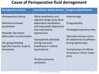
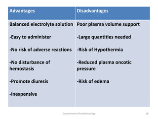
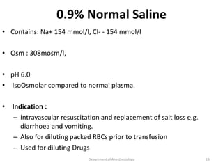
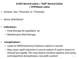
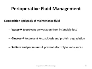
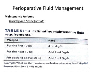
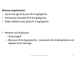
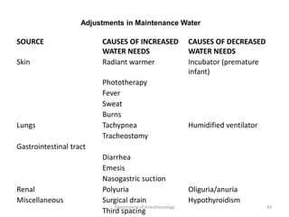
2. Objectives
Introduction
Physiology
Surgical fluid requirement
Fluid Replacement
Assessment and Monitoring
Crystalloid & Colloid
Department of Anesthesiology 2
3. Why do we need Fluid?
• Avoid dehydration
• Maintain adequate effective circulating volume
• Prevent inadequate tissue perfusion
• Maintain distribution of electrolytes
Department of Anesthesiology 3
4. Introduction
• Venous access
• Assess & Correction
• Errors leads to morbidity or mortality.
Department of Anesthesiology 4
7. • Osmolality: 285-290 mOsm/kg (both ECF and ICF)
• ECF: Sodium, Bicarbonate and Chloride
• ICF: Potassium and Magnesium
• ECF: volume controlled mainly by- Sodium
– ( Sensors:- Baroreceptors, Atrial stretch receptors,
Juxtaglomerular apparatus)
• Osmolarity
– controlled mainly by:- varying water intake and excretion
– ( Sensors:- Hypothalamus)
7
Department of Anesthesiology
8. Evaluation of Intravascular volume
• Intravascular volume can be estimated using
patient history
physical examination
laboratory evaluation
often with the aid of sophisticated hemodynamic
monitoring techniques.
Department of Anesthesiology 8
9. Patient History
• Oral intake
• Persistent vomiting or diarrhea
• Gastric suction
• Significant blood loss or wound drainage
• Intravenous fluid and blood administration
• Recent hemodialysis
Department of Anesthesiology 9
10. Physical Examination
• Indications of hypovolemia include
abnormal skin turgor
dehydration of mucous membranes
thready peripheral pulses
increased resting heart rate
decreased blood pressure
orthostatic heart rate and blood pressure changes from
the supine to sitting or standing positions
decreased urinary flow rate
Department of Anesthesiology 10
11. • Signs of excess extracellular water and likely
hypervolemia in patients with normal cardiac, hepatic,
and renal function
– Pitting edema
– Increased urinary flow
• Late signs of hypervolemia in settings such as congestive
heart failure may include
– Tachycardia
– Tachypnea
– Elevated jugular pulse pressure
– Pulmonary crackles,
– Wheezing
– Cyanosis
– Pink, frothy pulmonary secretions
Department of Anesthesiology 11
13. Laboratory Evaluation
• Laboratory signs of dehydration may include:
– Rising hematocrit and hemoglobin
– Progressive metabolic acidosis (including lactic acidosis)
– Urinary specific gravity greater than 1.010
– Urinary sodium less than 10 mEq/L
– Urinary osmolality greater than 450 mOsm/L
– Hypernatremia and
– BUN-to creatinine ratio greater than 10:1
• Radiographic indicators of volume overload include:
– Increased pulmonary vascular and interstitial markings (Kerley
“B” lines), diffuse alveolar infiltrates or both.
Department of Anesthesiology 13
14. Hemodynamic Measurements
Static measures Dynamic measures
Central venous pressure
Pulmonary artery occlusion
pressure
Lt ventricular end diastolic area
IVC diameter
Pulse pressure variation
Stroke volume variation(< 10 – 15 %
for pts on controlled ventilation)
Dynamic changes in aortic
flow velocity/sv assessed by echo
Positive pressure ventilation
changes in venacaval diameter
Department of Anesthesiology 14
15. Cause of Perioperative fluid derragement
Perioperative factors Anesthesia related factors Surgery related factors
•Peroperative fasting
•Mechanical bowel
preparation
•Disorder like bowel
obstruction or pancreatitis
•On going bleeding
typically requires surgical
hemostasis
•Most anesthetics and
adjuvant drugs cause dose
dependent vasodilation
and myocardial depression
that may lead to
hypotension
•Sympathetic blockade
during neuraxial
anesthesia in relative
hypovolemia
•Positive pressure
ventilation
•Haemorrage
•Coagulopathy
•Prolonged operative time
•Decrease venous return
d/t abdominal insufflation
during laparoscopy
•Compression of inferior
venacava or others major
veins
Department of Anesthesiology 15
17. Crystalloids
• Clear fluids made up of water and electrolyte solutions
• Will cross a semi-permeable membrane
• Grouped as isotonic, hypertonic, and hypotonic Eg:
– Normal saline 0.9%,3 %
– Dextrose solutions 5 %,10%,20%,25%
– DNS
– Ringer’s lactate
– Isolyte P
Department of Anesthesiology 17
18. Advantages Disadvantages
Balanced electrolyte solution
-Easy to administer
-No risk of adverse reactions
-No disturbance of
hemostasis
-Promote diuresis
-Inexpensive
Poor plasma volume support
-Large quantities needed
-Risk of Hypothermia
-Reduced plasma oncotic
pressure
-Risk of edema
Department of Anesthesiology 18
19. 0.9% Normal Saline
• Contains: Na+ 154 mmol/l, Cl- - 154 mmol/l
• Osm : 308mosm/l,
• pH 6.0
• IsoOsmolar compared to normal plasma.
• Indication :
– Intravascular resuscitation and replacement of salt loss e.g.
diarrhoea and vomiting.
– Also for diluting packed RBCs prior to transfusion
– Used for diluting Drugs
Department of Anesthesiology 19
20. • Distribution:
– Stays almost entirely in the extracellular space.
– Of 1 litre - 750ml extra vascular fluid; 250ml intravascular fluid
– 100ml blood loss – need to give 400ml N. saline [only 25%remains
intravascular
• Complications:
– When given in large volume can produces Hyperchloremic
metabolic acidosis because of highNa+ and Cl- content.
Department of Anesthesiology 20
21. 0.45% Normal saline = ‘Half’ Normal Saline
= HYPOtonic saline
• Cointais- Na+ 77mmol/l, Cl- 77mmol/l,
• Osmo 154mOsm/l
• Indications :
– Fluid therapy for paediatric pt
– Maintenance fluid therapy
• Complications :
– Leads to HYPOnatraemia if plasma sodium is normal
– May cause rapid reduction in serum sodium if used in excess or
infused too rapidly. This may lead to cerebral oedema and rarely,
centralpontine demyelinosis ; Use with caution!
Department of Anesthesiology 21
22. 3.0 % Saline = Hypertonic
saline
• 3% contain 513 mmol/l of Na+ and Cl- each,
• osmol of 1026 mOsm/l
• pH 5.0
• Indications :
– Treatment of severe symptomatic hyponatremia(coma, seizure)
– To resuscitate hypovolemic shock
Department of Anesthesiology 22
23. • Leads to an osmotic gradient between the ECF and ICF, causing
passage of fluid into the Extracellular space.
• Must be administered slowly and preferably with CV line because it
carries risk of causing
– phlebitis
– Necrosis
– hemolysis.
• Complications :
– Precaution in pt. with CHF
– severe renal insufficiency, edema with sod. retention.
Department of Anesthesiology 23
24. Dextrose
5% Dextrose (often written D5W):
• 50g/l of glucose, 252mOsm/l, pH 4.5
• Regarded as ‘electrolyte free’ – contains NO Sodium,
Potassium,Chloride or Calcium
• Indication :
– Primarily used to maintain water balance in patients who are not
able to take anything by mouth;
– Commonly used post-operatively in conjunction with salt retaining
fluids ie saline
– Hypernatremia treatment
Department of Anesthesiology 24
25. • When infused is rapidly redistributed into the intracellular space
• Side effects:
– Iatrogenic hyponatraemia in surgical patient
– Hyperglycemia
– Not compatible with blood ,cause hemolysis
– less than 10% stays in the intravascular space therefore it is of
limited use in fluid resuscitation
Cocn 5% 10% 20% 25% plasma
osmolarity 225 505 1010 1262 290
Department of Anesthesiology 25
26. Ringer Lactate
• Most physiological solution
• Electrolyte composition similar to ECF
• One litre of lactated Ringer's solution contains:
– Sodium ion= 130 mmol/L
– Chloride ion = 109 mmol/L
– Lactate = 28 mmol/L
– Potassium ion = 4 mmol/L
– Calcium ion = 1.5 mmol/L
– Osmolarity of 273 , pH of 6.5
Department of Anesthesiology 26
27. • Lactate is converted to bicarbonate in liver
• Indications :
– Deficit ,Intraoperative fluid loss
– Severe hypovolemia
• Precautions:
– Severe metabolic acidosis ( impaired lactate conversion)
– Don’t give with blood product ( Ca bind with citrate - reduced
anticoagulant activity )
Department of Anesthesiology 27
28. DNS
• 0.9% saline & 5% dextrose
• Contains - Na+ 154, Cl- 154, 5 gm, Glucose
• Osm : 432 mosm/L
• Indication :
– Maintenance solution
– Correction of fluid deficit with supply of energy
– Compatible with blood
Department of Anesthesiology 28
30. Colloids
• The colloid solutions contain particles which do not readily cross
semi-permeable membranes such as the capillary membrane.
• Thus the volume infused stays (initially) almost entirely within the
intravascular space .
• Stay intravascular for a prolonged period compared to crystalloids.
• However they leak out of the intravascular space when the capillary
permeability significantly changes e.g. Severe trauma or sepsis.
Department of Anesthesiology 30
31. • Because of their gelatinous properties they cause platelet
dysfunction and interfere with fibrinolysis and coagulation factors
(factor VIII) – thus they can cause significant coagulopathy in large
volumes.
• Natural : Albumin
• Artificial : Gelatin and Dextran , Hydroxyethyl starches(HES)
Department of Anesthesiology 31
32. colloids
Avantages Disadvantages
-Prolonged plasma volume support
-Moderate volume needed
-minimal risk of tissue edema
-enhances microvascular flow
-Risk of volume overload
-Adverse effect on haemostasis
-Anaphylactic reaction
- Expensive
Department of Anesthesiology 32
33. ALBUMIN
• Principal natural colloid comprising of 50-60% of all plasma proteins.
• Synthesized only in liver and has a half life of app. 20 days.
• 5% soln is iso oncotic and leads to 80% initial vol expansion ,25%
soln leads to 200-400% increase in vol.
• Used
– For emergency treatment of shock especially due to loss of plasma
– acute management of burns
– fluid resuscitation in ICU
– Hypoalbumineamia
Department of Anesthesiology 33
34. • Side effects :
– Pruritis
– anaphylactoid reactions and
– coagulation abnormalities as compared to synthetic
colloids.
• Disadvantages
– cost effectiveness
– volume overload (in septic shock pt albumin add to
interstitial edema)
Department of Anesthesiology 34
35. Colloid or Crystalloid Resuscitation
• Recommendations:
– Colloid should NOT be used as the sole fluid replacement
in resuscitation ,volumes infused should be limited
because of side effects and lack of evidence for their
continued use in the acutely ill.
• In severely ill patients –
– principally use crystalloid and blood products
– Colloid may be used in limited volume to reduce volume
of fluids required or until blood products are available.
Department of Anesthesiology 35
36. • In elective surgical patients
– Replace fluid loss with ‘physiological Ringer’s solutions.
– Blood products and colloid may be needed to replace
intravascular volume acutely.
Department of Anesthesiology 36
38. • Figure 51-2 morgan
Department of Anesthesiology 38
39. Perioperative Fluid Management
• Goals
– Replace pre-operative deficits
– Provide normal maintenance requirements
– Replace any intraoperative loss
39
Department of Anesthesiology
40. Perioperative Fluid Management
Composition and goals of maintenance fluid
– Water to prevent dehydration from insensible loss
– Glucose to prevent ketoacidosis and protein degradation
– Sodium and potassium prevent electrolyte imbalances
Department of Anesthesiology 40
42. Glucose requirement:
• Up to the age of 8 years 6 mg/kg/min
• Premature neonates 6–8 mg/kg/min.
• Older children and adults 2 mg/kg/min
• Routine use of glucose
– discouraged
– Because of its Hypotonicity , increased risk of dehydration and
hypoxic brain damage.
42
Department of Anesthesiology
43. SOURCE CAUSES OF INCREASED
WATER NEEDS
CAUSES OF DECREASED
WATER NEEDS
Skin Radiant warmer Incubator (premature
infant)
Phototherapy
Fever
Sweat
Burns
Lungs Tachypnea Humidified ventilator
Tracheostomy
Gastrointestinal tract
Diarrhea
Emesis
Nasogastric suction
Renal Polyuria Oliguria/anuria
Miscellaneous Surgical drain Hypothyroidism
Third spacing
Adjustments in Maintenance Water
Department of Anesthesiology 43
44. Deficit
• Deficit = number of hours NPO x maintenance fluid requirement.
• 50% replaced in first hour and 50% in next 2 hours
• Deficit due to fever, vomiting, blood loss, diarrhea
• Degree of dehydration should be assessed and fluid replaced
• Eg:-
• 70 kg pt fasting for 8 hrs
• Deficit : 8 X 110 = 880 ml
Department of Anesthesiology 44
45. Intra Operative Fluid Management
Intraoperative losses
• Third space loss and blood loss
• Third space loss
– isotonic transfer of fluid from the ECF to a non-
functional interstitial compartment
– surgical trauma, burn, infection
– The volume lost is impossible to measure
– Estimated by the extent of surgery and the
clinical response to appropriate fluid
replacement 45
Department of Anesthesiology
46. Intra Operative Fluid Management
• Type of fluid NS or RL
• Maintain adequate blood pressure, heart rate, and urine
output of 1-2ml/kg/min
46
Department of Anesthesiology
British Journal of Anaesthesia | CEPD Reviews | Volume 3 Number 1 2006
47. Blood loss
47
Department of Anesthesiology
British Journal of Anaesthesia | CEPD Reviews | Volume 3 Number 1 2003
48. Intra Operative Fluid Management
• Allowable blood loss
(MABL-Maximum allowable blood loss)
(EBV- Estimated blood volume)
• Initial replacement should be with crystalloid in a ratio of 3:1 or
with colloid in a ratio of 1:1
• Platelets and fresh frozen plasma, 10–15 mL/kg, should be given
when blood loss exceeds 1–2 blood volumes
48
Department of Anesthesiology
49. • The transfusion point can be determined preoperatively from
the hematocrit and by estimating blood loss.
• Patient with normal hematocrit should only be transfused
after loss greater than 10 – 20 % of their blood loss.
• The exact point is based on patient condition and the surgical
procedure.
Department of Anesthesiology 49
50. • The amount of blood loss necessary for the hematocrit to fall to 30%
can be calculated as follows:-
– Estimate blood volume
– Estimate the RBC volume at the preoperative hematocrit
– Estimate the RBC volume at the hematocrit of 30%, assuming
normal blood volume maintained.
– Calculate the RBCV lost when the hematocrit is 30%:
• RBCVlost = RBCVpreop – RBCV30%
– Alloiwable blood loss = RBCVlost X 3
Department of Anesthesiology 50
51. • Example:
An 85 kg women has a preoperative hematocrit of 35%. How much
blood loss will decrease her hematocrit to 30% ?
Here,
estimated blood volume = 65ml/kg X 85 kg = 5525 ml
RBCV35% = 5525 X 35% = 1934 ml
RBCV30% = 5525 X 30% = 11658 ml
RBCVlost at 30% = 1934 – 1658 = 276 ml
allowable blood loss = 3 X 276 ml = 828 ml
• There fore, transfusion should be considered only when this patient’s
blood loss exceeds 800 ml.
• Increasingly, transfusion is not recommended until the hematocrit
decreases to 24% of lower (Hb < 8 g/dl), but it is necessary to note
the rate of blood loss and comorbid conditions.
Department of Anesthesiology 51
52. • Clinical guideline commonly used include:
– One unit of RBC raises hemoglobin by 1g/dL (or raises HCT 3%)
– 10 mL/kg transfusion of RBC will Increases the hemoglobin by
about 3 g/dL and hematocrit by 10%
Department of Anesthesiology 52
A reduction in ECF volume causes non-osmotic ADH release, stimulation of the sympathetic nervous system to cause vasoconstriction, activation of the renin-angiotensin-aldosterone system
Increase in blood volume and atrial pressure – release of ANP- vasodilatation and decrease sodium reabsorption at collecting tubes. Dilates afferent renal arterioles, constrict efferent arterioles promoting diuresis and contributing aldosterone escape mechanism.
A rise in ECF osmolality triggers the sensation of thirst and causes release of ADH, which increases water re-absorption at the renal collecting ducts.
Pulmonary artery occlusion pressure monitoring
< 8 mm Hg indicate hypovolemia
< 15 mm Hg may be associated with relative hypovolemia in patients with poor venticular compliance
> 18 mm Hg generally imply left venticular volume overload
Lactated Ringer’s solute on with 1% dextrose is sufficient to prevent both hypoglycemia and hyperglycemia in most children
Propranolol-related hypoglycaemia All these patients should have glucose-containing maintenance fluids and intra-operative checks of blood sugar
10–15% increase in maintenance water needs for each 1°C increase in temperature above 38°C.