Download to read offline
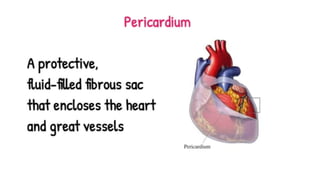
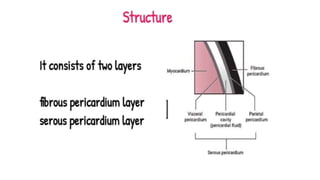
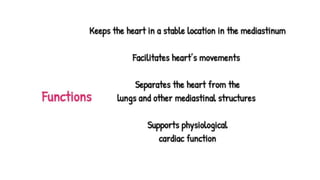
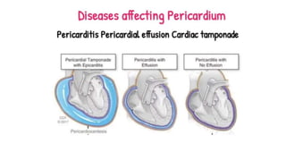
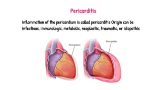

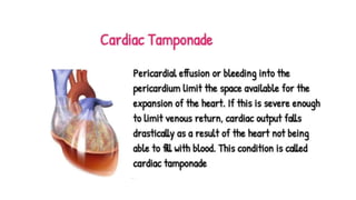


Introduction The pericardium is a fibrous sac that encloses the heart and great vessels. It keeps the heart in a stable location in the mediastinum, facilitates its movements, and separates it from the lungs and other mediastinal structures. It also supports physiological cardiac function. Structure and Function The pericardium consists of two layers: the fibrous and the serous. The fibrous pericardium is a conical-shaped sac. Its apex is fused with the roots of the great vessels at the base of the heart. Its broad base overlies the central fibrous area of the diaphragm with which it is fused. Weak sterno-pericardial ligaments connect the anterior aspect of the fibrous pericardium to the sternum. The serous pericardium is a layer of serosa that lines the fibrous pericardium (parietal layer), which is reflected around the roots of the great vessels to cover the entire surface of the heart (visceral layer). Between the parietal and visceral layers is a potential space that may be filled with a small amount of fluid. The part of the visceral layer that covers the heart, but not the great vessels is called the epicardium. As the serous pericardium reflects off various cardiac structures, it forms two sinuses: the transverse sinus and the oblique sinus. The oblique sinus is a cul-de-sac extending superiorly from the inferior vena cava between the two left pulmonary veins on one side and the two right pulmonary veins on the other. Its anterior wall is formed by the posterior wall of the left atrium, between the four pulmonary veins. The oblique sinus provides expansion space for the left atrium. The transverse sinus is open at both ends and formed by the reflection of visceral serosal pericardium from the posterior aspects of the aortic and pulmonary trunks over to the anterior aspect of the atrium. Thus, a finger in the transverse sinus will pass behind the aortic and pulmonary trunks, in front of the superior vena cava on the right, and the left atrial appendage on the left. The pericardial sac positions the heart in the mediastinum and limits its motion while providing a lubricated slippery surface for the heart to beat inside and the lungs to move outside. The pericardium prevents the excessive dilatation of the heart, and in pathological states, it can limit the overfilling of the heart, which would result in low cardiac output. It also influences the pressure-volume relationships of cardiac chambers by providing limited space for the heart as a whole. The pericardium also equalizes hydrostatic, inertial, and gravitational forces maintain the geometry of the left ventricle, and acts as a mechanical barrier to infection. Pericarditis Inflammation of the pericardium is called pericarditis. Its origin can be infectious, immunologic, metabolic, neoplastic, traumatic, or idiopathic. A myocardial infarction can also cause localized pericarditis of the area overlying the infarct.
![QMS SOP [QUALITY MANAGEMENT SYSTEM - STANDARD OPERATING PROCEDURE]](https://cdn.slidesharecdn.com/ss_thumbnails/qmssop-231128160542-8db367a9-thumbnail.jpg?width=640&height=640&fit=bounds)


