1. Unraveling Prion Function
and Disease Pathogenesis:
Prion Proteins, The Unfolded Protein Response,
and Metal Ions.
Daniel Twohig
#2205807
Supervisor: Dr. J.J.M. Hoozemans, PhD
Second Assessor: Prof. Dr. J.M. Rozemuller, MD, PhD
VU University, Amsterdam
August 2013
2. 2
Abstract
The prion protein (PrP) is a small molecule implicated in novel forms of
neurodegenerative disease. Revealing the pathogenesis of prion related diseases is
further complicated by the fact that the native function of PrP is unclear. Two areas of
inquiry have helped further our basic understanding of PrP pathogenesis and its native
function being: i) the endoplasmic reticulum’s unfolded protein response (UPR), and ii)
the interactions of PrP with divalent transition metal cations. Herein we review the
current literature pertaining to PrP pathogenesis and the UPR, as well as PrP metal ion
interactions, while also attempting to establish a bridge between the two subjects.
3. 3
INDEX
i. Abstract 2
1. Introduction 4
2. The Prion Protein Genetics and Structure 6
3. The ER and the UPR 7
4. UPR mechanisms 9
4.1. IRE1 9
4.2. PERK 10
4.3. ATF6 11
5. The UPR and Prions 13
5.1. Caspase-12 13
5.2. Protein Disulfide Isomerase&Grp 14
5.3. XBP1/Xbp1s 15
5.4. eIF2α 17
5.5. Snord 3A 18
5.6. ORs and the UPR 19
6. Metal Ions and Prions 19
6.1 The Ors and Cu(II)/Cu2+ Binding 21
6.2 Zn(II)/Zn2+ 25
6.3 Metal Ions, The UPR, and Prions 26
7. Conclusions 28
8. References 28
4. 4
1. Introduction
The prion protein (PrP) is a small, highly conserved protein expressed in vertebrates,
invertebrates, yeast, and fungi1. Prion diseases, also known as transmissible spongiform
encephalopathies (TSEs) or prionopathies, are utterly fatal neurodegenerative disorders
which arise either sporadically, genetically, or are acquired via direct contact with
infectious PrP species2. TSEs are thought to propagate via an unknown, unprecedented,
and nucleic acid free mechanism in which the primarily α-helical native form of PrP
(PrPC) becomes misfolded in the presence of a robust, misfolded, and protease resistant
β-sheet enriched conformer of PrPC named PrPSc1. Theoretically, prion biology resides
in an ethereal mist, in that current scientific dogmas and assays cannot explain the
native function of PrPC or the pathogenesis of PrPSc. Powerful tools such as nuclear
magnetic resonance spectroscopy (NMR) have allowed for the structure of PrPC to be
determined3, but we still fail to understand the most basic mechanistic principals of this
small chain of amino acids.
TSEs affect humans as Creutzfeldt-Jakob disease (CJD) of which there are
familial (fCJD)4, iatrogenic (iCJD)5, sporadic (sCJD)6 and variant (vCJD) forms7; kuru8,9;
fatal familial insomnia (FFI)4; and Gerstmann-Sträussler-Scheinker syndrome (GSS)10.
A wide variety of animals also contract TSEs such as cattle (bovine spongiform
encephalopathy (BSE))11, sheep (scrapie)12, deer and elk (chronic wasting disease)13, cats
(feline spongiform encephalopathy, minks (transmissible mink encephalopathy), and
some antelope species (exotic ungulate encephalopathy)14. Clinically TSEs are enigmatic
in that they display heterogeneous clinical presentations and pathogenesis15–18, which is
further complicated by the fact that 30 different mutations have been linked to inherited
forms of the TSEs19,20. If this isn’t intimidating enough, the Aguzzi group reported
5. 5
100% of mice exposed to 60 seconds of aerosol mists enriched with PrPSc develop
prionopathies21. Additionally, the report found a troubling correlation between PrPSc
aerosol exposure and the onset of TSE, simply stated, mice exposed to longer durations
of PrPSc aerosol develop TSEs faster21.
Currently there is a compelling line of research focused on the endoplasmic
reticulum’s (ER’s) unfolded protein response (UPR) as a possible mediator of TSE
pathogenesis22,23. The UPR is a robust system which is able to strongly influence
protein synthesis, modification, quality control, and degradation when disruptions in
protein production/degradation arise24,25. Another convincing body of literature
suggests that the native role of PrP is to chelate metal ions in order to mediate metal
ion trafficking across the plasma membrane, or to co-interact with metals ions and
receptors26,27.
The following analysis provides a basis of prion genetics and biology, and the
current findings regarding; i) the UPR in TSEs, and ii) the binding of PrP to copper and
zinc ions.
2. Genetics & Structure
Mature PrPC is abundantly expressed in both neurons and astrocytes and encoded by a
single open reading frame of one exon (exon 3) on the 16kB PRNP gene located on the
short arm of chromosome 20 at position 13 (20p13)28. Translation of the PRNP exon
results in a 253-residue precursor protein. Subsequent post-translational modifications
within the ER remove the last 22 N-terminal residues, the last 24 C-terminal residues,
6. 6
and installs a C-terminal glycosyl phosphatidylinositol (GPI) anchor, resulting in a
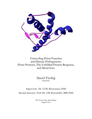
mature 208-209 residue protein which is bound to the plasma membrane (Fig. 1)29.
The secondary structure of PrPC can be divided between the unstructured N-
terminal from residues 23 to 126, and the primarily α-helical C-terminal between
residues 126 to 231 (Fig. 1B-D). The unstructured N-terminal’s most striking feature
are four identical repeats of the eight residue sequence PHGGGWGQ found between
residues 51 and 91 called the octarepeat (OR) domain (Fig. 1A-C). A charged cluster of
residues between T95 and K110 lies beside a hydrophobic domain between H111 and
M134, after which the C-terminal begins (Fig. 1A-B,D). The globular C-terminal has
three α-helices, two small β-sheets, a disulfide bond (between C179 and C 214), two
asparagine residues (N181 and N197) which can express varying degrees of
glycosylation (being either un-, mono-, or di-glycosylated), and a terminal GPI anchor
(Fig. 1A-B,D)30–32.
The difficulty in assaying prions poses another conundrum. Prions are severely
hydrophobic, yet easily disintegrate when being purified. Currently, the only acceptable
assay for PrPSc utilizes their insolubility in detergents, and the fragmentation induced
by proteinase K (PK) digestion1. PK exposure removes 60-70 N-terminal residues from
PrP leaving a PK-resistant core of approximately 142 residues denoted as PrP 27-302.
However, correct identification and PrPSc and PrP 27-30 are troublesome because, i)
PrPSc has yet to be structurally characterized because it has yet to be isolated, ii) there
are four distinct sCJD and iCJD sub-classifications correlating to four PrPSc species
with different sensitivities to PK-digestion33; iii) some pathologically typical sCJD cases
show no observable PK-resistant PrPSc34,35; and iii) the co-existence of different PK-
7. 7
resistant strains in the same sCJD individual36. Together these protease sensitive TSEs
have been termed variably protease-sensitive prionopathies34.
The variability to PK digestion exemplifies the general complexity of prion
research, furthermore, there is a uniform lack of consensus about i) a standard
lexicon/terminology for PrP researchers, ii) correlating TSE genotypes to their
respective phenotypes, or iii) the correct model systems to use for TSE research which
all serve to hinder the progression of this field37–40.
3. The ER and the UPR
The ER is an key organelle involved in protein folding, protein translation, protein
degradation, phospholipid and sterol synthesis41. The ER possess a rigorous quality
control system termed the unfolded protein response (UPR) that can respond in many
ways to perturbations in ER homeostasis (a.k.a. ER stress)42. Activation of the UPR can
induce mechanisms that: i) attenuate transcription and translation, ii) upregulate the
expression of folding enzymes (e.g. isomerases) and molecular chaperones, iii) activate
the ER associated protein degradation response (ERAD), iv) induce new phospholipid
synthesis to increase the volume of the ER, and v) upregulate pro-apoptosis genes43.
Clinically, mechanisms of the UPR have been suggested to be involved in Parkinson’s
disease44, Alzheimer’s disease45, Amyotrophic lateral sclerosis46, and TSEs22.
The following sections will summarize the mechanisms of the UPR, the links
between the UPR and PrPC, and identify components of the UPR that have been studied
in TSE research.
8. 8
4. UPR mechanisms
The ER has three inherent transmembrane stress sensors (Fig. 2): i) the inositol-
requiring transmembrane kinase/endoribonuclease 1 (IRE1), ii) the double-stranded
RNA (PKR)-activated protein kinase-like eukaryotic initiation factor 2α kinase (PERK),
and iii) the activating transcription factor-6 (ATF6). Activation of the UPR sensors
occurs when the binding immunoglobulin protein (BiP or glucose-regulated protein 78
(Grp78)), an ER luminal chaperone, dissociates from the stress sensors and binds to
misfolded proteins47. Part of the 70 kilodalton heat shock protein family, BiP strongly
binds to misfolded and improperly modified proteins which are not able to translocate
9. 9
out of the ER48. Although BiP-misfolded protein binding induces UPR activation,
recent evidence has also shown that lipids can also activate modified IRE1 and PERK
lacking a luminal sensor module49.
4.1. IRE1
Of all the UPR sensors, IRE1 is the most highly conserved50,51. Two forms of
mammalian IRE1 exist, IRE1α and IRE1β52. IRE1α is ubiquitously expressed in all
cells, while IRE1β is only found in intestinal epithelial cells, thus, for the purposes of
this review, we will only focus on IRE1α. The structure of IRE1α consists of a ER
lumenal domain which binds to BiP, and a bifunctional cytoplasmic domain possessing
both a kinase and anendoribonuclease (Fig. 2A)50,53 . Upon lumenal activation of IRE1α
it oligomerizes and autophosphorylates itself, termed trans-autophosphorylation54. The
trans-autophosphorylation activates the endoribonuclease module, inducing the cleavage
of 26-nucleotides from the X-box binding protein-1 (XBP1 or HAC1) mRNA within the
cytoplasm (Fig. 2A)55. Translation of this truncated form of XBP1 produces a potent
transcription factor known as Xbp1s, which is transported to the nucleus where it is
able to upregulate genes involved in ER protein folding, protein secretion from the ER,
ER membrane synthesis, and the ERAD (Fig. 2A)24.
4.2. PERK
PERK resembles IRE1 in that it has a phylogenetically and structurally related luminal
domain that is activated by BiP dissociation, and also a cytosolic kinase module 56.
Upon activation of the luminal domain, PERK oligomerizes and undergoes trans-
autophosphorylation of its cytosolic domains, while additionally phosphorylating the
10. 10
important translation initiation factor eIF2α (Fig. 2B)24. The phosphorylation of eIF2α
(eIF2α-P) greatly reduces global protein translation, effectively reducing the volume of
new proteins being trafficked to the ER57. In spite of this, some mRNAs show an
increase in translation when elF2α is phosphorylated such as the transcription factor
ATF4. ATF4 initiates the upregulation of a host of genes58 such as: 1) the apoptotic
gene transcription factor C/EBP homologous protein (CHOP/GADD153)59, and 2) the
growth arrest and DNA damage inducible 34 (GADD34) gene which leads to de-
phosphorylation of eIF2α (Fig. 2B)60. The upregulation of CHOP is important during
ER-stress because it activates the transcription pro-apoptotic components like BIM61
and PUMA62. The actions of ATF4 thus initiate a negative regulatory feedback loop
where pro-apoptotic genes (CHOP) are upregulated at the same time GADD34 induces
elF2α–P desphosphorylation to allow for the synthesis of CHOP targeted genes. This
feedback loop ensures that cells can still remain viable even under stress by maintaining
a basal level of protein synthesis required to make essential and pro-UPR proteins.
4.3. ATF6
The transmembrane protein ATF6 possesses a large and unique luminal domain at its
C-terminal, that when activated via BiP dissociation, causes the vesicular translocation
of ATF6 to the Golgi apparatus (Fig. 2C)63. Within the Golgi, ATF6 is cleaved by two
proteases, site-1 protease (S1P) cleaves the luminal domain, and site-2 protease (S2P)
removes the transmembrane anchor64,65. The freed cytosolic N-terminal portion
(ATF6(N)) then travels to the nucleus where it acts as a transcription factor for XBP1
and chaperones such as BiP, glucose-related protein 94 (GRP94), and protein disulfide
isomerase (PDI)25,55.
11. 11
5. The UPR & Prions
5.1. Caspase-12
One of the first experiments to implicate the UPR in TSEs used i) neuroblastoma cells
cultured with nanomolar amounts of PrPSc, and ii) post-mortem samples from vCJD and
fCJD patients. They found a significant upregulation of caspase-12 (C12) (a ER cysteine
protease), and ER chaperones with PDI activity called the glucose regulated family
proteins (Grps)66, all of which function as part of the UPR. Subsequent investigations
by the same group found C12 was only active during the terminal phase of the disease in
which neuronal loss occurs suggesting that C12 is not an early mediator of TSE
neurotoxicity67, which was also confirmed by a recent report22. Furthermore, studies
done using C12 knockout mice (C12-/-) inoculated with PrPSc found no significant
differences in behavior, survival, pathology, or accumulation of PrPSc compared to wild-
type animals, thus questioning the notion that C12 could mediate prion
neurodegeneration68. It is worth noting that the latter study used mice exposed to the
RML (Rocky Mountain Laboratory Chandler strain) strain of PrPSc, while the former
study used the 139A strain. Although these two strains were once thought to be the
same, a recent report using the extended cell panel assay (ECPA) strongly suggests
otherwise69 (for a comprehensive review of transgenic mouse models of prion diseases
see Groschup et al.70).
5.2. Protein Disulfide Isomerase and Grps
The expression of PDI and BiP/Grp78 have been shown to be elevated in the pre-
symptomatic stages of hamster TSE67. Further investigation into PDI and PDI-like
proteins found elevated levels in murine models of TSE, and overexpression of PDI in
12. 12
HEK cells reduced ER-stress induced by an ER-localized PrP construct71. In the same
study it was also found that a knockdown of endogenous PDI rescued cellular apoptosis
caused by a PrP mutant with 10 extra ORs. The authors concluded that PDI/PDI-like
proteins have complex and pleiotropic effects which could be neuroprotective at the
beginning stages of disease, yet possibly apoptosis inducing during the terminal
stages71, this is not very consoling because in general the UPR is thought to work in the
same way (e.g. protect cells during the initial disease stages, and destroy cells at the end
stages)25. Although interesting targets for study have arose from this paper, it
highlights the inherent differences between PrP mutants, and the difficulty that these
structural differences theoretically pose when assessing their varying responses.
The activation of a chaperone called Grp58 (a.k.a. PDIA3) which is a structural
homolog to PDI has been strongly correlated with prion disease pathogenesis67,72. The
upregulation of Grp58 been observed in neuroblastoma cells infected with PrPSc,
rodents inoculated with scrapie, and humans patients with vCJD and sCJD. Using the
139A PrPSc analog to inoculate wild type mice and infect N2a cells, a 2005 article by
Hetzet. al. found early activation of the UPR via expression of Grp5867. Grp58 may
have a neuroprotective role in that blocking Grp58 expression using siRNA in N2a cells
increased PrPSc toxicity, and overexpression of Grp58 reduced PrPSc toxicity and C12
activation. Other members of the Grp family were observed (Grp78, and -94) but were
only transiently expressed, with no correlation to PrPSc accumulation.
13. 13
.
Thus it seems that Grp58 could be an important protein for the reduction of PrPSc
toxicity, however, attempts to utilize it as a clinical marker have met with less than
conclusive results73.
14. 14
5.3. XBP1/Xbp1s
The role of the evolutionary conserved IREα-Xbp1s UPR pathway has been studied
extensively and been shown to be actively engaged in models of neurodegeneration and
nervous system injury including Parkinson’s disease74, Huntington’s disease75, ALS76,
brain ischemia77, brain trauma78, and spinal cord injury79.
Studies by Orsi and co-workers implicated the activation of the IREα-Xbp1s
pathway in maintaining proper translocation of PrP to the ER80. Due to its weak
targeting signal, PrPC can be improperly post-translationally modified and become an
un-anchored cytosolic species during periods of ER-stress81. Mouse models
demonstrate that a lack of Xbp1 induces a higher fraction of aggregation prone,
cytosolic PrPC, which can be rescued by an overexpression of active Xbp180.
Hetz et al. reported complementary findings by suggesting that ER-stress
increased the replication of PrPSc-prone PrPC (e.g. PrPC forms PrPSc more easily under
ER-stress conditions), which could be ameliorated by overexpression of XBP1 (or
ATF6)82. Thus both above studies offered the tantalizing notion that ATF6-Xbp1
signaling may mediate PrPC replication, PrPC translocation, and PrPSc formation.
In a follow up study, Hetz et al. were confronted by confounding results that
contradicted their earlier report by using murine models with a brain-specific XBP1
knock-out (KO) (XBP-1Nes-/-)83. Upon exposure to murine PrPSc, XBP-1Nes-/- mice had
no significant differences in stress response, survival, neuronal loss, or PrPSc
aggregation compared to wild-type mice. This suggests that although this arm of the
UPR is evolutionary important, disruption of ATF6-XBP1/IREα-XBP1 signaling
during prion disease propagation does not help directly mediate TSE pathogenesis, but
instead might enact other (unknown) compensatory mechanisms.
15. 15
Recently, (June 2013), a paper has shown that when ER-stress is chemically
induced in HEK293 cells, Xbp1s binds to a regulatory promoter called ERSE-26 within
the PRNP gene to induce transcription84,85. This evidence seems to point to PRNP
expression having a protective role, because another study released by the same group
earlier this year showed that in human breast cancer tissue high levels of PrP mRNA
correlated to high levels of BiP mRNA, which then both correlated to increased severity
of the tumor84. This evidence points to PRNP expression as a general way for cells to
maintain homeostasis when under duress, which is therefore positive for neurons yet
detrimental when cells are cancerous.
5.4. elF2α
Using a mouse model in which PRNP is post-natally knocked out it was discovered that
the levels of phosphorylated PERK (PERK-P) and eIF2α (eIF2α-P) increased as the
total levels of PrPSc increased, and as the disease symptomology advanced (Fig. 3 (3))22.
Sudden rises in eIF2α-P paralleled reduced ribosome activity resulting in a significant
reduction in protein synthesis (50% reduction), while mRNA levels remained
unchanged. This strongly suggests that in TSEs the UPR compensates by lowering
translation, not transcription, which can have dire consequences when beneficial
proteins (e.g. SNARE proteins) remain un-translated. To strengthen their case the
same study then looked to see if dephosphorylating eIF2α-P would be neuroprotective,
surprisingly, when infected mice were inoculated with a lentivirus containing the
GADD34 transcript their synaptic protein levels, number of synapses, and synaptic
16. 16
transmission, were the same as wild type, while also increasing their survival by ~10
days (Fig. 3 (3))22.
This elegant study found what many in the prion field were looking for,
candidate molecules (besides PrP/PrPSc itself) from known mechanistic pathways that
could strongly mediate TSE pathogenesis. As an interesting side note, in studying the
pathology of TSE infected mice, the authors could not find evidence of necrosis,
autophagy, or apoptosis to explain neuronal loss, even though there was a notable rise
in CHOP and C12 expression during the latter stages of the disease22.
5.5 Snord 3A
A small non-coding RNA called Snord 3A (small nucleolar RNA, C/D box 3A) has
shown the potential to be an interesting topic for further study (Fig. 3 (2)). Levels of
Snord 3A where found to be consistently elevated in blood samples from fCDJ sufferers
compared to healthy controls23. The fCDJ group studied were those carrying the
common E200K mutation which shares a similar clinical presentation to sCDJ, therefore
this study thoughtfully attempted to encompass a large proportion TSE sufferers with
one model. The findings were also reproduced in i) a E200K mouse model
(TgMHuME199K mice) where Snord 3A expression was increased in a disease and age
dependent manner, and ii) scrapie infected mice in which the levels of PrPSc correlated
to Snord 3A expression23.
The E200K mouse also exhibited elevated levels of ATF6(N) as PrPSc levels
rose, however levels of BiP remained unchanged which led the authors to speculate that
Snord 3A itself triggers the UPR, or somehow interferes with the expression of BiP
17. 17
downstream of ATF6 activation (Fig. 3 (2)a1-a4). Two other theories were also put
forth, the first being that the accumulation of PrPSc activates the ATF6 UPR pathway
leading to Snord 3A transcription (Fig. 3 (2)b1-b2) . The second being that PrPSc
accumulation causes the transcription of Snord 3A which then activates the ATF6
pathway (Fig. 3 (2)c1-c2)23. Snord 3A is currently not well understood, therefore this
article provides possibilities for additional research.
5.6. OR’s and the UPR
A dynamic relationship seems to exist between PrP and the ER in that the number of
OR’s significantly alters the UPR response. It was found that when 4 and 7 OR’s were
added to PrP a significant increase in UPR proteins Grp94, Grp78, Xbp1, and CHOP
were found, while deletion of all OR’s did not lead to any detectable ER stress in human
neuroblastoma cell cultures (Fig. 3.(4))86. Interestingly these results seem to parallel
the cellular effects found in fCJD sufferers which also have additional OR inserts. A
deletion of one OR does not induce disease, two to seven additional OR inserts share
similar pathologies but vary considerably in their clinical phenotypes depending on the
number of extra ORs, whereas eight or nine extra OR’s exhibit GSS pathology87. The
activation of the UPR was suggested to be due to increased oxidative stress possibly
due to oxidation of histidine residues, or alterations in metal cation homeostasis (by
some unknown mechanism)86. To understand the root cause of TSEs it may also be
important to understand how metal ions interact with PrP. In the following section the
role of metal ions in prion disease will discussed.
18. 18
6. Metal Ions and Prions
Redox active metal ions can induce highly neurotoxic free radicals which have been
implicated in Alzheimer’s disease (AD), Huntington’s disease (HD), Parkinson’s disease
(PD), amyotrophic lateral sclerosis (ALS), and TSE neurodegeneration88,89. The OR
region of PrP has been shown to have particularly interesting interactions with, and
affinities for both divalent (+2 oxidized species of) copper and zinc ions written as
Cu(II)/Cu2+ and Zn(II)/Zn2+. Substantial evidence suggests that PrPC functions as a
metal regulatory protein (moreover, the prion-cation binding kinetics for divalent
transition metal cations is very sensitive93–96: i) the PRNP gene is a descendant of the
ZIP family transmembrane cation transport proteins90, ii) the addition of Cu(II) alone
triggers the upregulation of the PRNP gene91, iii) mouse models expressing differing
amounts of PrPC (wild-type, PrP-/-, and PrPC overexpressing) had significantly altered
regional brain distributions of Cu(II) and Zn(II)92, iv) the prion-cation binding kinetics
for divalent transition metal cations is very sensitive93–96 and v) binding of both Cu(II)
and Zn(II) to PrPC induces the endocytosis of PrP97, all of which will be discussed in the
following sections.
6.1. The ORs and Cu(II) / Cu2+
binding
Numerous studies have pointed to PrPC having a role in Cu(II) regulation98–101.
Binding assays have shown that PrPC can bind multiple Cu(II) in with Kds ranging from
femto- to nanomolar at physiological pH96. The primary metal binding area are the
octarepeats (OR) within the unstructured N-terminal which can bind
19. 19
1-4 Cu2+ ions via their histidine residues. The ORs coordinate to Cu(II) which are Lewis
bases at physiological pH, and secondarily via glycine residues and water molecules
(Fig. 4)102–104. A 1:1 ratio of Cu:protein causes four OR histidines to chelate one Cu2+
and is called the low occupancy binding mode, denoted OR-Cu2+ (Fig. 4C). As the ratio
20. 20
of Cu2+ to protein increases the OR domain can accommodate up to four Cu2+ ions,
called the high occupancy binding mode (denoted as OR-Cu2+
4), at which point each Cu2
is chelated by a single histidine, two deprotonated nitrogens from neighboring glycines,
and a carbonyl or water molecule (Fig. 4A)94,99,102. In general, PrP binds to Cu(II) with
femtomolar sensitivity similar to that of other divalent cation binding proteins like
superoxide dismutase104,105.
Two other non-OR mononuclear binding sites within the N-terminal have also
been shown to bind one equivalent of Cu(II) each at His96 and His111 which are in the
N-terminal region essential for TSE propagation, however the Kds of these sights are
considerably higher (four –five times )105,106.
Experiments with cultured neuroblastoma cells suggested that PrPC participates
in a ~1hr cycle in which Cu2+ binding induces endocytosis from the cell surface to
internal endocytic compartments, followed by subsequent re-secretion and re-
21. 21
attachment to the plasma membrane107. When PrPC is expressed with nine extra OR’s,
Cu(II) induced endocytosis is arrested suggesting that PrPC endocytosis is somehow
activated or blocked by the conformational changes induced by the binding of Cu(II)
ions to the OR region.
In regards to TSEs, the insertion of one to nine extra OR’s or the deletion of
two OR’s leads to fCJD with both the clinical and pathological presentation being
strongly correlated to number of ORs108. A noteworthy study found that the number of
additional ORs has been correlated to the onset of fCJD with five to nine extra ORs
reducing the onset of symptoms from ~60 y/old to ~30 y/old109, with another group
reporting that each extra OR-insertion induces a proportional increase in PrP
aggregation110.
In murine models, studies done with PrPC null mutants (Prn-p0/0) have shown
that dramatically (80%) less Cu2+ is incorporated into their synaptosomes and crude-
membrane fractions26. Prn-p0/0 mice also incorporate less Cu(II) (and Zn(II)) into the
important free radical scavenging enzyme superoxide dismutase (SOD) correlating with
a simultaneous decrease in SOD activity111, suggesting that the native function of PrP is
related to Cu(II) (and Zn(II)) trafficking, possibly transporting cations into cellular
compartments before becoming incorporated into membranes and metalloenzymes.
Although Prn-p0/0 models are viable, they have shown a heightened sensitivity to
oxidative stress112 which may be in part due to a deregulation of ion trafficking by PrP
possibly either reducing the activity of SOD, or perhaps increasing oxidative stress due
to increased concentrations of free transition metal ions.
Recent literature has suggested that NMDA receptors (NMDAR) can be
regulated by PrPC in a copper-dependent manner27. Previous reports from the same
22. 22
group demonstrated that slices from PrP0/0 mice had hyperexcitable NMDAR
exhibiting enhanced amplitude and duration of whole cell and miniature synaptic
currents, and that PrPC specifically co-precipitates with the NR2D subunit of
NMDARs113. Their current study adds to these findings by suggesting that PrPC
directly interacts with NMDARs when bound to Cu(II) leading to a reduced affinity of
NMDAR for their co-agonist glycine thus desensitizing NMDARs. When Cu(II)
chelators are added, the NMDARs become hyper-excitable again similar to PrP0/0
models, resulting in a toxic increase of Ca2+ released through the sensitized NMDARs27.
These results suggest that PrP may participate in a myriad of copper dependent
events. Because copper (and PrPC) are common in vivo and used for different cellular
processes, the true purpose of the PrP-Cu relationship is hard to untangle. In the future
model organisms such a zebrafish may help to provide clarity because one could observe
real time changes in copper transport and distributions in vivo. Currently there are PrP
knockout models114 and PrP knockdown methods115 available for zebrafish as well as
fluorescent copper sensors for use in vivo116.
6.2. Zn(II) / Zn2+
Zinc (Zn) is the second most abundant metal found in the body (besides iron) and has
been shown to have many important and diverse uses. Zn acts as a structural element
for important protein motifs (e.g. zinc finger proteins), is an essential catalytic cofactor
for >300 enzymes (e.g. carbonic anhydrase and alcohol dehydrogenase)117, a
neurotransmitter118, and a non-neuronal intra- and intercellular signaling ion119.
23. 23
The OR domain of PrPC also has a strong affinity for Zn(II) ions with a Kdin the
micromolar range100, only Cu(II) has a lower Kd. Zn(II) competes strongly for His
residues bound to Cu(II) within the OR region and the addition of Zn(II) can cause OR-
Cu2+ coordination to switch to OR-Cu2+
2 at low concentrations of Cu(II)120. However,
there are conflicting reports regarding how the OR’s bind to Cu(II) and Zn(II)
depending on [Cu2+] and [Zn2+] (see Walter et al. and Shearer et al. for more explicit
discussions100,121.)
Besides Cu(II), Zn(II) is the only other cation that induces endocytosis of PrPC
although a host of other divalent metal ions bind to the OR region or PrP (e.g. Ni(II),
Co(II), and Mn(II))97. Interestingly, the Prnp gene is a descendant of genes encoding
ZIP proteins with are transmembrane cation transporters90 further supporting the
notion that native PrPC primarily acts as a metal regulation protein. Zn, like Cu, also
facilitates PrP-PrP interactions via the OR region, however zinc is nearly three times
more powerful in promoting these interactions122. This would make one wonder if
prion amyloids are enriched with Zn(II), to date however one study has shown that
PrPSc plaques are low in Cu(II) yet high in Mn(II)123, which strongly warrants a look
into the content of other divalent metal cations in PrPSc deposits.
Zn uptake from synapses occurs mainly through NMDARs, AMPA receptors
(AMPARs), and voltage gated calcium channels (VDCC, D=dependent)124. Recently
published data has shown that AMPAR uptake of Zn(II) is enhanced by PrPC which was
shown to interact with the AMPAR subunit GluA using immunoprecipitation assays.
While the OR region was required for Zn(II) uptake, it does not directly interact with
AMPARs, but instead it was found that the polybasic region of the N-terminus is
responsible125. As the authors note, these findings highlight the question as to if TSEs
24. 24
are the result of PrPSc toxicity or a loss of PrPC function, highlighted by findings from
Alzheimer’s research which show that Zn(II) promotes the formation of toxic amyloid-
beta plaques (Aβ-plaques)126, and attachment of Aβ-plaques to NMDA receptors127.
Thus, if PrPC loses its ability to bind and sequester Zn(II), higher physiological
concentrations of Zn(II) could mediate the formation of Aβ-plaques. (Fascinatingly,
PrPC also has a high affinity for Aβ-oligomers, making it a very sticky situation
indeed128.)
6.3. Metal ions, the UPR, and Prions
The evidence that PrP may regulate Cu(II) and other divalent metal cations is strong.
What’s not clear is how this is important to the normal function of the brain. Do PrPSc
plaques sequester ions leading to a shortage of ions for ER-associated metalloenzymes
(?)(Fig 5a), or does a mutation in PrP reduce the ability of PrPC to translocate ions to
the ER (Fig. 5B)? If PrPC cannot bind to ions this may also lead to oxidative stress due
to a build up of oxidizing metal ions which can also trigger the UPR (Fig. 5B).
Evidence also suggests that PrPC interacts with AMPA and NMDA receptors thus a
loss of homeostasis may result in oxidative stress and also trigger the UPR or lead to
cell death by other mechanisms (Fig. 5B).
7. Conclusion
To understand the pathogenesis of prion diseases it is pertinent to realize the native
function of PrPC, i.e. how does one determine if a protein is malfunctioning without
knowing its function. It is reasonable to postulate that the pathogenesis of TSEs and the
25. 25
triggering of the UPR could be linked to dysfunctional prion-metal interactions based
on the findings of current literature.
Investigating the role of the UPR in TSEs has also uncovered interesting
targets like Xbp1s, elF2α, and Snord 3A. Further investigation into these topics may
help to clarify the mechanisms of TSE pathogenesis, and may also help to resolve
mechanisms of other neurodegenerative diseases that trigger the UPR. Currently, the
UPR is the most promising area in which to potentially identify clinical markers and
therapeutics for TSEs.
.
26. 26
8. References
1. Prusiner, S. B. Prions. Proceedings of the National Academy of Sciences 95 , 13363–13383 (1998).
2. Colby, D. W. & Prusiner, S. B. Prions. Cold Spring Harbor perspectives in biology3, a006833
(2011).
3. Riek, R., Hornemann, S., Wider, G., Glockshuber, R. & Wüthrich, K. NMR characterization of the
full-length recombinant murine prion protein, mPrP(23–231). FEBS Letters413, 282–288 (1997).
4. Parchi, P., Petersen, R. B., Chen, S. G., Lugaresi, E. & Gambetti, P. Fatal Familial Insomnia and
Familial Creutzfeldt-Jakob Disease: Clinical, Pathological and Molecular Features. Brain
Pathology5, 43–51 (1995).
5. Brown, P. et al. Iatrogenic Creutzfeldt-Jakob disease: An example of the interplay between ancient
genes and modern medicine. Neurology44, 291–291 (1994).
6. Parchi, P. et al. Classification of Sporadic Creutzfeldt-Jakob Disease Based on Molecular and
Phenotypic Analysis of 300 Subjects. 224–233 (1999).
7. Wadsworth, J. D. Molecular and clinical classification of human prion disease. British Medical
Bulletin66, 241–254 (2003).
8. Liberski, P. P. & Gajdusek, D. C. Kuru: Forty Years Later, A Historical Note. Brain Pathology7,
555–560 (1997).
9. Hainfellner, J. A. et al. Pathology and Immunocytochemistry of a Kuru Brain. Brain Pathology7,
547–553 (1997).
10. Hsiao, K. K. et al. A prion protein variant in a family with the telencephalic form of
Gerstmann‐Sträussler‐Scheinker syndrome. Neurology 41 , 681–684 (1991).
11. Brown, P., Will, R. G., Bradley, R., Asher, D. M. & Detwiler, L. Bovine spongiform
encephalopathy and variant Creutzfeldt-Jakob disease: background, evolution, and current
concerns. Emerging infectious diseases7, 6–16
12. Baylis, M. & Goldmann, W. The Genetics of Scrapie in Sheep and Goats. Current Molecular
Medicine4, 385–396 (2004).
13. Sigurdson, C. J. & Aguzzi, A. Chronic wasting disease. Biochimica et biophysica acta1772, 610–8
(2007).
14. Sigurdson, C. J. Other animal prion diseases. British Medical Bulletin66, 199–212 (2003).
15. Head, M. W. Human prion diseases: Molecular, cellular and population biology. Neuropathology :
official journal of the Japanese Society of Neuropathology33, 221–36 (2013).
16. Johnson, D. Y. et al. Sporadic Jakob-Creutzfeldt disease presenting as primary progressive aphasia.
JAMA neurology70, 254–7 (2013).
17. Matsuo, K. et al. Sjögren syndrome presenting with encephalopathy mimicking Creutzfeldt-Jakob
disease. Journal of the neurological sciences326, 100–3 (2013).
18. Mader, E. C. et al. Sporadic Creutzfeldt-Jakob disease with focal findings: caveats to current
diagnostic criteria. Neurology international5, e1 (2013).
19. Schelzke, G. et al. Report about four novel mutations in the prion protein gene. Dementia and
geriatric cognitive disorders35, 229–37 (2013).
20. Mead, S. Prion disease genetics. European journal of human genetics : EJHG14, 273–81 (2006).
21. Haybaeck, J. et al. Aerosols transmit prions to immunocompetent and immunodeficient mice. PLoS
pathogens7, e1001257 (2011).
22. Moreno, J. A. et al. Sustained translational repression by elF2a-P mediates prion
neurodegeneration. Nature485, 507–511 (2012).
23. Cohen, E. et al. Snord 3A: a molecular marker and modulator of prion disease progression. PloS
one8, e54433 (2013).
24. Ron, D. & Walter, P. Signal integration in the endoplasmic reticulum unfolded protein response.
Nature reviews. Molecular cell biology8, 519–29 (2007).
25. Walter, P. & Ron, D. The unfolded protein response: from stress pathway to homeostatic
regulation. Science (New York, N.Y.)334, 1081–6 (2011).
26. Brown, D. R. et al. letters to nature The cellular prion protein binds copper in vivo. 390, 23–26
(1997).
27. 27
27. Stys, P. K., You, H. & Zamponi, G. W. Copper-dependent regulation of NMDA receptors by
cellular prion protein: implications for neurodegenerative disorders. The Journal of physiology590,
1357–68 (2012).
28. Sparkes, R. S. Assignment of the Human and Mouse Prion Protein Genes to Homologous
Chromosomes. Proceedings of the National Academy of Sciences83, 7358–7362 (1986).
29. Yusa, S., Sugita-konishi, Y. & Kikuchi, Y. Cellular Prion Protein: From Physiology to Pathology.
3109–3131 (2012). doi:10.3390/v4113109
30. Gasset, M. et al. Predicted alpha-helical regions of the prion protein when synthesized as peptides
form amyloid. Proceedings of the National Academy of Sciences of the United States of America89,
10940–4 (1992).
31. Baldwin, M. a et al. Spectroscopic characterization of conformational differences between PrPC
and PrPSc: an alpha-helix to beta-sheet transition. Philosophical transactions of the Royal Society
of London. Series B, Biological sciences343, 435–41 (1994).
32. Riek, R. et al. NMR solution structure of the human prion protein. (1999).
33. Uro-Coste, E. et al. Beyond PrP9res) type 1/type 2 dichotomy in Creutzfeldt-Jakob disease. PLoS
pathogens4, e1000029 (2008).
34. Gambetti, P. et al. ORIGINAL ARTICLE A Novel Human Disease with Abnormal Prion Protein
Sensitive to Protease. 697–708 (2008).
35. Head, M. W. et al. A case of protease sensitive prionopathy in a patient in the UK. Neuropathology
and applied neurobiology35, 628–32 (2009).
36. Rodríguez-Martínez, A. B. et al. Coexistence of protease sensitive and resistant prion protein in
129VV homozygous sporadic Creutzfeldt-Jakob disease: a case report. Journal of medical case
reports6, 348 (2012).
37. Brown, P. & Cervenakova, L. A prion lexicon (out of control). The Lancet365, 2005 (2005).
38. Head, M. W. & Ironside, J. W. Review : Creutzfeldt – Jakob disease : prion protein type , disease
phenotype and agent strain. 296–310 (2012). doi:10.1111/j.1365-2990.2012.01265.x
39. Martin, H. G. & Anne, B. Review article Rodent models for prion diseases. (2008).
doi:10.1051/vetres
40. Telling, G. Transgenic mouse models and prion strains. Prion Proteins 79–99 (2011).
doi:10.1007/128
41. Wang, S. & Kaufman, R. J. The impact of the unfolded protein response on human disease. 197,
857–867 (2012).
42. Szegezdi, E., Logue, S. E., Gorman, A. M. & Samali, A. Mediators of endoplasmic reticulum
stress-induced apoptosis. EMBO Rep7, 880–885 (2006).
43. Walter, P. & Ron, D. The Unfolded Protein Response : 1081, (2013).
44. Hoozemans, J. J. M., Rozemuller, J. M. & Scheper, W. Activation of the unfolded protein response
in Parkinson ’ s disease. 354, 707–711 (2007).
45. Hoozemans, J. J. M. et al. The unfolded protein response is activated in Alzheimer ’ s disease. Acta
Neuropathologica 165–172 (2005). doi:10.1007/s00401-005-1038-0
46. Kanekura, K., Suzuki, H. & Aiso, S. ER Stress and Unfolded Protein Response in Amyotrophic
Lateral Sclerosis. 81–89 (2009). doi:10.1007/s12035-009-8054-3
47. Lee, A. S. The ER chaperone and signaling regulator GRP78/BiP as a monitor of endoplasmic
reticulum stress. Methods35, 373–381 (2005).
48. Gething, M. Role and regulation of the ER chaperone BiP. Seminars in cell & developmental
biology10, (1999).
49. Volmer, R., Ploeg, K. Van Der & Ron, D. Membrane lipid saturation activates endoplasmic
reticulum unfolded protein response transducers through their transmembrane domains. (2013).
doi:10.1073/pnas.1217611110/-/DCSupplemental.www.pnas.org/cgi/doi/10.1073/pnas.1217611110
50. Calfon, M. et al. IRE1 couples endoplasmic reticulum load to secretory capacity by processing the
XBP-1 mRNA. 415, 1–6 (2002).
51. Back, S. H., Schröder, M., Lee, K., Zhang, K. & Kaufman, R. J. ER stress signaling by regulated
splicing: IRE1/HAC1/XBP1. Methods (San Diego, Calif.)35, 395–416 (2005).
52. Niwa, M., Sidrauski, C., Kaufman, R. J. & Walter, P. A role for presenilin-1 in nuclear
accumulation of Ire1 fragments and induction of the mammalian unfolded protein response. Cell99,
691–702 (1999).
28. 28
53. Cox, J. S., Shamu, C. E. & Walter, P. Transcriptional Induction of Genes Encoding Endoplasmic
Reticulum Resident Proteins Requires a Transmembrane Protein Kinase. 73, 1197–1206 (1993).
54. Korennykh, A. V et al. The unfolded protein response signals through high-order assembly of Ire1.
Nature457, 687–93 (2009).
55. Yoshida, H., Matsui, T., Yamamoto, A., Okada, T. & Mori, K. XBP1 mRNA Is Induced by ATF6
and Spliced by IRE1 in Response to ER Stress to Produce a Highly Active Transcription Factor.
107, 881–891 (2001).
56. Bertolotti, A., Zhang, Y., Hendershot, L. M., Harding, H. P. & Ron, D. Dynamic interaction of BiP
and ER stress transducers in the unfolded- protein response. 2, (2000).
57. Harding, H. P., Zhang, Y. & Ron, D. Protein translation and folding are coupled by an resident
kinase letters to nature Protein translation and folding are coupled by an resident kinase. 398,
(1999).
58. Harding, H. P. et al. An Integrated Stress Response Regulates Amino Acid Metabolism and
Resistance to Oxidative Stress National Institute of Environmental Health Sciences. 11, 619–633
(2003).
59. Ma, Y. & Hendershot, L. M. Delineation of a Negative Feedback Regulatory Loop That Controls
Protein Translation during Endoplasmic Reticulum Stress *. 278, 34864–34873 (2003).
60. Novoa, I. et al. Feedback Inhibition of the Unfolded Protein Response by GADD34 -mediated
Dephosphorylation of eIF2 ␣ . 153, 1011–1021 (2001).
61. Puthalakath, H. et al. ER stress triggers apoptosis by activating BH3-only protein Bim. Cell129,
1337–49 (2007).
62. Galehdar, Z. et al. Neuronal apoptosis induced by endoplasmic reticulum stress is regulated by
ATF4-CHOP-mediated induction of the Bcl-2 homology 3-only member PUMA. The Journal of
neuroscience : the official journal of the Society for Neuroscience30, 16938–48 (2010).
63. Schindler, A. J. & Schekman, R. In vitro reconstitution of ER-stress induced ATF6 transport in
COPII vesicles. 106, 17775–17780 (2009).
64. Haze, K., Yoshida, H., Yanagi, H., Yura, T. & Mori, K. Mammalian Transcription Factor ATF6 Is
Synthesized as a Transmembrane Protein and Activated by Proteolysis in Response to Endoplasmic
Reticulum Stress. 10, 3787–3799 (1999).
65. Ye, J. et al. of Membrane-Bound ATF6 by the Same Proteases that Process SREBPs. 6, 1355–1364
(2000).
66. Hetz, C., Russelakis-carneiro, M., Maundrell, K., Castilla, J. & Soto, C. Caspase-12 and
endoplasmic reticulum stress mediate neurotoxicity of pathological prion protein. 22, 5435–5445
(2003).
67. Hetz, C. et al. The Disulfide Isomerase Grp58 Is a Protective Factor against Prion Neurotoxicity.
25, 2793–2802 (2005).
68. Steele, A., Hetz, C. & Caroline, H. Prion pathogenesis is independent of caspase-12. Prion 243–
247 (2007). at <http://www.landesbioscience.com/journals/3/article/5551/>
69. Oelschlegel, A. M., Fallahi, M. & Weissmann, C. The Extended Cell Panel Assay Characterizes the
Relationship of Prion Strains RML , 79A , and 139A and Reveals Conversion of 139A. (2012).
doi:10.1128/JVI.00181-12
70. Groschup, M. H. & Buschmann, A. Rodent models for prion diseases. Veterinary research39, 32
71. Wang, S. et al. Protein Disulfide Isomerase Regulates Endoplasmic Reticulum Stress and the
Apoptotic Process during Prion Infection and PrP Mutant-Induced Cytotoxicity. 7, (2012).
72. Yoo, B. C. et al. Overexpressed protein disulfide isomerase in brains of patients with sporadic
Creutzfeldt-Jakob disease. Neuroscience letters334, 196–200 (2002).
73. Quaglio, E. et al. Expression of Mutant or Cytosolic PrP in Transgenic Mice and Cells Is Not
Associated with Endoplasmic Reticulum Stress or Proteasome Dysfunction. 6, (2011).
74. Sado, M., Yamasaki, Y., Iwanaga, T., Onaka, Y. & Ibuki, T. Protective effect against Parkinson ’ s
disease-related insults through the activation of XBP1. Brain Research1257, 16–24 (2008).
75. Vidal, R. L. et al. Targeting the UPR transcription factor XBP1 protects against Huntington ’ s
disease through the regulation of FoxO1 and autophagy. 21, 2245–2262 (2012).
76. Hetz, C. et al. XBP-1 deficiency in the nervous system protects against amyotrophic lateral
sclerosis by increasing autophagy. Genes & development23, 2294–306 (2009).
29. 29
77. Urban, P. et al. Molecular analysis of endoplasmic reticulum stress response after global forebrain
ischemia/reperfusion in rats: effect of neuroprotectant simvastatin. Cellular and molecular … 181–
192 (2009). doi:10.1007/s10571-008-9309-7
78. Paschen, W., Yatsiv, I., Shohamà, S. & Shohami, E. Brain trauma induces X-box protein 1
processing indicative of activation of the endoplasmic reticulum unfolded protein response. 983–
992 (2004). doi:10.1046/j.1471-4159.2003.02218.x
79. Valenzuela, V. et al. Activation of the unfolded protein response enhances motor recovery after
spinal cord injury. Cell Death and Disease3, 272–279 (2012).
80. Orsi, A., Fioriti, L., Chiesa, R. & Sitia, R. Conditions of Endoplasmic Reticulum Stress Favor the
Accumulation of Cytosolic Prion Protein *. 281, 30431–30438 (2006).
81. Rane, N. S., Yonkovich, J. L. & Hegde, R. S. Protection from cytosolic prion protein toxicity by
modulation of protein translocation. 23, 4550–4559 (2004).
82. Hetz, C., Castilla, J. & Soto, C. Perturbation of endoplasmic reticulum homeostasis facilitates prion
replication. The Journal of biological chemistry282, 12725–33 (2007).
83. Soto, C. et al. Unfolded protein response transcription factor XBP-1 does not influence prion
replication or pathogenesis. (2007).
84. Déry, M. et al. Endoplasmic reticulum stress induces PRNP prion protein gene expression in breast
cancer. Breast Cancer Research15, R22 (2013).
85. Misiewicz, M. et al. Gene Regulation : Identification of a novel Endoplasmic Reticulum Stress
Response Element regulated by XBP1. 0–25 (2013). doi:10.1074/jbc.M113.457242
86. An, R. et al. PrP mutants with different numbers of octarepeat sequences are more susceptible to
the oxidative stress. Science in China. Series C, Life sciences / Chinese Academy of Sciences51,
630–9 (2008).
87. Mastrianni, J. Genetic Prion Diseases. (2010). at <http://www.ncbi.nlm.nih.gov/books/NBK1229/>
88. Shukla, V., Mishra, S. K. & Pant, H. C. Oxidative Stress in Neurodegeneration. 2011, (2011).
89. Milhavet, O. & Lehmann, S. Oxidative stress and the prion protein in transmissible spongiform
encephalopathies. Brain research. Brain research reviews38, 328–39 (2002).
90. Schmitt-Ulms, G., Ehsani, S., Watts, J. C., Westaway, D. & Wille, H. Evolutionary descent of
prion genes from the ZIP family of metal ion transporters. PloS one4, e7208 (2009).
91. Varela-Nallar, L. et al. Induction of cellular prion protein gene expression by copper in neurons.
American journal of physiology. Cell physiology290, C271–81 (2006).
92. Pushie, M. J. et al. Prion protein expression level alters regional copper, iron and zinc content in the
mouse brain. Metallomics : integrated biometal science3, 206–14 (2011).
93. Hodak, M., Chisnell, R., Lu, W. & Bernholc, J. Functional implications of multistage copper
binding to the prion protein. (2009).
94. Walter, E. D., Chattopadhyay, M. & Millhauser, G. L. The affinity of copper binding to the prion
protein octarepeat domain: evidence for negative cooperativity. Biochemistry45, 13083–92 (2006).
95. Kramer, M. L. et al. Prion protein binds copper within the physiological concentration range. The
Journal of biological chemistry276, 16711–9 (2001).
96. Treiber, C., Thompsett, A. R., Pipkorn, R., Brown, D. R. & Multhaup, G. Real-time kinetics of
discontinuous and highly conformational metal-ion binding sites of prion protein. Journal of
biological inorganic chemistry : JBIC : a publication of the Society of Biological Inorganic
Chemistry12, 711–20 (2007).
97. Perera, W. S. & Hooper, N. M. Ablation of the metal ion-induced endocytosis of the prion protein
by disease-associated mutation of the octarepeat region. Current biology : CB11, 519–23 (2001).
98. Viles, J. H., Klewpatinond, M. & Nadal, R. C. Copper and the structural biology of the prion
protein. Biochemical Society transactions36, 1288–92 (2008).
99. Chattopadhyay, M. et al. The octarepeat domain of the prion protein binds Cu(II) with three distinct
coordination modes at pH 7.4. Journal of the American Chemical Society127, 12647–56 (2005).
100. Walter, E. D., Stevens, D. J., Visconte, M. P. & Millhauser, G. L. The Prion Protein is a Combined
Zinc and Copper Binding Protein : Zn 2 + Alters the Distribution of Cu 2 + Coordination Modes.
15440–15441 (2007).
101. Emwas, A.-H. M., Al-Talla, Z. a, Guo, X., Al-Ghamdi, S. & Al-Masri, H. T. Utilizing NMR and
EPR spectroscopy to probe the role of copper in prion diseases. Magnetic resonance in chemistry :
MRC51, 255–68 (2013).
30. 30
102. Protein, P. et al. Molecular Features of the Copper Binding Sites in the Octarepeat Domain of the.
3991–4001 (2002).
103. Davies, P., Mchugh, P. C., Hammond, V. J., Marken, F. & Brown, D. R. Contribution of Individual
Histidines to Prion Protein Copper Binding. (2011).
104. Arena, G., La, D., Pappalardo, G., Sóvágó, I. & Rizzarelli, E. Interactions of Cu 2 + with prion
family peptide fragments : Considerations on affinity , speciation and coordination. Coordination
Chemistry Reviews256, 2202–2218 (2012).
105. Jackson, G. S. et al. Location and properties of metal-binding sites on the human prion protein.
Proceedings of the National Academy of Sciences of the United States of America98, 8531–5
(2001).
106. Klewpatinond, M. & Viles, J. H. Fragment length influences affinity for Cu2+ and Ni2+ binding to
His96 or His111 of the prion protein and spectroscopic evidence for a multiple histidine binding
only at low pH. The Biochemical journal404, 393–402 (2007).
107. Shyng, S. L., Huber, M. T. & Harris, D. A. A prion protein cycles between the cell surface and an
endocytic compartment in cultured neuroblastoma cells. J. Biol. Chem.268, 15922–15928 (1993).
108. Li, B., Qing, L., Yan, J. & Kong, Q. Instability of the octarepeat region of the human prion protein
gene. PloS one6, e26635 (2011).
109. Stevens, D. J. et al. Early onset prion disease from octarepeat expansion correlates with copper
binding properties. PLoS pathogens5, e1000390 (2009).
110. Yu, S. et al. Aggregation of prion protein with insertion mutations is proportional to the number of
inserts. The Biochemical journal403, 343–51 (2007).
111. Urso, E. et al. Role of the cellular prion protein in the neuron adaptation strategy to copper
deficiency. Cellular and molecular neurobiology32, 989–1001 (2012).
112. Steele, A. D., Lindquist, S. & Aguzzi, A. The prion protein knockout mouse: a phenotype under
challenge. Prion1, 83–93
113. Khosravani, H. et al. Prion protein attenuates excitotoxicity by inhibiting NMDA receptors. The
Journal of cell biology181, 551–65 (2008).
114. Fleisch, V. C. et al. Targeted mutation of the gene encoding prion protein in zebrafish reveals a
conserved role in neuron excitability. Neurobiology of disease55, 11–25 (2013).
115. Nourizadeh-Lillabadi, R. et al. Early embryonic gene expression profiling of zebrafish prion
protein (Prp2) morphants. PloS one5, e13573 (2010).
116. Hirayama, T., Van de Bittner, G. C., Gray, L. W., Lutsenko, S. & Chang, C. J. Near-infrared
fluorescent sensor for in vivo copper imaging in a murine Wilson disease model. Proceedings of
the National Academy of Sciences of the United States of America109, 2228–33 (2012).
117. Mccall, K. A., Huang, C. & Fierke, C. A. Zinc and Health : Current Status and Future Directions
Function and Mechanism of Zinc Metalloenzymes 1. (2000).
118. Sensi, S. L., Paoletti, P., Bush, A. I. & Sekler, I. Zinc in the physiology and pathology of the CNS.
Nature reviews. Neuroscience10, 780–91 (2009).
119. Frederickson, C. J., Koh, J.-Y. & Bush, A. I. The neurobiology of zinc in health and disease.
Nature reviews. Neuroscience6, 449–62 (2005).
120. Stellato, F. et al. Zinc modulates copper coordination mode in prion protein octa-repeat
subdomains. European biophysics journal : EBJ40, 1259–70 (2011).
121. Shearer, J., Rosenkoetter, K. E., Callan, P. E. & Pham, C. One octarepeate expansion to the human
prion protein alters both the Zn2+ and Cu2+ coordination environments within the octarepeate
domain. Inorganic chemistry50, 1173–5 (2011).
122. Kenward, A. G., Bartolotti, L. J. & Burns, C. S. Copper and zinc promote interactions between
membrane-anchored peptides of the metal binding domain of the prion protein. Biochemistry46,
4261–71 (2007).
123. Johnson, C. J. et al. Low copper and high manganese levels in prion protein plaques. Viruses5,
654–62 (2013).
124. Watt, N. T., Whitehouse, I. J. & Hooper, N. M. The role of zinc in Alzheimer’s disease.
International journal of Alzheimer’s disease2011, 971021 (2010).
125. Watt, T. et al. Prion protein facilitates uptake of zinc into neuronal cells. Nature Communications3,
1112–1134 (2012).
126. Bush, A. I. et al. Rapid induction of Alzheimer A beta amyloid formation by zinc. Science (New
York, N.Y.)265, 1464–7 (1994).
31. 31
127. Deshpande, A., Kawai, H., Metherate, R., Glabe, C. G. & Busciglio, J. A role for synaptic zinc in
activity-dependent Abeta oligomer formation and accumulation at excitatory synapses. The Journal
of neuroscience : the official journal of the Society for Neuroscience29, 4004–15 (2009).
128. Laurén, J., Gimbel, D. a, Nygaard, H. B., Gilbert, J. W. & Strittmatter, S. M. Cellular prion protein
mediates impairment of synaptic plasticity by amyloid-beta oligomers. Nature457, 1128–32 (2009).