1. Abstract
The exon junction complex (EJC) is conserved across eukaryotes and is involved in
metabolism of post-transcriptional mRNAs, including mRNA localization, alternative
splicing, nonsense mediated decay (NMD), and nuclear export. It consists of a core protein
cluster including Mago, Y14, eIF4AIII, and Btz. Mago and Y14 form a heterodimer, and
UPF3, a protein triggering NMD, binds to the C-terminus of Y14 in the heterodimer. This
signals the degradation of mRNAs with premature termination codons (PTCs). A mutation,
hap1-2, was created by a T-DNA insertion (sail_269_C02) in the Arabidopsis Mago protein
coding gene, which introduced a PTC. This alternative transcript can be translated into a
truncated protein, AtMagoΔC. In vitro pull-down assay showed that the C-terminal domain
of AtMago is required for the Y14-Mago heterodimer. Heterozygous mutants (qrt;hap1-2/+)
exhibited abnormal nuclear distribution or lack of nuclei in pollen grains and the mutant
pollen eventually aborted. The phenotypic changes due to the hap1-2 mutation were most
prominent in haploid cells because the genotype is no longer masked as in the heterozygous
form. A chromosome spread technique was used to examine meiosis in both wild-type and
mutant anthers with DAPI. Preliminary results found an abnormal chromosome distribution
pattern during metaphase II in some mutants, but not in the wild-type. In conclusion, we
hypothesize that the hap1-2 mutation prevents the formation of the Mago-Y14 heterodimer,
which hinders UPF3 from tethering to the EJC. This causes PTC-containing transcripts to
escape from surveillance and leads to faulty proteins disturbing critical bioprocesses, such as
meiosis. Our goal is to determine the mechanism that causes only some of the chromosomes
to lose their ability to separate correctly during meiosis. We are in the process of quantifying
meiotic abnormality associated with the hap1-2 mutation.
Effects of AtMago∆C mutation on EJC function during
Arabidopsis thaliana pollen development
Sarah Metcalfe, Zachary Mazanek, Meera Babu, Kevin Cilano,
Aubrie Russell, Xiao-Ning Zhang
Department of Biology, St. Bonaventure University, St. Bonaventure, N.Y. 14778
Conclusions and Future Work
• There is an abnormal distribution of chromosomes in metaphase II of the hap1-2/+ male
gametes
• The hap1-2 mutation prevents the formation of the Mago-Y14 heterodimer, which may stop
UPF3 from binding to the EJC. This results in transcripts with PTCs being translated and not
degraded. These misfolded proteins could disrupt normal processes like meiosis.
• Our goal is to find out the mechanism of this mutation that causes only some of the proteins
to separate incorrectly.
• We also would like to find any other defects caused by the truncated AtMago protein.
References
• Bono, F., Ebert, J., Lorentzen, E., & Conti, E. (2006). Cell, 126,
713-725. http://dx.doi.org/10.1016/j.cell.2006.08.006.
• Buchwald, G., Ebert, J., Basquin, C., Sauliere, J., Jayachandran, U.,
Bono, F.,Conti,E.(2010).PNAS,107(22), 10050-10055.
• Ross, K. J., Fransz, P., & Jones, G. H. (1996). 4, 507-516.
• Shi, H. & Xu, R.M. (2003). Genes Dev. 17, 971–976.
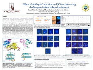
Figure 3. AtY14 only binds to full length AtMago.
Figure 1. Structure of the EJC. A. The core complex of the EJC binding to RNA
and ATP (Bono, et al., 2006). B. UPF3b binding to the Mago-Y14 heterodimer
(Buchwald et al. 2010) . C. Full length AtMago protein with 6 β-sheets and 3 α-
helices. D. AtMagoΔC protein with the C terminus end truncated leaving 4β-sheets
and one α-helix (Shi and Xu 2003).
B.
C. D.
Figure 4. Meiosis in wildtype (qrt) Arabidopsis Thaliana. Prepared by a
chromosome spread technique and stained with DAPI. Bar = 10μm
Figure 5. Meiosis in mutant (qrt;hap1-2/+) Arabidopsis thaliana. Prepared
by a chromosome spread technique and stained with DAPI. Bar = 10μm
A.
Figure 2. A. Location of hap1-2 mutation. B. Pollen grains in bright
field (left panel) and pollen grains stained with DAPI (right panels).
Bar = 20μm. Abnormal nuclei indicated by *.
T-DNA insertion (sail_269_C02)
Normal
splicing
Splicing of hap1-
2 mutant
Normal length
protein
Truncated
Protein
5’ 3’
5’ 5’3’ 3’
A. B.
Editor's Notes
Technique used for figure 2 and 3??? I will explain to you next time when you come in.
Comments:
Your figures need to go with the abstract. This means: you don’t have a figure to support the statement “Heterozygous mutants (qrt;hap1-2/+) exhibited abnormal nuclear distribution or lack of nuclei in pollen grains and the mutant pollen eventually aborted”. And you didn’t mention Mago-eIF4AIII binding at all in the abstract, so there is no need for the current figure 2. You could change figure 2 to a different figure showing the results supporting the statement above.
Figure 1 has images from different papers. You need to cite all of them in the figure legend and references.
Move Telophase I in Fig. 4 to next to Anaphase I and make the layout of Figure 4 and 5 more comparable.
The scale bar in Anaphase I for WT is a lot bigger than others. Check to see if it is correct.
Your conclusion bullet points should go with the order of the figure.
I deleted the unnecessary references. You only need to show the ones that your poster content actually cites.