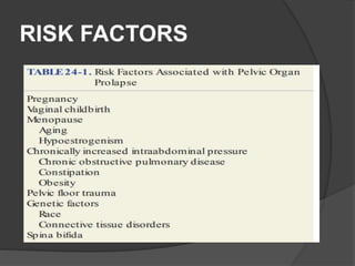
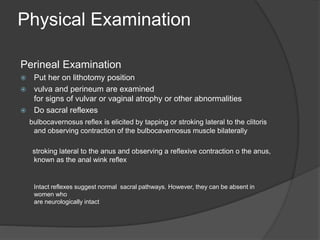
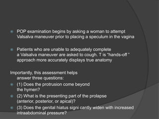
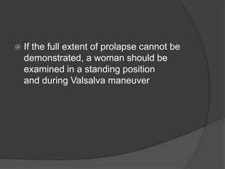
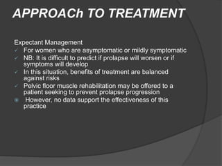
 Pelvic organ prolapse is a common
condition that can lead to genital tract
dysfunction and diminished quality of life
 Signs: descent of one or more of the
following
 the anterior vaginal wall, posterior
vaginal wall, uterus and cervix, vaginal
apex, or the perineum
Symptoms
 vaginal bulging,
 pelvic pressure, and
 splinting or digitation
 For pelvic organ prolapse to be considered a
disease state in a given individual, symptoms
should be attributable to pelvic organ descent
such that surgical or nonsurgical reduction relieves
the symptoms, restores function, and improves
quality of life.
RISK FACTORS
 Currently, two obstetric interventions-
elective forceps delivery to shorten second-
stage labor and elective episiotomy are not
advocated
 Elective cesarean delivery to prevent pelvic
floor disorders such as POP and urinary
incontinence is controversial
DESCRIPTION AND
CLASSIFICATION
 The terms cystocele, cystourethrocele, uterine prolapse, uterine
procidentia, rectocele, and enterocele have traditionally been
used to describe the structures behind the vaginal wall
thought to be prolapsed
 However, these terms are imprecise and misleading
b/c they focus on what is presumed to be prolapsed rather than what is objectively
noted to be prolapsed.
 it is more clinically useful to describe prolapse in terms of what
one actually sees:
 anterior vaginal wall prolapse, apical prolapse, cervical
prolapse, posterior vaginal wall prolapse, rectal prolapse, or
perineal descent.
Pelvic Organ Prolapse
Quantification POP Q
 This system allows clinicians and researchers to
report findings in a standardized, easily
reproducible fashion
 Six points are located with reference to the plane
of the hymen:
 All POP-Q points, except TVL, are measured
during patient Valsalva and should reflect
maximum protrusion.
Anterior vaginal wall
 Point Aa : is 3 cm proximal to the external urethral meatus
 In relation to the hymen, this point’s position ranges from –3
(normal support) to + 3 cm (maximum prolapse of point Aa)
 Point Ba : represents the most distal position of any part o
the upper anterior vaginal wall
 It is –3 cm in the absence of prolapse
 Point C defines a point that is at either the most distal edge of the
cervix or the leading edge of the vaginal cuff after total
hysterectomy.
 Point D defines a point that represents the location of the posterior
fornix in a woman who still has a cervix , It is omitted in the absence
of a cervix
 In a woman with total vaginal eversion
post-hysterectomy, Ba / Bp would have
a positive value equal to the position of
the cuff from the hymen
Posterior Vaginal Wall
Points
 Point Ap :defines a point in the midline of the posterior
vaginal wall that lies 3 cm proximal to the hymen
 this point’s range of position is by definition –3 (normal
support) to + 3 cm (maximum prolapse of point Ap)
 Point Bp represents the most distal position of any part of the upper
posterior vaginal wall , –3 cm in the absence o prolapse
Genital Hiatus and Perineal
Body
 Gh : is measured from the middle of the
external urethral meatus to the midline
of the posterior hymenal ring
 Pb :is measured from the posterior
margin of the genital hiatus to the
midanal opening
Baden Walker halfway
System
 Although not as informative as the POP-Q, it is adequate for clinical
use if each compartment (anterior, apical, and posterior) is
evaluated
PATh OPh YSIOLOGY
Pelvic organ support : by complex
interaction of
 Pelvic floor muscle
 Pelvic floor connective tissue
 Vaginal wall
Levator Ani muscle : consist of 3
muscle,ilococcygus,pubococcygus,puborectalis
 Connective tissue cover superior and inferior surface
 Resting contraction elevates pelvic floor and compress
vagina, urtethra and rectum towards pubic bone ; this narrow
the Gh and Prevents POP
Upper Vaginal wall
 Lies nearly horizontal in standing female
 Increased Intraabdominal pressure put tension on the levator
ani loos of support of vagina which become semi vertical
 Which widen GH and lead to prolpase
The Defect Theory of Pelvic
Organ Prolapse
 attenuation of the vaginal wall without loss of fascial
attachments is called a distention cystocele or rectocele
 With distention-type prolapse, the vaginal wall appears
smooth and without rugae, due to abdominal contents
pressed against the vagina from within
 In contrast, anterior and posterior wall defects due to loss o
the connective tissue attachment of the lateral vaginal wall to
the pelvic sidewall are described as displacement
(paravaginal) cystocele or rectocele
 With displacement-type prolapse, vaginal rugae are visible.
Both defect types could result from the stretching or tearing
of support tissues during second-stage labor
Levels of Vaginal Support
Level 1 support :
 suspends the upper or proximal vagina
 consists of the cardinal and uterosacral ligaments
attachment to the cervix and upper vagina
 The cardinal ligaments fan out laterally and attach to the parietal fascia of the obturator internus and piri ormis
muscles, the anterior border o the greater sciatic oramen, and the ischial spines.
 The e uterosacral ligaments are posterior bers that attach to the presacral region at the level o S2 through S4
 maintains vaginal length and horizontal axis.
 Defects in this support complex may lead to apical
prolapse
 This is frequently associated with small bowel
herniation into the vaginal wall, that is, enterocele
2411/8/2020
Level II support
 consists of the paravaginal attachments that are
contiguous with the cardinal/uterosacral complex at the
ischial spine
 These are the connective tissue attachments of the lateral
vagina anteriorly to the arcus tendineus fascia pelvis and
posteriorly to the arcus tendineus rectovaginalis.
 Detachment of this connective tissue from the arcus
tendineus fascia pelvis leads to lateral or paravaginal
anterior vaginal wall prolapse
Level III support
 is composed of the perineal body, super- fcial and deep
perineal muscles, and fibromuscular connective tissue
 these support the distal one third of the vagina and introitus
 The perineal body is essential for distal vaginal support and
proper function of the anal canal.
 Damage to level III support contributes to anterior and
posterior vaginal wall prolapse, gaping introitus, and perineal
descent
PATIENT EVALUATION
symptom
 Commonly assted with GI,GU and MS
system
 Assess carefully the symptom if they r
caused by prolapse
 Symptom asst can be done using
questionner : Pelvic Floor Distress Inventory (PFDI) / the Pelvic
Floor Impact Questionnaire (PFIQ)
 If bulge symptoms are the primary complaint, successful
replacement of the prolapse with nonsurgical or surgical
therapy will usually provide adequate symptom relief .
 Urinary symptoms often accompany POP and may include
stress urinary incontinence (SUI), urgency urinary incontinence,
requency, urgency, urinary retention, recurrent urinary tract in ection, or
voiding dys unction
 Although these symptoms may be caused or exacerbated by POP, it
should not be assumed that surgical or nonsurgical correction of
prolapse will be curative.
 In contrast, urinary retention has been found to improve with
prolapse treatment if the symptom is due to an obstructed urethra
Constipation
 often present in women with POP, although
it is generally not caused by POP
 surgical repair or treatment with a pessary will not usually cure
constipation and may actually worsen it
 Therefore, if a patient’s primary symptom is constipation, treatment
of prolapse may not be indicated.
 Constipation should be viewed as a problem distinct from prolapse
and evaluated separately
 Digital decompression of the posterior vaginal wall, the
perineal body, or the distal rectum to evacuate the rectum is
the most common defecatory symptom associated with
posterior vaginal wall prolapse
 Surgical approaches to this problem provide variable
success, and symptom resolution rates range from 36 to 70
percent
Female sexual dysfunction
 some prolapse procedures such as
posterior repair with levator plication and
vaginal placement of mesh may contribute
to postoperative dyspareunia.
 Therefore, care is taken in planning
appropriate surgical procedures for women
with concomitant sexual dysfgtunction
 Pelvic and back pain is another complaint
in women with POP, but little evidence
supports a direct association.
 POP is associated with varied complaints,
symptoms and their severity do not
always correlate well with advancing
stages of prolapse.
 Thus, when planning surgical or nonsurgical therapy,
realistic expectations should be set with regard to
symptom relief
 A patient is informed that symptoms directly related
to prolapse such as vaginal bulge and pelvic
pressure are likely to improve with a successful
anatomic repair.
 However, other associated symptoms such as
constipation, back pain, and urinary urgency and
frequency may or may not improve
Physical Examination
Perineal Examination
 Put her on lithotomy position
 vulva and perineum are examined
for signs of vulvar or vaginal atrophy or other abnormalities
 Do sacral reflexes
bulbocavernosus reflex is elicited by tapping or stroking lateral to the clitoris
and observing contraction of the bulbocavernosus muscle bilaterally
stroking lateral to the anus and observing a reflexive contraction o the anus,
known as the anal wink reflex
Intact reflexes suggest normal sacral pathways. However, they can be absent in
women who
are neurologically intact
 POP examination begins by asking a woman to attempt
Valsalva maneuver prior to placing a speculum in the vagina
 Patients who are unable to adequately complete
a Valsalva maneuver are asked to cough. T is “hands-off ”
approach more accurately displays true anatomy
Importantly, this assessment helps
answer three questions:
 (1) Does the protrusion come beyond
the hymen?
 (2) What is the presenting part of the prolapse
(anterior, posterior, or apical)?
 (3) Does the genital hiatus signi cantly widen with increased
intraabdominal pressure?
 If the full extent of prolapse cannot be
demonstrated, a woman should be
examined in a standing position
and during Valsalva maneuver
APPROACh TO TREATMENT
Expectant Management
 For women who are asymptomatic or mildly symptomatic
 NB: It is difficult to predict if prolapse will worsen or if
symptoms will develop
 In this situation, benefits of treatment are balanced
against risks
 Pelvic floor muscle rehabilitation may be offered to a
patient seeking to prevent prolapse progression
 However, no data support the effectiveness of this
practice
For significant prolpase or bothersome sx :
option of mgt
 Surgical
 Non surgical
Choice depends on
 the type and severity of symptoms,
 age and medical comorbidities,
 desire for future sexual function and/or
fertility, and
 Risk factors for recurrence.
Non surgical treatment
Pessary
 usually made of silicone or inert plastic, and they are
safe and simple to manage.
Indication
 most common indication for vaginal pessary is POP
 Other indication POP associated urinary
incontinence
 Diagnostically i.e whether the sx is related to POP
 Traditionally pessary was for those unfit or unwilling
to go for surgery
Pessary type: can be
 Support (e.g ring)
 Space filing (e.g Cube)
Of all pessaries, the two most commonly
used and studied devices are
the ring and the Gellhorn pessaries
Pessary placement
 Vaginal atoprhy should be treated first if there is
 Ideally, a pessary is removed nightly to weekly,
washed in soap and water, and replaced the next
morning
 After initial placement, a return visit may follow in 1
to 2 weeks.
 For patients comfortable with their pessary
management, return visits may be semiannual
 For those unable or unwilling to remove and
replace a device themselves
 pessary may be removed and the patient’s
vagina inspected at
the provider’s of ce every 2 or 3 months
 Delaying visits longer than this may lead to
problematic discharge and odor
Pessary complication
 Pessary Ulcer
are treated by changing the pessary type or size to alleviate pressure points or by
removing the pessary completely until healed. T
treatment of vaginal atrophy with local estrogen is commonly required. Alternatively,
wate rbased lubricants applied to the pessary may help prevent these complications
Prolapse ulcers :
 have the same appearance as pessary ulcers,
 result from the prolapsed bulge rubbing against patient clothing.
 T ese are treated by replacing
the prolapse either with a pessary or by surgery
 Pelvic pain with pessary use is not normal. T is usually indicates that the size is too large, and a smaller pessary would be
more suitable.
 Abnormal odor : managed by encouraging more requent nighttime
device removal, washing, and reinsertion the next day
Pelvic floor muscle exercise
 also known as Kegel exercises
 suggested as a therapy that might
limit progression and alleviate prolapse
symptoms
 Unfortunately, high-quality scientific
evidence supporting pelvic exercise for
prevention and treatment of prolapse is
lacking
 Exercise sets are performed numerous
times during the day, with some reporting
up to 50 or 60 times each day
 However, specific details in performance of
these exercises a subject to provider pref
erence and clinical setting.
SURGICAL TREATMENT
 Thus, the surgeon and the patient must agree on the desired
results
 surgical success be defined as absence of bulge symptoms in
addition to anatomic criteria.
Can be
OBLITERATIVE PREOCDURES
_ Include Lefort colpocleisis and complete colpocleisis
RECONSTRUCTIVE PROCEDURES
- Procedures include sacrocolpopexy, paravaginal repair, and vaginal
vault suspension to Uterosacral ligament
Obliterative Procedures
 include Lefort colpocleisis and complete colpocleisis
 These procedures involve removing vaginal epithelium, suturing anterior
and posterior vaginal walls together, obliterating the vaginal vault, and
effectively
closing the vagina
 are only appropriate for elderly or medically compromised patients who
have no desire for future coital activity
 are technically easier, require less operative time, and offer
superior success rates compared withreconstructive
procedures.

 Success rates : range from 91 to 100 percent
 after colpocleisis, fewer than 10 percent of patients
express regret, often due to loss of coital activity
 If ther is uterus prior to procedure, hystrectomy can be
done before colpocleisis or
 If uterus retention is planned , neoplasia excluded
preoperatively
Reconstructive Procedures
 More commonly performed
 Has vaginal, abdominal,laparascopic and robotic approach
 Procedures include sacrocolpopexy, paravaginal repair, and vaginal
vault suspension to uterosacral ligament
Abdominal :
 preffered for those with recurrent prolpase
 Those believed to be @ high risk for recurrence
Vaginal
 typically offers shorter operative time and a quicker return to
daily activities

Surgical treatment
In preparing for prolapse correcting surgery
 Each pt should understand anticpated result
Surgeon should factor
 A pt’s goals and expectations
 Symptoms and quality of issues
 best estimates of goal attainment based
on scientific evidence and the surgeon's
personal experience
 the surgeon and the patient must agree on
desired endpoints.
Anterior Compartment
anterior colporrhaphy
 has been the most common operation,
 yet long-term anatomic success rates are poor
 symptom relief may be acceptable.
 Mesh or biomaterial may also be used in conjunction
 Mesh is used to reinforce the vaginal wall and is sutured in
place laterally.
 However, the use o mesh and mesh kits or anterior vaginal wall
prolapse remains controversial
 If its due to anterior apical segment defect : do
apical suspension procedure (sacrocolpopexy or
uterosacral ligament vaginal vault suspension) will
resuspend the anterior vaginal wall to the apex and
reduce anterior wall prolapse
 Also prevent enterocele formation
 if a lateral defect is suspected, paravaginal
repair can be performed
Vaginal Apex
 can be resuspended with several
procedures which include
 abdominal sacrocolpopexy
 sacrospinous ligament fixation,
 uterosacral ligament vaginal vault
suspension
Abdomnial sacrocolpopexy
 suspends the vaginal vault to the sacrum
using synthetic mesh
Advantages
 the procedure’s durability over time and
conservation o normal vaginal anatomy
 offers greater vaginal apex mobility and
avoids vaginal shortening.
 When hysterectomy is performed in conjunction
with sacrocolpopexy, consideration is given to performing a
supracervical rather than a total abdominal hysterectomy.
 With the cervix left in situ, the risk of postoperative mesh
erosion at the vaginal apex is believed to be diminished
 In this case, the mesh is not exposed to vaginal bacteria,
which occurs when the vagina is opened with total
hysterectomy
 In addition, the strong connective tissue o the
cervix allows for an additional anchoring point or the permanent
mesh.
sacrospinous ligament fixation
(SSLF)
 vaginal apex is suspended to the sacrospinous
ligament unilaterally or bilaterally using a vaginal
extraperitoneal approach
 After SSLF, recurrent apical prolapse is
uncommon
 anterior vaginal wall prolapse develops
postoperatively in 6 to 28 percent of patients and
is thought to develop from redirection of
abdominal forces anteriorly
Complications (SSLF)
 buttock pain
 vascular injury
 Uterosacral ligament vaginal vault suspension is
another apical surgery. With this procedure, the
vaginal apex is attached to remnants o the
uterosacral ligament at the level o the ischial
spines or highe

Pelvic organ prolapse

  • 2.
     Pelvic organprolapse is a common condition that can lead to genital tract dysfunction and diminished quality of life  Signs: descent of one or more of the following  the anterior vaginal wall, posterior vaginal wall, uterus and cervix, vaginal apex, or the perineum
  • 3.
    Symptoms  vaginal bulging, pelvic pressure, and  splinting or digitation
  • 4.
     For pelvicorgan prolapse to be considered a disease state in a given individual, symptoms should be attributable to pelvic organ descent such that surgical or nonsurgical reduction relieves the symptoms, restores function, and improves quality of life.
  • 5.
  • 6.
     Currently, twoobstetric interventions- elective forceps delivery to shorten second- stage labor and elective episiotomy are not advocated  Elective cesarean delivery to prevent pelvic floor disorders such as POP and urinary incontinence is controversial
  • 7.
    DESCRIPTION AND CLASSIFICATION  Theterms cystocele, cystourethrocele, uterine prolapse, uterine procidentia, rectocele, and enterocele have traditionally been used to describe the structures behind the vaginal wall thought to be prolapsed  However, these terms are imprecise and misleading b/c they focus on what is presumed to be prolapsed rather than what is objectively noted to be prolapsed.  it is more clinically useful to describe prolapse in terms of what one actually sees:  anterior vaginal wall prolapse, apical prolapse, cervical prolapse, posterior vaginal wall prolapse, rectal prolapse, or perineal descent.
  • 8.
    Pelvic Organ Prolapse QuantificationPOP Q  This system allows clinicians and researchers to report findings in a standardized, easily reproducible fashion  Six points are located with reference to the plane of the hymen:  All POP-Q points, except TVL, are measured during patient Valsalva and should reflect maximum protrusion.
  • 9.
    Anterior vaginal wall Point Aa : is 3 cm proximal to the external urethral meatus  In relation to the hymen, this point’s position ranges from –3 (normal support) to + 3 cm (maximum prolapse of point Aa)  Point Ba : represents the most distal position of any part o the upper anterior vaginal wall  It is –3 cm in the absence of prolapse  Point C defines a point that is at either the most distal edge of the cervix or the leading edge of the vaginal cuff after total hysterectomy.  Point D defines a point that represents the location of the posterior fornix in a woman who still has a cervix , It is omitted in the absence of a cervix
  • 10.
     In awoman with total vaginal eversion post-hysterectomy, Ba / Bp would have a positive value equal to the position of the cuff from the hymen
  • 11.
    Posterior Vaginal Wall Points Point Ap :defines a point in the midline of the posterior vaginal wall that lies 3 cm proximal to the hymen  this point’s range of position is by definition –3 (normal support) to + 3 cm (maximum prolapse of point Ap)  Point Bp represents the most distal position of any part of the upper posterior vaginal wall , –3 cm in the absence o prolapse
  • 12.
    Genital Hiatus andPerineal Body  Gh : is measured from the middle of the external urethral meatus to the midline of the posterior hymenal ring  Pb :is measured from the posterior margin of the genital hiatus to the midanal opening
  • 17.
    Baden Walker halfway System Although not as informative as the POP-Q, it is adequate for clinical use if each compartment (anterior, apical, and posterior) is evaluated
  • 18.
    PATh OPh YSIOLOGY Pelvicorgan support : by complex interaction of  Pelvic floor muscle  Pelvic floor connective tissue  Vaginal wall
  • 19.
    Levator Ani muscle: consist of 3 muscle,ilococcygus,pubococcygus,puborectalis  Connective tissue cover superior and inferior surface  Resting contraction elevates pelvic floor and compress vagina, urtethra and rectum towards pubic bone ; this narrow the Gh and Prevents POP Upper Vaginal wall  Lies nearly horizontal in standing female  Increased Intraabdominal pressure put tension on the levator ani loos of support of vagina which become semi vertical  Which widen GH and lead to prolpase
  • 21.
    The Defect Theoryof Pelvic Organ Prolapse  attenuation of the vaginal wall without loss of fascial attachments is called a distention cystocele or rectocele  With distention-type prolapse, the vaginal wall appears smooth and without rugae, due to abdominal contents pressed against the vagina from within  In contrast, anterior and posterior wall defects due to loss o the connective tissue attachment of the lateral vaginal wall to the pelvic sidewall are described as displacement (paravaginal) cystocele or rectocele
  • 22.
     With displacement-typeprolapse, vaginal rugae are visible. Both defect types could result from the stretching or tearing of support tissues during second-stage labor
  • 23.
    Levels of VaginalSupport Level 1 support :  suspends the upper or proximal vagina  consists of the cardinal and uterosacral ligaments attachment to the cervix and upper vagina  The cardinal ligaments fan out laterally and attach to the parietal fascia of the obturator internus and piri ormis muscles, the anterior border o the greater sciatic oramen, and the ischial spines.  The e uterosacral ligaments are posterior bers that attach to the presacral region at the level o S2 through S4  maintains vaginal length and horizontal axis.  Defects in this support complex may lead to apical prolapse  This is frequently associated with small bowel herniation into the vaginal wall, that is, enterocele
  • 24.
  • 25.
    Level II support consists of the paravaginal attachments that are contiguous with the cardinal/uterosacral complex at the ischial spine  These are the connective tissue attachments of the lateral vagina anteriorly to the arcus tendineus fascia pelvis and posteriorly to the arcus tendineus rectovaginalis.  Detachment of this connective tissue from the arcus tendineus fascia pelvis leads to lateral or paravaginal anterior vaginal wall prolapse
  • 26.
    Level III support is composed of the perineal body, super- fcial and deep perineal muscles, and fibromuscular connective tissue  these support the distal one third of the vagina and introitus  The perineal body is essential for distal vaginal support and proper function of the anal canal.  Damage to level III support contributes to anterior and posterior vaginal wall prolapse, gaping introitus, and perineal descent
  • 28.
    PATIENT EVALUATION symptom  Commonlyassted with GI,GU and MS system  Assess carefully the symptom if they r caused by prolapse  Symptom asst can be done using questionner : Pelvic Floor Distress Inventory (PFDI) / the Pelvic Floor Impact Questionnaire (PFIQ)
  • 29.
     If bulgesymptoms are the primary complaint, successful replacement of the prolapse with nonsurgical or surgical therapy will usually provide adequate symptom relief .  Urinary symptoms often accompany POP and may include stress urinary incontinence (SUI), urgency urinary incontinence, requency, urgency, urinary retention, recurrent urinary tract in ection, or voiding dys unction  Although these symptoms may be caused or exacerbated by POP, it should not be assumed that surgical or nonsurgical correction of prolapse will be curative.
  • 30.
     In contrast,urinary retention has been found to improve with prolapse treatment if the symptom is due to an obstructed urethra Constipation  often present in women with POP, although it is generally not caused by POP  surgical repair or treatment with a pessary will not usually cure constipation and may actually worsen it  Therefore, if a patient’s primary symptom is constipation, treatment of prolapse may not be indicated.  Constipation should be viewed as a problem distinct from prolapse and evaluated separately
  • 31.
     Digital decompressionof the posterior vaginal wall, the perineal body, or the distal rectum to evacuate the rectum is the most common defecatory symptom associated with posterior vaginal wall prolapse  Surgical approaches to this problem provide variable success, and symptom resolution rates range from 36 to 70 percent
  • 32.
    Female sexual dysfunction some prolapse procedures such as posterior repair with levator plication and vaginal placement of mesh may contribute to postoperative dyspareunia.  Therefore, care is taken in planning appropriate surgical procedures for women with concomitant sexual dysfgtunction
  • 33.
     Pelvic andback pain is another complaint in women with POP, but little evidence supports a direct association.  POP is associated with varied complaints, symptoms and their severity do not always correlate well with advancing stages of prolapse.
  • 34.
     Thus, whenplanning surgical or nonsurgical therapy, realistic expectations should be set with regard to symptom relief  A patient is informed that symptoms directly related to prolapse such as vaginal bulge and pelvic pressure are likely to improve with a successful anatomic repair.  However, other associated symptoms such as constipation, back pain, and urinary urgency and frequency may or may not improve
  • 35.
    Physical Examination Perineal Examination Put her on lithotomy position  vulva and perineum are examined for signs of vulvar or vaginal atrophy or other abnormalities  Do sacral reflexes bulbocavernosus reflex is elicited by tapping or stroking lateral to the clitoris and observing contraction of the bulbocavernosus muscle bilaterally stroking lateral to the anus and observing a reflexive contraction o the anus, known as the anal wink reflex Intact reflexes suggest normal sacral pathways. However, they can be absent in women who are neurologically intact
  • 36.
     POP examinationbegins by asking a woman to attempt Valsalva maneuver prior to placing a speculum in the vagina  Patients who are unable to adequately complete a Valsalva maneuver are asked to cough. T is “hands-off ” approach more accurately displays true anatomy Importantly, this assessment helps answer three questions:  (1) Does the protrusion come beyond the hymen?  (2) What is the presenting part of the prolapse (anterior, posterior, or apical)?  (3) Does the genital hiatus signi cantly widen with increased intraabdominal pressure?
  • 37.
     If thefull extent of prolapse cannot be demonstrated, a woman should be examined in a standing position and during Valsalva maneuver
  • 38.
    APPROACh TO TREATMENT ExpectantManagement  For women who are asymptomatic or mildly symptomatic  NB: It is difficult to predict if prolapse will worsen or if symptoms will develop  In this situation, benefits of treatment are balanced against risks  Pelvic floor muscle rehabilitation may be offered to a patient seeking to prevent prolapse progression  However, no data support the effectiveness of this practice
  • 39.
    For significant prolpaseor bothersome sx : option of mgt  Surgical  Non surgical
  • 40.
    Choice depends on the type and severity of symptoms,  age and medical comorbidities,  desire for future sexual function and/or fertility, and  Risk factors for recurrence.
  • 41.
    Non surgical treatment Pessary usually made of silicone or inert plastic, and they are safe and simple to manage. Indication  most common indication for vaginal pessary is POP  Other indication POP associated urinary incontinence  Diagnostically i.e whether the sx is related to POP  Traditionally pessary was for those unfit or unwilling to go for surgery
  • 42.
    Pessary type: canbe  Support (e.g ring)  Space filing (e.g Cube) Of all pessaries, the two most commonly used and studied devices are the ring and the Gellhorn pessaries
  • 43.
    Pessary placement  Vaginalatoprhy should be treated first if there is  Ideally, a pessary is removed nightly to weekly, washed in soap and water, and replaced the next morning  After initial placement, a return visit may follow in 1 to 2 weeks.  For patients comfortable with their pessary management, return visits may be semiannual
  • 44.
     For thoseunable or unwilling to remove and replace a device themselves  pessary may be removed and the patient’s vagina inspected at the provider’s of ce every 2 or 3 months  Delaying visits longer than this may lead to problematic discharge and odor
  • 45.
    Pessary complication  PessaryUlcer are treated by changing the pessary type or size to alleviate pressure points or by removing the pessary completely until healed. T treatment of vaginal atrophy with local estrogen is commonly required. Alternatively, wate rbased lubricants applied to the pessary may help prevent these complications Prolapse ulcers :  have the same appearance as pessary ulcers,  result from the prolapsed bulge rubbing against patient clothing.  T ese are treated by replacing the prolapse either with a pessary or by surgery  Pelvic pain with pessary use is not normal. T is usually indicates that the size is too large, and a smaller pessary would be more suitable.  Abnormal odor : managed by encouraging more requent nighttime device removal, washing, and reinsertion the next day
  • 46.
    Pelvic floor muscleexercise  also known as Kegel exercises  suggested as a therapy that might limit progression and alleviate prolapse symptoms  Unfortunately, high-quality scientific evidence supporting pelvic exercise for prevention and treatment of prolapse is lacking
  • 47.
     Exercise setsare performed numerous times during the day, with some reporting up to 50 or 60 times each day  However, specific details in performance of these exercises a subject to provider pref erence and clinical setting.
  • 48.
    SURGICAL TREATMENT  Thus,the surgeon and the patient must agree on the desired results  surgical success be defined as absence of bulge symptoms in addition to anatomic criteria. Can be OBLITERATIVE PREOCDURES _ Include Lefort colpocleisis and complete colpocleisis RECONSTRUCTIVE PROCEDURES - Procedures include sacrocolpopexy, paravaginal repair, and vaginal vault suspension to Uterosacral ligament
  • 49.
    Obliterative Procedures  includeLefort colpocleisis and complete colpocleisis  These procedures involve removing vaginal epithelium, suturing anterior and posterior vaginal walls together, obliterating the vaginal vault, and effectively closing the vagina  are only appropriate for elderly or medically compromised patients who have no desire for future coital activity  are technically easier, require less operative time, and offer superior success rates compared withreconstructive procedures. 
  • 50.
     Success rates: range from 91 to 100 percent  after colpocleisis, fewer than 10 percent of patients express regret, often due to loss of coital activity  If ther is uterus prior to procedure, hystrectomy can be done before colpocleisis or  If uterus retention is planned , neoplasia excluded preoperatively
  • 51.
    Reconstructive Procedures  Morecommonly performed  Has vaginal, abdominal,laparascopic and robotic approach  Procedures include sacrocolpopexy, paravaginal repair, and vaginal vault suspension to uterosacral ligament Abdominal :  preffered for those with recurrent prolpase  Those believed to be @ high risk for recurrence Vaginal  typically offers shorter operative time and a quicker return to daily activities 
  • 52.
    Surgical treatment In preparingfor prolapse correcting surgery  Each pt should understand anticpated result Surgeon should factor  A pt’s goals and expectations  Symptoms and quality of issues  best estimates of goal attainment based on scientific evidence and the surgeon's personal experience  the surgeon and the patient must agree on desired endpoints.
  • 53.
    Anterior Compartment anterior colporrhaphy has been the most common operation,  yet long-term anatomic success rates are poor  symptom relief may be acceptable.  Mesh or biomaterial may also be used in conjunction  Mesh is used to reinforce the vaginal wall and is sutured in place laterally.  However, the use o mesh and mesh kits or anterior vaginal wall prolapse remains controversial
  • 54.
     If itsdue to anterior apical segment defect : do apical suspension procedure (sacrocolpopexy or uterosacral ligament vaginal vault suspension) will resuspend the anterior vaginal wall to the apex and reduce anterior wall prolapse  Also prevent enterocele formation  if a lateral defect is suspected, paravaginal repair can be performed
  • 55.
    Vaginal Apex  canbe resuspended with several procedures which include  abdominal sacrocolpopexy  sacrospinous ligament fixation,  uterosacral ligament vaginal vault suspension
  • 56.
    Abdomnial sacrocolpopexy  suspendsthe vaginal vault to the sacrum using synthetic mesh Advantages  the procedure’s durability over time and conservation o normal vaginal anatomy  offers greater vaginal apex mobility and avoids vaginal shortening.
  • 57.
     When hysterectomyis performed in conjunction with sacrocolpopexy, consideration is given to performing a supracervical rather than a total abdominal hysterectomy.  With the cervix left in situ, the risk of postoperative mesh erosion at the vaginal apex is believed to be diminished  In this case, the mesh is not exposed to vaginal bacteria, which occurs when the vagina is opened with total hysterectomy  In addition, the strong connective tissue o the cervix allows for an additional anchoring point or the permanent mesh.
  • 58.
    sacrospinous ligament fixation (SSLF) vaginal apex is suspended to the sacrospinous ligament unilaterally or bilaterally using a vaginal extraperitoneal approach  After SSLF, recurrent apical prolapse is uncommon  anterior vaginal wall prolapse develops postoperatively in 6 to 28 percent of patients and is thought to develop from redirection of abdominal forces anteriorly
  • 59.
    Complications (SSLF)  buttockpain  vascular injury  Uterosacral ligament vaginal vault suspension is another apical surgery. With this procedure, the vaginal apex is attached to remnants o the uterosacral ligament at the level o the ischial spines or highe