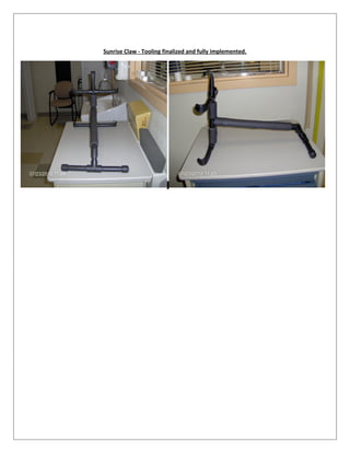
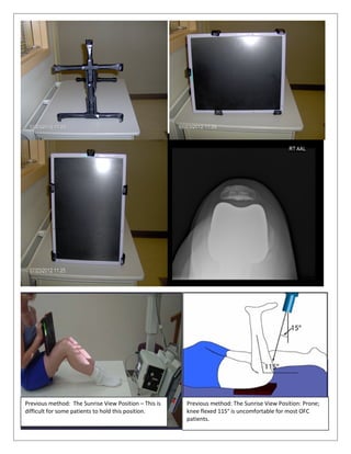
PDSA-OFC-RADIOLOGY TOOLING
Name of PDSA: OFC - Radiology Tooling for Sunrise Projections Start Date: 10/01/2009
PDSA cycle number: 1 Resource owners: Al Lopez
______________________________________________________________________________________
Plan: Analyze process to be improved and determine what changes to make
Goal: What are we trying to accomplish?
 Improve patient comfort level and satisfaction while capturing sunrise projection.
 Improve provider satisfaction with patient flow.
 Improve imaging productivity and imaging workflow.
 Improve the Radiology Technologist position speed and work satisfaction.
 Improve Image quality of the sunrise projection.
 Reduce radiation expose to the Radiology Technologist who often holds the cassette for poorly conditioned
or otherwise physically compromised patient.
 Reduce the frequency of using two technologist or support staff to hold the cassette for capturing the
sunrise projections.
What change(s) will we make to improve the process?
 Generate an inexpensive light weight easy to clean tool to meet goals.
What improvement(s) do we expect to happen?
 Improve patient comfort level and satisfaction while capturing sunrise projection.
 Improve provider satisfaction with patient flow and image quality.
 Improve imaging productivity and imaging workflow.
 Improve Radiology Technologist satisfaction.
 Improve Image quality of the sunrise projection.
 Reduce radiation expose to the Radiology Technologist who often holds the cassette for poorly conditioned
patient.
 Reduce the frequency of using two technologist or support staff to hold the cassette for sunrise projections.
What measurement(s) will we use to monitor the change? How will we collect and share the data?
 Image quality of the sunrise projection.
 Tooling set-up cycle times vs. set-up time without tooling.
 Patient comments.
 Radiology Technologists’ comments.
 Provider comments.
 Support staff comments.
 Radiologist comments.
 Radiology QC department notification.
 Orthopedics knee surgeon comments.
 Occupational Radiation Exposure Report.
 Monitor the frequency of using two technologist or support staff to hold the cassette for sunrise projections.
Do: Implement and test the changes
What were the experiences, problems, surprises that occurred?
Study: Monitor and analyze the results of the doing stage
What were the improvements that occurred from the change? How did the measurements change?
Act: Fully implement, revise or abandon the improvement changes
Is the change ready to be implemented? If no, what modifications need to be made to continue the improvement
process and start the PDSA again?
PDSA-OFC-RADIOLOGY TOOLING
Name of PDSA: OFC - Radiology Tooling for Sunrise Projections Start Date: 11/12/2009 - 05/03/2012
PDSA cycle number: 2 Resource owners: Al Lopez
_______________________________________________________________________________________
Plan: Analyze process to be improved and determine what changes to make
Goal: What are we trying to accomplish?
 Improve patient comfort level and satisfaction while capturing sunrise projection.
 Improve provider satisfaction with patient flow and image quality.
 Improve imaging productivity and imaging workflow.
 Improve the Radiology Technologist position speed and work satisfaction.
 Improve Image quality of the sunrise projection.
 Reduce radiation expose to the Radiology Technologist who often holds the cassette for poorly conditioned
or otherwise physically compromised patient.
 Reduce the frequency of using two technologist or support staff to hold the cassette for capturing the
sunrise projections.
What change(s) will we make to improve the process?
 I have fabricated an inexpensive PVC light weight easy to clean simple to load cassette tooling to meet the goals.
What improvement(s) do we expect to happen?
 Improved patient comfort level and satisfaction while capturing sunrise projection.
 Improved provider satisfaction with patient flow and image quality.
 Improved imaging productivity and imaging workflow.
 Improved Radiology Technologist satisfaction.
 Improved Image quality of the sunrise projection.
 Reduced radiation expose to the Radiology Technologist who often holds the cassette for poorly conditioned
patient.
 Reduced the frequency of using two technologist or support staff to hold the cassette for sunrise
projections.
What measurement(s) will we use to monitor the change? How will we collect and share the data?
 Image quality of the sunrise projection.
 Tooling set-up cycle times vs. set-up time without tooling.
 Patient comments.
 Radiology Technologists’ comments.
 Provider comments.
 Support staff comments.
 Radiologist comments.
 Radiology QC department notification.
 Orthopedics knee surgeon comments.
 Occupational Radiation Exposure Report.
 Monitor the frequency of using two technologist or support staff to hold the cassette for sunrise projections.
Do: Implement and test the changes
What were the experiences, problems, surprises that occurred?
 Our observations at this point are that the prototype design achieves many of the above goals;
nevertheless, some of the problems with the prototype include, tooling weight, design the tool with an
adjustable positional handle to better capture Rt or Lt knee in the center of the cassette (unilateral
sunrise), design with an adjustable positional handle to capture Rt and Lt knee in the center of the
cassette (bilateral sunrise), modify the design to be more aesthetically pleasing and improve tooling
durability.
Study: Monitor and analyze the results of the doing stage
What were the improvements that occurred from the change? How did the measurements change?
The improvements include enhanced image quality of the sunrise projection; improved patient comfort;
reduced occurrences that a Radiology Technologist had to hold the cassette for patients; decreased cassette
motion or patient motion (reduction in repeats); reduction in occupational radiation exposure, and improved
Radiology Technologist set-up time. The set-up time analysis was performed by stop watch and the results are
as follow:
0
1
2
3
4
5
6
7
W/o tool patient average imaging
time (mins)
W/o tool number of repeats due to
motion or patient “dropping” the
cassette
W/o tool number of times
technologist held cassette for the
patient
Without tooling: n = 30; patient average imaging time = 2.0 mins., number of repeats due to motion or patient
“dropping” the cassette = 4, number of times technologist held cassette for the patient = 6.
0
0.2
0.4
0.6
0.8
1
1.2
With tool patient average imaging
time (mins)
With tool number of repeats due to
motion or patient “dropping” the
cassette
With tool number of times
technologist held cassette for the
patient
With tooling: n = 30; patient average imaging time = 1.00 mins., number of repeats due to motion or patient
“dropping” the cassette = 0; number of times technologist held cassette for the patient = 0.
682
40
y = -642x + 1324
0
100
200
300
400
500
600
700
800
without tooling with tooling
Occupation Radiation Exposure
Lifetime to Date (Deep)
Without tooling: occupational radiation exposure report = 8/01/2006 (0 Deep) to 11/23/2011 (682 Deep) = 682
With tooling: occupational radiation exposure report = 11/23/2011 (682 Deep) to 06/05/2012 (722 Deep) = 40
Without tooling: Radiology department QC notification = 0
With tooling: Radiology department QC notification = 0
0%
10%
20%
30%
40%
50%
60%
70%
80%
90%
100%
Without tooling: percentage of images in center of IP With tooling: percentage of images in center IP
Without tooling: percentage of images in center of IP = 12%
With tooling: percentage of images in center IP = 88%
Without tooling:
number of positive
patient comments, 45
With tooling:
number of positive
patient comments ,
100
Without tooling:
number of positive
comments from
Technologistfull time &
PRN comments , 65
With tooling: number of
positivecomments
from Technologistfull
time & PRN comments,
100
Without tooling:
number of positive
comments from
Orthopedic Knee
replacement surgeon ,
0
With tooling: number of
positivecomments
from Orthopedic Knee
replacement surgeon ,
100
Comparison- Comments Analysis
percent
Without tooling: number of positive patient comments = 45%
With tooling: number of positive patient comments = 100%
Without tooling: number of positive comments from Technologist full time & PRN comments = 65%
With tooling: number of positive comments from Technologist full time & PRN comments = 100%
Without tooling: number of positive comments from Orthopedic Knee replacement surgeon = 0%
With tooling: number of positive comments from Orthopedic Knee replacement surgeon = 100%
We experienced faster, easier setup for the projection with improved image quality, improved Radiology
Technologist satisfaction, reduction in occupational radiation exposure, improved patient satisfaction without
any Radiology QC notification.
Act: Fully implement, revise or abandon the improvement changes
Is the change ready to be implemented? If no, what modifications need to be made to continue the improvement
process and start the PDSA again?
Before I finalize the tooling design I am going to address the problems with the prototype design which include, tooling
weight, design the tool with an adjustable positional handle to better capture Rt or Lt knee in the center of the cassette
(unilateral sunrise), design with an adjustable positional handle to capture Rt and Lt knee in the center of the cassette
(bilateral sunrise), modify the design to be more aesthetically pleasing and improve tooling durability. The prototype
images are below.
PDSA-OFC-RADIOLOGY TOOLING
Name of PDSA: OFC - Radiology Tooling for Sunrise Projections Start Date: 11/12/2009- 06/05/2012
PDSA cycle number: 3 Finalized Resource owners: Al Lopez
_______________________________________________________________________________________
Plan: Analyze process to be improved and determine what changes to make
Goal: What are we trying to accomplish?
 Improve patient comfort level and satisfaction while capturing sunrise projection.
 Improve provider satisfaction
 Improve productivity and workflow
 Improve employee satisfaction
 Improve Image quality of the sunrise projection
 Reduce expose to patient t and Radiology tech.
 Improve the impact resistance of the tool
 Decrease the weight of the tool
 Improve the appearance of the tool
What change(s) will we make to improve the process?
 I have completed a modified inexpensive aesthetic formed PVC light weight easy to clean simple to load cassette
tool to meet the goals.
What improvement(s) do we expect to happen?
 Improve patient comfort level and satisfaction while capturing sunrise projection.
 Improve provider satisfaction
 Improve productivity and workflow
 Improve employee satisfaction
 Improve Image quality
 Reduce expose to patient and Radiology tech.
 Improve the impact resistance of tool
 Decrease the weight of the tool
 Improve the appearance of the tool
What measurement(s) will we use to monitor the change? How will we collect and share the data?
 Set-up and examination cycle times
 Patient comments
 Provider comments
 Staff observations and input
Do: Implement and test the changes
What were the experiences, problems, surprises that occurred?
No surprises or problems.
Study: Monitor and analyze the results of the doing stage
What were the improvements that occurred from the change? How did the measurements change?
We experienced quicker, easier setup for the projection, improved image quality, improved Radiology
Technologist satisfaction, and improved patient satisfaction. The tool affords swifter exanimation room
turnaround without negatively influencing patient continuity of care.
Act: Fully implement, revise or abandon the improvement changes
Is the change ready to be implemented? If no, what modifications need to be made to continue the improvement
process and start the PDSA again?
FULLY IMPLEMENTED
The tool was designed and manufactured with the following objectives: to permit a single technologist to perform
quality imaging of the patella in the coronal view (sunrise view); improve work flow by significantly reducing interruption
of clinic support staff to hold cassette thus improving patient flow; improve Radiology Technologist satisfaction; improve
patient comfort therefore positively affecting patient satisfaction.
There have been occasions that a technologist requests assistance with holding of the cassette as the radiology
technologist performs the examination. All patients historically were requested to hold the cassette but this method
was cumbersome for some patients because some patients have difficulty holding the cassette in a steady position for
numerous reason (poor core control, large panus, L-spine pain, L-spine fusion, age, deconditioning etc…); consequently,
without tooling it has been my observation that this condition increases the probability of generating a suboptimal
image requiring repeating the projection. The alternative positioning (Prone; knee flexed 115° - CR directed toward
patella with 15° cephalic angulation) method for capturing the sunrise is equally difficult for many patients due to
obesity, L-spine pain etc… More than one-third of U.S. adults (35.7%) are obese. The percentage is presently 25.1% in
New Mexico and as high as 30% in some regions of New Mexico and obesity is exponentially increasing according to the
CDC 2012 trend line analysis.
The sunrise tool has successfully reducing exposure to employees (they are no longer holding the cassette), reduces the
probability of repeating the examination, allows one technologist to reliably perform the examination, improves the
image quality, improves patient comfort, improves technologist efficiency, and improves technologist satisfaction.
Sunrise Claw - Tooling finalized and fully implemented.
Previous method: The Sunrise View Position – This is
difficult for some patients to hold this position.
Previous method: The Sunrise View Position: Prone;
knee flexed 115° is uncomfortable for most OFC
patients.
PDSA- Orthopedics Faculty Clinic (OFC)-RADIOLOGY SUNRISE TOOLING
It was reported in Today (2012) that total knee replacements are increasing-Over 600,000 Yearly in US! Compared to
1999, total knee replacements have doubled (100% increase), to more than 600,000 per year. “On average, we take 1
[million] to 2 million steps per year. That’s a lot of back and forth on that hinge. And patients who are younger and
active can put significant force on that,” said Dr. Hanosh orthopedic surgery at the University of New Mexico in
Albuquerque. “But in terms of the value that you get from the surgery in terms of improvement in quality of life, this is
one of the best ways we can use our health care dollars.”
The sunrise view is a commonly requested x-ray view at Orthopedics Faculty Clinic (OFC). All new and follow-up total
knee arthroplasty (TKA) patients at Orthopedics Faculty Clinic require AP, Lateral, and Sunrise x-ray views. Due to the
increasing patient total knee arthroplasty patient volume, and a directive to improve clinic flow and to reduce operating
cost, tooling was selected to aid in improving radiology imaging services to meet these demands.
The Sunrise Claw tool
The Sunrise View demonstrates the inverted mountain of the patella riding in the valley of the trochlear groove in an
approximately 45 degree flexion of the knee. The positioning of the patient is similar to that of a Merchant's View except
that the locations of the x-ray tube and the cassette are horizontally rotated approximately 180 degrees so that the x-ray
beam is directed toward the patient's patella from the caudal aspect to the cranial aspect. As used hereinafter, "Sunrise
View" is defined as the view that demonstrates the inverted mountain of the patella riding in the valley of the trochlear
groove. The Sunrise Extender a tool used for capturing the sunrise view cost $378.00. The cost of the Sunrise Claw is
$15.00. This is a cost saving of $363.00. Tru-Vue Pillow for Knee Radiography cost $670.00/unit. Comparatively, this is a
cost saving of $655.00. The Sunrise Claw is a simple, low cost, easy to use alternative compared to other tooling options.
The Sunrise Claw is a foam covered PVC molded pipe with two evenly spaced side fingers. One end of claw clips onto the
cassette for correct cassette orientation and the opposite end of the cassette is open for easy loading. The Sunrise Claw
effectively extending the patient’s reaches by approximately 12 inches. The Sunrise claw is easy to clean, and works
with any 10 x 12 size cassette.
The Sunrise Claw is designed to capture a unilateral knee or bilateral knees. It can function like an alternative to the
Merchant (so-called Merchant view, named after Alan Merchant, MD from California who first described it). The
Merchant's View demonstrates the anatomy of the distal femoral sulcus and the patella (knee cap) in an approximately
45 degree flexion of the knee and allows measurement of the depth of the distal femoral sulcus in the functional range
of the patellofemoral joint. Other views may show the shape of the patella but do not show the important relationship
between the patella and its articulation with the distal femoral sulcus in flexion where dislocations or subluxations
occur. The distal femoral sulcus can have dysplasia which can only be seen in the knee flexion position of the Merchant's
View. To obtain this view, an x-ray beam is projected horizontally 10 to 15 degrees below the horizontal axis of the
patient. The x-ray beam is preferably directed toward the patient's patella from the cranial aspect to caudal the aspect
(the cranial aspect of a bone is the portion that is toward the head while the caudal aspect of a bone is the portion that
is toward the feet). The x-ray beam is projected onto a cassette containing a radiation receptor, such as an x-ray film or
an imaging plate, or any other form of radiation-absorbing media (hereinafter "cassette"). The cassette is in a vertical
position, generally perpendicular to the x-ray beam. As used hereinafter, "Merchant's View" is defined as the view that
demonstrates the anatomy of the distal femoral sulcus and the patella in an approximately 45 degree flexion of the
knee. The Sunrise Claw tool demonstrates: tangential view of patella; femoropatellar articulation and is helpful for:
Patellofemoral Arthritis, Patellar Fracture, Patellofemoral Pain, Patellar alignment. The cost of Merchant table is $
544.00. This is a cost savings of $529.00.
Prior to the sunrise tool development and use, the sunrise view was difficult to obtain at OFC due to a variety of reasons.
Some of the reasons include:
 Patient significantly decondition state,
 Patient has had recent lower back fusion,
 Patient suffers from lower back pain,
 Patient comorbidity factors (e.g. Parkinson) cassette movement,
 Patient is destabilization and demonstrates poor coordination,
 Patient is an elderly Geriatric patients with dementia ,
 Patient is gross obesity.
Due to the aforementioned reasons, a second person occasionally had to be located and requested to hold the cassette
for the patient. This has negative implications concerning radiation exposure to the staff member holding the cassette
and the clinic flow is interrupted. Using another staff member to hold the cassette is counterproductive concerning
PDSA-OFC -Ideal Process-Fast Track.
This PDSA is designed to monitor a method and inexpensive device for positioning a patient in order to obtain x-ray
Sunrise views of one or both of the patient's knees while making the image attainment easy for the patient.

PDSA Sunrise tooling 10

  • 1.
    PDSA-OFC-RADIOLOGY TOOLING Name ofPDSA: OFC - Radiology Tooling for Sunrise Projections Start Date: 10/01/2009 PDSA cycle number: 1 Resource owners: Al Lopez ______________________________________________________________________________________ Plan: Analyze process to be improved and determine what changes to make Goal: What are we trying to accomplish?  Improve patient comfort level and satisfaction while capturing sunrise projection.  Improve provider satisfaction with patient flow.  Improve imaging productivity and imaging workflow.  Improve the Radiology Technologist position speed and work satisfaction.  Improve Image quality of the sunrise projection.  Reduce radiation expose to the Radiology Technologist who often holds the cassette for poorly conditioned or otherwise physically compromised patient.  Reduce the frequency of using two technologist or support staff to hold the cassette for capturing the sunrise projections. What change(s) will we make to improve the process?  Generate an inexpensive light weight easy to clean tool to meet goals. What improvement(s) do we expect to happen?  Improve patient comfort level and satisfaction while capturing sunrise projection.  Improve provider satisfaction with patient flow and image quality.  Improve imaging productivity and imaging workflow.  Improve Radiology Technologist satisfaction.  Improve Image quality of the sunrise projection.  Reduce radiation expose to the Radiology Technologist who often holds the cassette for poorly conditioned patient.  Reduce the frequency of using two technologist or support staff to hold the cassette for sunrise projections. What measurement(s) will we use to monitor the change? How will we collect and share the data?  Image quality of the sunrise projection.  Tooling set-up cycle times vs. set-up time without tooling.  Patient comments.  Radiology Technologists’ comments.  Provider comments.  Support staff comments.  Radiologist comments.  Radiology QC department notification.  Orthopedics knee surgeon comments.  Occupational Radiation Exposure Report.  Monitor the frequency of using two technologist or support staff to hold the cassette for sunrise projections.
  • 2.
    Do: Implement andtest the changes What were the experiences, problems, surprises that occurred? Study: Monitor and analyze the results of the doing stage What were the improvements that occurred from the change? How did the measurements change? Act: Fully implement, revise or abandon the improvement changes Is the change ready to be implemented? If no, what modifications need to be made to continue the improvement process and start the PDSA again?
  • 3.
    PDSA-OFC-RADIOLOGY TOOLING Name ofPDSA: OFC - Radiology Tooling for Sunrise Projections Start Date: 11/12/2009 - 05/03/2012 PDSA cycle number: 2 Resource owners: Al Lopez _______________________________________________________________________________________ Plan: Analyze process to be improved and determine what changes to make Goal: What are we trying to accomplish?  Improve patient comfort level and satisfaction while capturing sunrise projection.  Improve provider satisfaction with patient flow and image quality.  Improve imaging productivity and imaging workflow.  Improve the Radiology Technologist position speed and work satisfaction.  Improve Image quality of the sunrise projection.  Reduce radiation expose to the Radiology Technologist who often holds the cassette for poorly conditioned or otherwise physically compromised patient.  Reduce the frequency of using two technologist or support staff to hold the cassette for capturing the sunrise projections. What change(s) will we make to improve the process?  I have fabricated an inexpensive PVC light weight easy to clean simple to load cassette tooling to meet the goals. What improvement(s) do we expect to happen?  Improved patient comfort level and satisfaction while capturing sunrise projection.  Improved provider satisfaction with patient flow and image quality.  Improved imaging productivity and imaging workflow.  Improved Radiology Technologist satisfaction.  Improved Image quality of the sunrise projection.  Reduced radiation expose to the Radiology Technologist who often holds the cassette for poorly conditioned patient.  Reduced the frequency of using two technologist or support staff to hold the cassette for sunrise projections. What measurement(s) will we use to monitor the change? How will we collect and share the data?  Image quality of the sunrise projection.  Tooling set-up cycle times vs. set-up time without tooling.  Patient comments.  Radiology Technologists’ comments.  Provider comments.  Support staff comments.  Radiologist comments.  Radiology QC department notification.  Orthopedics knee surgeon comments.  Occupational Radiation Exposure Report.
  • 4.
     Monitor thefrequency of using two technologist or support staff to hold the cassette for sunrise projections. Do: Implement and test the changes What were the experiences, problems, surprises that occurred?  Our observations at this point are that the prototype design achieves many of the above goals; nevertheless, some of the problems with the prototype include, tooling weight, design the tool with an adjustable positional handle to better capture Rt or Lt knee in the center of the cassette (unilateral sunrise), design with an adjustable positional handle to capture Rt and Lt knee in the center of the cassette (bilateral sunrise), modify the design to be more aesthetically pleasing and improve tooling durability. Study: Monitor and analyze the results of the doing stage What were the improvements that occurred from the change? How did the measurements change? The improvements include enhanced image quality of the sunrise projection; improved patient comfort; reduced occurrences that a Radiology Technologist had to hold the cassette for patients; decreased cassette motion or patient motion (reduction in repeats); reduction in occupational radiation exposure, and improved Radiology Technologist set-up time. The set-up time analysis was performed by stop watch and the results are as follow: 0 1 2 3 4 5 6 7 W/o tool patient average imaging time (mins) W/o tool number of repeats due to motion or patient “dropping” the cassette W/o tool number of times technologist held cassette for the patient Without tooling: n = 30; patient average imaging time = 2.0 mins., number of repeats due to motion or patient “dropping” the cassette = 4, number of times technologist held cassette for the patient = 6.
  • 5.
    0 0.2 0.4 0.6 0.8 1 1.2 With tool patientaverage imaging time (mins) With tool number of repeats due to motion or patient “dropping” the cassette With tool number of times technologist held cassette for the patient With tooling: n = 30; patient average imaging time = 1.00 mins., number of repeats due to motion or patient “dropping” the cassette = 0; number of times technologist held cassette for the patient = 0. 682 40 y = -642x + 1324 0 100 200 300 400 500 600 700 800 without tooling with tooling Occupation Radiation Exposure Lifetime to Date (Deep) Without tooling: occupational radiation exposure report = 8/01/2006 (0 Deep) to 11/23/2011 (682 Deep) = 682 With tooling: occupational radiation exposure report = 11/23/2011 (682 Deep) to 06/05/2012 (722 Deep) = 40 Without tooling: Radiology department QC notification = 0 With tooling: Radiology department QC notification = 0
  • 6.
    0% 10% 20% 30% 40% 50% 60% 70% 80% 90% 100% Without tooling: percentageof images in center of IP With tooling: percentage of images in center IP Without tooling: percentage of images in center of IP = 12% With tooling: percentage of images in center IP = 88% Without tooling: number of positive patient comments, 45 With tooling: number of positive patient comments , 100 Without tooling: number of positive comments from Technologistfull time & PRN comments , 65 With tooling: number of positivecomments from Technologistfull time & PRN comments, 100 Without tooling: number of positive comments from Orthopedic Knee replacement surgeon , 0 With tooling: number of positivecomments from Orthopedic Knee replacement surgeon , 100 Comparison- Comments Analysis percent Without tooling: number of positive patient comments = 45% With tooling: number of positive patient comments = 100% Without tooling: number of positive comments from Technologist full time & PRN comments = 65% With tooling: number of positive comments from Technologist full time & PRN comments = 100% Without tooling: number of positive comments from Orthopedic Knee replacement surgeon = 0% With tooling: number of positive comments from Orthopedic Knee replacement surgeon = 100%
  • 7.
    We experienced faster,easier setup for the projection with improved image quality, improved Radiology Technologist satisfaction, reduction in occupational radiation exposure, improved patient satisfaction without any Radiology QC notification. Act: Fully implement, revise or abandon the improvement changes Is the change ready to be implemented? If no, what modifications need to be made to continue the improvement process and start the PDSA again? Before I finalize the tooling design I am going to address the problems with the prototype design which include, tooling weight, design the tool with an adjustable positional handle to better capture Rt or Lt knee in the center of the cassette (unilateral sunrise), design with an adjustable positional handle to capture Rt and Lt knee in the center of the cassette (bilateral sunrise), modify the design to be more aesthetically pleasing and improve tooling durability. The prototype images are below.
  • 8.
    PDSA-OFC-RADIOLOGY TOOLING Name ofPDSA: OFC - Radiology Tooling for Sunrise Projections Start Date: 11/12/2009- 06/05/2012 PDSA cycle number: 3 Finalized Resource owners: Al Lopez _______________________________________________________________________________________ Plan: Analyze process to be improved and determine what changes to make Goal: What are we trying to accomplish?  Improve patient comfort level and satisfaction while capturing sunrise projection.  Improve provider satisfaction  Improve productivity and workflow  Improve employee satisfaction  Improve Image quality of the sunrise projection  Reduce expose to patient t and Radiology tech.  Improve the impact resistance of the tool  Decrease the weight of the tool  Improve the appearance of the tool What change(s) will we make to improve the process?  I have completed a modified inexpensive aesthetic formed PVC light weight easy to clean simple to load cassette tool to meet the goals. What improvement(s) do we expect to happen?  Improve patient comfort level and satisfaction while capturing sunrise projection.  Improve provider satisfaction  Improve productivity and workflow  Improve employee satisfaction  Improve Image quality  Reduce expose to patient and Radiology tech.  Improve the impact resistance of tool  Decrease the weight of the tool  Improve the appearance of the tool What measurement(s) will we use to monitor the change? How will we collect and share the data?  Set-up and examination cycle times  Patient comments  Provider comments  Staff observations and input Do: Implement and test the changes
  • 9.
    What were theexperiences, problems, surprises that occurred? No surprises or problems. Study: Monitor and analyze the results of the doing stage What were the improvements that occurred from the change? How did the measurements change? We experienced quicker, easier setup for the projection, improved image quality, improved Radiology Technologist satisfaction, and improved patient satisfaction. The tool affords swifter exanimation room turnaround without negatively influencing patient continuity of care. Act: Fully implement, revise or abandon the improvement changes Is the change ready to be implemented? If no, what modifications need to be made to continue the improvement process and start the PDSA again? FULLY IMPLEMENTED The tool was designed and manufactured with the following objectives: to permit a single technologist to perform quality imaging of the patella in the coronal view (sunrise view); improve work flow by significantly reducing interruption of clinic support staff to hold cassette thus improving patient flow; improve Radiology Technologist satisfaction; improve patient comfort therefore positively affecting patient satisfaction. There have been occasions that a technologist requests assistance with holding of the cassette as the radiology technologist performs the examination. All patients historically were requested to hold the cassette but this method was cumbersome for some patients because some patients have difficulty holding the cassette in a steady position for numerous reason (poor core control, large panus, L-spine pain, L-spine fusion, age, deconditioning etc…); consequently, without tooling it has been my observation that this condition increases the probability of generating a suboptimal image requiring repeating the projection. The alternative positioning (Prone; knee flexed 115° - CR directed toward patella with 15° cephalic angulation) method for capturing the sunrise is equally difficult for many patients due to obesity, L-spine pain etc… More than one-third of U.S. adults (35.7%) are obese. The percentage is presently 25.1% in New Mexico and as high as 30% in some regions of New Mexico and obesity is exponentially increasing according to the CDC 2012 trend line analysis. The sunrise tool has successfully reducing exposure to employees (they are no longer holding the cassette), reduces the probability of repeating the examination, allows one technologist to reliably perform the examination, improves the image quality, improves patient comfort, improves technologist efficiency, and improves technologist satisfaction.
  • 10.
    Sunrise Claw -Tooling finalized and fully implemented.
  • 11.
    Previous method: TheSunrise View Position – This is difficult for some patients to hold this position. Previous method: The Sunrise View Position: Prone; knee flexed 115° is uncomfortable for most OFC patients.
  • 12.
    PDSA- Orthopedics FacultyClinic (OFC)-RADIOLOGY SUNRISE TOOLING It was reported in Today (2012) that total knee replacements are increasing-Over 600,000 Yearly in US! Compared to 1999, total knee replacements have doubled (100% increase), to more than 600,000 per year. “On average, we take 1 [million] to 2 million steps per year. That’s a lot of back and forth on that hinge. And patients who are younger and active can put significant force on that,” said Dr. Hanosh orthopedic surgery at the University of New Mexico in Albuquerque. “But in terms of the value that you get from the surgery in terms of improvement in quality of life, this is one of the best ways we can use our health care dollars.” The sunrise view is a commonly requested x-ray view at Orthopedics Faculty Clinic (OFC). All new and follow-up total knee arthroplasty (TKA) patients at Orthopedics Faculty Clinic require AP, Lateral, and Sunrise x-ray views. Due to the increasing patient total knee arthroplasty patient volume, and a directive to improve clinic flow and to reduce operating cost, tooling was selected to aid in improving radiology imaging services to meet these demands. The Sunrise Claw tool
  • 13.
    The Sunrise Viewdemonstrates the inverted mountain of the patella riding in the valley of the trochlear groove in an approximately 45 degree flexion of the knee. The positioning of the patient is similar to that of a Merchant's View except that the locations of the x-ray tube and the cassette are horizontally rotated approximately 180 degrees so that the x-ray beam is directed toward the patient's patella from the caudal aspect to the cranial aspect. As used hereinafter, "Sunrise View" is defined as the view that demonstrates the inverted mountain of the patella riding in the valley of the trochlear groove. The Sunrise Extender a tool used for capturing the sunrise view cost $378.00. The cost of the Sunrise Claw is $15.00. This is a cost saving of $363.00. Tru-Vue Pillow for Knee Radiography cost $670.00/unit. Comparatively, this is a cost saving of $655.00. The Sunrise Claw is a simple, low cost, easy to use alternative compared to other tooling options. The Sunrise Claw is a foam covered PVC molded pipe with two evenly spaced side fingers. One end of claw clips onto the cassette for correct cassette orientation and the opposite end of the cassette is open for easy loading. The Sunrise Claw effectively extending the patient’s reaches by approximately 12 inches. The Sunrise claw is easy to clean, and works with any 10 x 12 size cassette. The Sunrise Claw is designed to capture a unilateral knee or bilateral knees. It can function like an alternative to the Merchant (so-called Merchant view, named after Alan Merchant, MD from California who first described it). The Merchant's View demonstrates the anatomy of the distal femoral sulcus and the patella (knee cap) in an approximately 45 degree flexion of the knee and allows measurement of the depth of the distal femoral sulcus in the functional range of the patellofemoral joint. Other views may show the shape of the patella but do not show the important relationship between the patella and its articulation with the distal femoral sulcus in flexion where dislocations or subluxations occur. The distal femoral sulcus can have dysplasia which can only be seen in the knee flexion position of the Merchant's View. To obtain this view, an x-ray beam is projected horizontally 10 to 15 degrees below the horizontal axis of the patient. The x-ray beam is preferably directed toward the patient's patella from the cranial aspect to caudal the aspect (the cranial aspect of a bone is the portion that is toward the head while the caudal aspect of a bone is the portion that is toward the feet). The x-ray beam is projected onto a cassette containing a radiation receptor, such as an x-ray film or an imaging plate, or any other form of radiation-absorbing media (hereinafter "cassette"). The cassette is in a vertical position, generally perpendicular to the x-ray beam. As used hereinafter, "Merchant's View" is defined as the view that demonstrates the anatomy of the distal femoral sulcus and the patella in an approximately 45 degree flexion of the knee. The Sunrise Claw tool demonstrates: tangential view of patella; femoropatellar articulation and is helpful for: Patellofemoral Arthritis, Patellar Fracture, Patellofemoral Pain, Patellar alignment. The cost of Merchant table is $ 544.00. This is a cost savings of $529.00. Prior to the sunrise tool development and use, the sunrise view was difficult to obtain at OFC due to a variety of reasons. Some of the reasons include:  Patient significantly decondition state,  Patient has had recent lower back fusion,  Patient suffers from lower back pain,  Patient comorbidity factors (e.g. Parkinson) cassette movement,  Patient is destabilization and demonstrates poor coordination,  Patient is an elderly Geriatric patients with dementia ,  Patient is gross obesity. Due to the aforementioned reasons, a second person occasionally had to be located and requested to hold the cassette for the patient. This has negative implications concerning radiation exposure to the staff member holding the cassette and the clinic flow is interrupted. Using another staff member to hold the cassette is counterproductive concerning PDSA-OFC -Ideal Process-Fast Track.
  • 14.
    This PDSA isdesigned to monitor a method and inexpensive device for positioning a patient in order to obtain x-ray Sunrise views of one or both of the patient's knees while making the image attainment easy for the patient.