
Recommended
More Related Content
Similar to PDF document.pdf
Similar to PDF document.pdf (20)
Recently uploaded
Recently uploaded (20)
PDF document.pdf
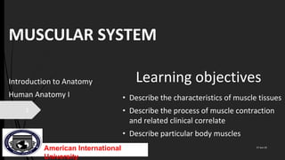
- 1. MUSCULAR SYSTEM Introduction to Anatomy Human Anatomy I • Describe the characteristics of muscle tissues • Describe the process of muscle contraction and related clinical correlate • Describe particular body muscles Learning objectives 17-Jun-22 1 American International University
- 2. 17-Jun-22 2 Smooth muscle Non-straited; mononucleated; Spindle-shaped (fusiform) cells; Innervated by the ANS; involuntary, weak, slow and sustained contraction Skeletal muscle Straited; multinucleated; cylindrical cells; innervated by the motor nerve; voluntary, quick, and strong contraction; 40% 0f body weight Cardiac muscle Straited; mononucleated; short cylindrical cells; branched fibers; cardiac conducting system causes its contraction; ANS, hormones regulate the heartrate; involuntary, quick, strong and continuous contraction
- 3. Arrangement of skeletal muscle Endomysium wraps muscle fiber Perimysium wraps muscle bundle/fasciculus Epimysium wraps whole muscle Tendon – unity of these connective tissues 17-Jun-22 3
- 4. Histology of a skeletal muscle fiber Organelles: multinucleated; sarcolemma; sarcoplasm; numerous myofibrils (contractile proteins); numerous mitochondria; sarcoplasmic reticulum (Ca2+ storage; wraps round myofibril); glycogen; myoglobin, transverse (T) tubules;. Myofibrils Contains numerous myofilaments (protein microfilaments). 17-Jun-22 4
- 5. Histology cont. Thick filaments - made of numerous myosin molecules Thin filaments – actin (2 twisted strands of G-actin monomers), tropomyosin (2 polypeptide chains in the groove of actin helix) and troponin (3 polypeptides- TnT, TnC, Tni). Thick and thin filaments overlap to form Dark (A, anisotropic) band, H zone, Light (I, isotropic) band. Z line to another Z line – sarcomere (smallest unit of myofibril) 17-Jun-22 5
- 6. Transverse (T) tubule system The system is the invagination of the sarcolemma that surrounds the myofibril. It is in close relationship with the terminal cisterna of sarcoplasmic reticulum The T tubules runs between 2 terminal cisternae, forming a triad (T tubule and 2 terminal cisternae) at the A-I junction Function: it conducts each impulse to the deepest regions of the muscle fiber, this ensures deeper myofibrils contract at the same time as the superficial ones. As the impulse travels, Ca2+ are released for contraction. 17-Jun-22 6
- 7. Neuromuscular Junction The point an axon terminal of a motor neuron communicates with a muscle fiber. Action potential gets to presynaptic terminal Voltage-gated calcium channels Ca2+ influx Synaptic vesicles at the synaptic knob move distally to eject neurotransmitter into the synaptic cleft. Acetylcholine (Ach), excitatory in action, binds to nicotinic acetylcholine receptors, nAchRs on the sarcolemma. nAChRs are ionotropic receptors and they function as ligand-gated channels. Na+ influx → depolarize muscle fiber NMJ is enclosed by basal lamina to prevent excess leakage 17-Jun-22 7
- 8. Myasthenia gravis Autoimmune disorder caused by autoantibodies against the postsynaptic Ach receptors Most common NMJ disorder and it is characterized by weakness and easy fatigue of muscles that worsens with muscle use, ptosis, diplopia. It tends to become fatal if respiratory muscles are affected. Ach receptors works for like 10 days and then they degraded by lysosomes and replaced with new ones produced by golgi apparatus. The attack of autoantibodies on the receptors reduces their half-life to about 2 days, leaving very few receptors available to propagate and sustain muscle contraction. Administration of AChE inhibitors (edrophonium, pyridostigmine) is used for diagnosis and treatment. AChE inhibitors ↓rate of ACh degradation thereby ↑ the binding time of Ach with the little available receptors. This improves muscle strength. Myasthenia gravis is associated with thymoma and thymic hyperplasia 17-Jun-22 8
- 9. Lambert-Eaton myasthenic syndrome Autoimmune disorder caused by autoantibodies against the presynaptic Ca2+ channels leading to low Ach release Uncommon NMJ disorder; characterized by proximal muscle weakness and autonomic symptoms (dry mouth and impotence) The features improves with the use of the muscles and it is associated with small cell lung cancer Administration of AChE inhibitors has little effect on reversing the symptoms 17-Jun-22 9 Spastic paralysis: caused by some pesticides that contain cholinesterase inhibitors that prevent degradation of ACh thereby increasing the binding-time of ACh making the muscle contract excessively. Flaccid paralysis: caused by venom or poison (curare)that competes with Ach on the nACh receptors, causing inhibited contraction of muscle
- 10. Muscle excitation, contraction and relaxation Muscle excitation: Na+ pumps open allowing Na+ influx into the muscle fiber (depolarization of the muscle fibre). Immediately, Na+ gates close and K+ gates open. K+ rush out so the inside becomes negative again (repolarization). Depolarization and repolarization create an action potential that excites the muscle fiber (outside in). Muscle contraction: threshold; Ca2+ exits to bind to troponin → tropomyosin changing position exposing myosin-binding sites on the actin; myosin binds to this site → filaments slide over themselves → shortens sarcomere (sliding filament theory). Myosin–actin cross-bridge. Muscle relaxation: nerve stimulation stops; Ach release stops; Ca2+ withdraw; breaking of cross-bridge → sarcomere elongates. 17-Jun-22
- 11. Muscles of facial expression and mastication 17-Jun-22 11
- 12. BELL’S PALSY Paralysis of upper and lower facial muscles due to lesion of ipsilateral facial nucleus or facial n. along its course Sudden symptoms that peak within 2 days, but pain in or behind the ear can precede by a day or two Mostly idiopathic or may be due to Herpes simplex virus 17-Jun-22 12 Evident distorted face on the affected size (sagging corner of the mouth; drooping of the eyebrow; sagging of the lower eyelid; inability to close or blink the eye; deviation of the lower jaw [lesion of n. to digastric]); inability to smile, whistle, or blow; tingling around the lips; decreased lacrimation, loss of taste in ant. 2/3 of tongue [lesion of chorda tympani branch], hyperacusis [due to paralyzed stapedius]. Treatment: corticosteroids (prednisone [anti-inflammatory]) and/or anti-viral (acyclovir) drugs. Most patients gradually recover function if they also avoid cold, wind, and eyes drying out.
- 13. Muscles of the tongue and pharynx 17-Jun-22 13 Muscles of the neck
- 14. Muscles of anterior abdominal wall 17-Jun-22 14 Muscles of the back
- 15. 17-Jun-22 15 Muscles of the perineum
- 16. 17-Jun-22 16 Muscles that control the arm Muscles of the thorax
- 17. Muscles of the arm 17-Jun-22 17
- 18. 17-Jun-22 18 Muscles of the thigh
- 19. 17-Jun-22 19 Muscles of the leg
- 20. Duchenne muscular dystrophy (DMD), X-linked recessive disorder caused by frameshift or nonsense mutation causing dystrophin to be missing or truncated. Dystrophin connects the cytoskeleton (actin) to the transmembrane proteins α- and β-dystroglycan (cytoskeleton-transmembrane association), which are connected to the extracellular matrix (ECM). Contractions of muscles thus pull at the weakened connections of the sarcolemma to create a leaky-hole through which extracellular Ca2+ flood the muscle fiber causing cell damage and eventual cell death. DMD causes muscle weakness from the pelvis upward (respiratory and cardiac muscles) It evident at age 2-5 years and victims rarely live past 30 years. 3/29/2023 20