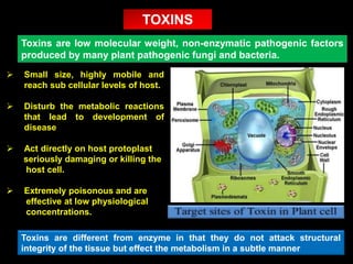
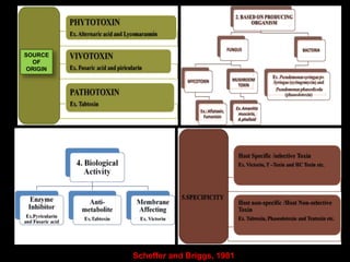
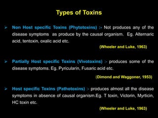
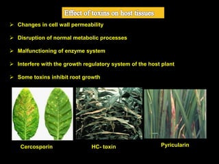
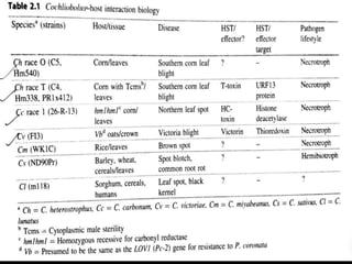
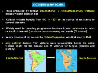
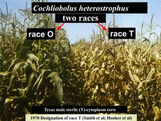
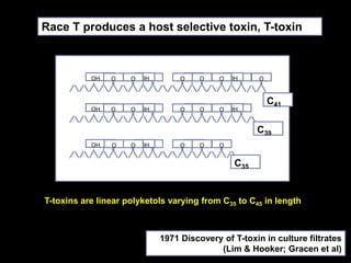
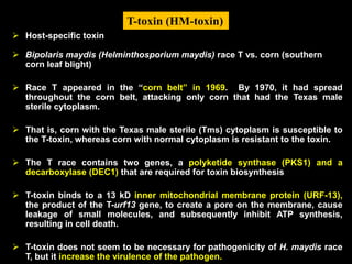
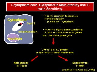
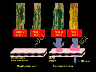
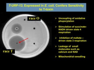
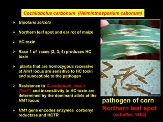
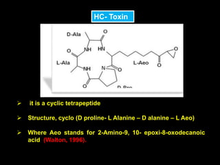
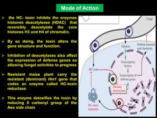
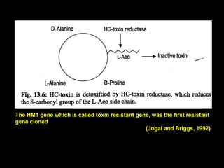
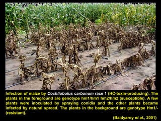
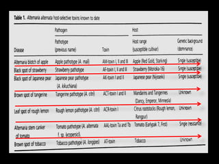
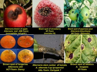
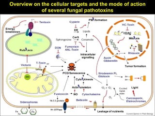
APM Welcome, APM North West Network Conference, Synergies Across Sectors
pathotoxin and Plant diseases
1. PATHOTOXINS AND PLANT DISEASES
Rathod Parshuram
Ph.D 1st year
Department of Plant Pathology
Indira Gandhi Krishi Vishwavidyalaya
Raipur (Chhattisgarh)
by
2. Toxins are low molecular weight, non-enzymatic pathogenic factors
produced by many plant pathogenic fungi and bacteria.
Small size, highly mobile and
reach sub cellular levels of host.
Disturb the metabolic reactions
that lead to development of
disease
Act directly on host protoplast
seriously damaging or killing the
host cell.
Extremely poisonous and are
effective at low physiological
concentrations.
TOXINS
Toxins are different from enzyme in that they do not attack structural
integrity of the tissue but effect the metabolism in a subtle manner
3. The idea that toxins play a causal role in disease causation
and death of cells was first enunciated by
De Bary (1886)
This is called the “toxin theory” of plant disease.
De Bary claimed that oxalic acid secreted by Sclerotinia sp,
was responsible for killing the cells.
Smith (1902) supported this for Botrytis cinerea
Brown (1915), working with the same fungus, disproved any such role for oxalic
acid.
Gaeumann (1954) belived that “ microorganisms are pathogenic only if they are
toxigenic”
The discovery of fusaric acid as a vivotoxin (1955), and victorin as a
pathotoxin (1963), received interest in toxin theory of disease, advanced by
De Bary.
History of Toxins
6. According to Wheeler and Luke, 1963 based on the source of
origin, toxins are divided into 3 broad classes namely.
Pathotoxins
Phytotoxins
Vivotoxins
(Wheeler and Luke, 1963)
Classification of toxins
7. Types of Toxins
Non Host specific Toxins (Phytotoxins) :- Not produces any of the
disease symptoms as produce by the causal organism. Eg. Alternaric
acid, tentoxin, oxalic acid etc.
Partially Host specific Toxins (Vivotoxins) :- produces some of the
disease symptoms. Eg. Pyricularin, Fusaric acid etc.
Host specific Toxins (Pathotoxins) :- produces almost all the disease
symptoms in absence of causal organism.Eg. T toxin, Victorin, Myrticin,
HC toxin etc.
(Wheeler and Luke, 1963)
(Dimond and Waggoner, 1953)
(Wheeler and Luke, 1963)
8. Changes in cell wall permeability
Disruption of normal metabolic processes
Malfunctioning of enzyme system
Interfere with the growth regulatory system of the host plant
Some toxins inhibit root growth
Cercosporin HC- toxin Pyricularin
9. Pathogen TP gene Host TS gene Result
+ + Disease
- + Resistance
+ - Resistance
- - Resistance
Host specific toxins (HSTs) are proteins or peptides that are directly
encoded by race specific, toxin producing gene (TP gene) of the pathogen.
The toxin are transported into cells by a highly coordinated delivery
system.
The HSTs are effective only on toxic sensitive cultivars, which harbor a
toxin sensitive (TS) gene.
The absence of either of both genes results in disease resistance, as
shown
10.
11. VICTORIN or HV TOXIN
Toxin produced by fungus Cochliobolus ( Helminthosporium) victoriae
causes victoria blight in oat
Cultivar victoria bought from SA in 1927 act as source of resistance to
several diseases
Widely used in breeding programme because it was resistance to most
races of crown rust (puccinia coronata avenae) and smuts (U. avenae)
A new disease of oat caused by Helminthosporium was first seen in 1944
only cultivar derived from victoria were susuceptible hence the name
victoria blight for the disease and H. victiriea for fungus (Meehan and
Murphy)
South America North America
12. Disease symptom
Toxin produced by Cochliobolus ( Helminthosporium) victoriae,
the fungus that causes victoria blight of oats.
Disease is characterized by necrosis of root and stem bases and
blighting of leaves
13. Low molecular weight Chlorinated,
Partially Cyclic Pentapeptide compound
Linked to a tricyclic amaine named
victotoxine (victaalanine)
Empirial formula of victotoxinine is
C17H29NO
The peptide is non toxic and contain
aspartic acid, glutamic acid, valine, and
leucine.
It is host specific toxin. Pentapeptide + victotoxinine
Therefore, characterization of the Vb gene and the underlying mechanisms of victorin
sensitivity may lead to understanding of both disease susceptibility and resistance.
Victorin is a peptide toxin that is required for the pathogenicity on oat cultivars carrying the
dominant Vb allele (Wolpert et al. 1985).
The Vb gene has not been separated genetically from Pc-2 a resistance gene against certain
races of crown rust fungi, Puccinia coronata f. sp. avenae , suggesting that both genes may
be identical.
Victorin- chemical structure
14. The current model of victorin-induced cellular responses
A. Victorin-sensitive cells B. Victorin-insensitive cells
15. Programmed cell death (apoptosis) and senescence
Curtis and Wolepert (2002) proved
that victorin causes PCD similar to
apoptosis in animal system
This change in mitochondria is due to
mitochondrial permeabiity transition
(MPT)
Victorin binds to the P-protein
component of glycine decarboxylase
enzyme complex (GDC) which plays
an important role in photorespiration
cycle
Victorin causes a loss of chlorophyll suggests that victorin triggers premature
senescence
Victorin also cleaves DNA forming ladder which is a very important indicator
of PDC
16. Ophiobolus heterostrophus
(Drechsler, 1925)
Cochliobolus heterostrophus
(Drechsler, 1934)
Anamorphs:
Helminthosporium maydis
(Nisikado & Miyake, 1926)
Bipolaris maydis
(Shoemaker, 1959)
Drechslera maydis
(Subramanian & P.C. Jain,1966)
Cochliobolus heterostrophus
Journal Of Agricultural Research
Vol. XXXI. 1925 by Charles Drechsler
asexual reproduction sexual reproduction
pathogen of corn
Southern Corn Leaf Blight
17. The host: Corn
One of the world’s three
most important cereal
crops
40% of the world
corn crop is produced
in the United States
(1968)
18.
19. T-cytoplasm corn
(genetic homogeneity)
Southern Corn
Leaf Blight
(1970)
Southern corn leaf blight (SCLB) is favored by warm temperatures
(68-90°F or 20-32°C) and high humidities.
Thus, it tends to be a problem in the Southern half of Illinois,
although it can be found farther north if weather conditions are
favourable.
Frequent rainy periods enhance disease development.
20. 1970 Designation of race T (Smith et al; Hooker et al)
two races
race T
Cochliobolus heterostrophus
race O
Texas male sterile (T)-cytoplasm corn
21. T-toxins are linear polyketols varying from C35 to C45 in length
OHOH O OOH OH O OOH OO O O
C41
OHOH O OOH OH O OOHO O O
C39
OHOH O OOH OH OO O O
C35
Race T produces a host selective toxin, T-toxin
1971 Discovery of T-toxin in culture filtrates
(Lim & Hooker; Gracen et al)
22. Host-specific toxin
Bipolaris maydis (Helminthosporium maydis) race T vs. corn (southern
corn leaf blight)
Race T appeared in the “corn belt” in 1969. By 1970, it had spread
throughout the corn belt, attacking only corn that had the Texas male
sterile cytoplasm.
That is, corn with the Texas male sterile (Tms) cytoplasm is susceptible to
the T-toxin, whereas corn with normal cytoplasm is resistant to the toxin.
The T race contains two genes, a polyketide synthase (PKS1) and a
decarboxylase (DEC1) that are required for toxin biosynthesis
T-toxin binds to a 13 kD inner mitochondrial membrane protein (URF-13),
the product of the T-urf13 gene, to create a pore on the membrane, cause
leakage of small molecules, and subsequently inhibit ATP synthesis,
resulting in cell death.
T-toxin does not seem to be necessary for pathogenicity of H. maydis race
T, but it increase the virulence of the pathogen.
T-toxin (HM-toxin)
23. URF13- a 13 kD protein
(mitochondrial inner membrane)
Male sterility
in T-corn
Sensitivity to
T- toxin
(modified from Wise et al, 1999)
nucleus
Cytoplasm
mitochondrion
T-urf13
T-cytoplasm corn, Cytoplasmic Male Sterility and T-
toxin Sensitivity
• T-corn: corn with Texas male
sterile cytoplasm
(T-cms, or T-cytoplasm)
• T-urf13- a hybrid gene consisting
of parts of 2 mitochondrial genes
and one chloroplast gene
24. race O
(Tox -)
race T
(Tox +)
race O
(Tox -)
race T
(Tox +)
N-cytoplasm corn T-cytoplasm corn
Mitochondrial
inner membrane
URF13
protein 600 m.w.
25. T-URF-13, Expressed in E. coli, Confers Sensitivity
to T-toxin
race T
race O Uncoupling of oxidative
phsporylation
Stimulation of succinate-
NADH driven state 4
respiration
inhibition of maltate –
driven state 3 respiration
Leakage of small
molecules such as
calcium and NAD
Mitochondrial swealling
26. Cochliobolus carbonum (Helminthosporium cabonum)
Bipolaris zeicola
Northern leaf spot and ear rot of maize
HC toxin
Race 1 of races (2, 3, 4) produces HC
toxin
plants that are homozygous recessive
at Hm1 locus are sensitive to HC toxin
and susceptible to the pathogen
Resistance to C. carbonum race 1
(Tox2+) and insensitivity to HC toxin are
determined by the dominant allele at the
HM1 locus
HM1 gene encodes enzymes corbonyl
reductase and HCTR
pathogen of corn
Northern leaf spot
(scheffer, 1965)
27. HC- Toxin
it is a cyclic tetrapeptide
Structure, cyclo (D proline- L Alanine – D alanine – L Aeo)
Where Aeo stands for 2-Amino-9, 10- epoxi-8-oxodecanoic
acid (Walton, 1996).
28. the HC- toxin inhibits the enzymes
histones deacetylease (HDAC) that
reversibly deacetylate the core
histones H3 and H4 of chromatin.
By so doing, the toxin alters the
gene structure and function.
Inhibition of deacetylases also affect
the expression of defense genes so
allowing fungal activities to progress
Resistant maize plant carry the
resistant (dominant) Hm1 gene that
codes an enzyme called HC-toxin
reductase
This enzyme detoxifies the toxin by
reducing 8 carbonyl group of the
Aeo side chain
Mode of Action
29. The HM1 gene which is called toxin resistant gene, was the first resistant
gene cloned
(Jogal and Briggs, 1992)
30. Infection of maize by Cochliobolus carbonum race 1 (HC-toxin-producing). The
plants in the foreground are genotype hm1/hm1 hm2/hm2 (susceptible). A few
plants were inoculated by spraying conidia and the other plants became
infected by natural spread. The plants in the background are genotype Hm1/-
(resistant).
(Baidyaroy et al., 2001)
31. Bipolaris (Helminthosporim)sacchari
Produce toxin called HS toxins
Causes eye spot disease of
sugarcane
Especially severe in seedling stage of
certain sugarcane cultivars
Host specific toxin produced by
fungus induces reddish brown
stripes when injected into
susceptible leaves.
The toxin bring about changes in
chloroplast and cell wall permeability
and is reported to bind a single
protein in susceptible host.
Structure of toxin is probably 2-
hydroxycyclopropyl-α- D-
galactopyranoside.
pathogen of sugarcane
Eye spot disease
32. Host selective toxin produced by the plant pathogenic
fungus Alternaria alternata
The genus Alternaria, the Hyphomycetes in the fungi imperfecti, includes
saprophytes on organic substrate and parasites on living organism
(Rotem, 1994; thomma, 2003)
The involvement of an HST in plant disease was first suggested in the
black spot disease of Japanese pear in 1933
(Tanaka, 1933)
This disease appeared after the new cultivar
Nijisseiki was introduced
Black spot disease of
Japanese pear
Beginning of 1970s, HST were identified from
other Alternaria pathogens , and there are
now seven disease caused by alterneria in
which HST are responsible for fungal
pathogenicity
33.
34. Alternaria blotch of apple
Alternaria mali (AM-Toxin)
(susceptible: red gold )
Black spot of strawberry
AF-Toxin
(morioka-16)
Black spot of japanese pear
Alterneria kikuchiania
AK-Toxin: (Nijisseiki)
Brown spot of tangerine
Alterneria citri
ACT-Toxin: Dancy
Alterneria stem canker of tomato
A. alternata f.sp lycopersici
AAL-Toxin: Earlypak 7
Brown spot of tobacco
A. longipes
AT-Toxin
35. Chemical structure of Alternaria HSTs
Epoxy- decatrieonic acid (EDA) family
Toxins of Japanese pear, strawberry and
tangerine pathotypes were found to be
structurally analogue metabolites
Esters of 9, 10-epoxy-8-hydroxy-9-
methyl-decatrionic acid (EDA)
AF- Toxin I, II, III- Strawberry cultivars
AK- Toxin I, II – Pear cultivars
ATC-Toxin I, II – mandarins & Tangerins
Host-selective toxins produced by Alternaria alternata 47
O EDA 2,3-DHIV
COOH O OH
36. Chemical structure of Alternaria HSTs
Cyclic depsipeptide (cyclic tetrapeptide)
Chemical structure of AM-Toxin I from
the apple pathotype was elucidated as
cyclic depsipeptide.
cause Alternaria blotch of apple
Aminopentol/polyketide
The tomato pathotype produces
aminopentol ester toxin called AAL toxin
Polyketide-ACR-toxin
the rough lemon pathotype produce
ACR-toxin
ACR-1 is a C19 poly alcohol with α-
dihydro pyrone ring
38. AAL-toxin and the mycotoxin fumonisin
are SAMs(sphinganine-analogue
mycotoxins).
Fumonisin is as selectively toxic to the
AAL- toxin-sensitive tomato genotypes
as AAL-toxin.
SAMs induce programmed cell death
in susceptible tomato cells and
mammalian cells by inhibiting ceramide
biosynthesis
When AAL-toxin-sensitive tomato tissue
was treated with SAMs, sphinganine
and phytosphingosine accumulated in
the tissue (Abbas et al., 1994).
AAL-toxin-induced cell death could be
avoided in sensitive tomato leaves by
feed-ing ceramide, indicating that a
ceramide imbalance is critical in
causing cell death
ALL-Toxin
39. Mechanism of ACR-toxin sensitivity controlled by
receptor transcript processing in mitochondria
Rough lemon (Citrus jambhiri)
40. HST works as an effector, having a suppressor function to HST-sensitive
plants as well as an elicitor function to HST-insensitive plants.
Yellow dots:
HST released from
germinated conidia
HST-sensitive plants
(Suppression of defense)
Suppression off defense
HST-insensitive
plants (Induction
of defense)
JA- dependent signaling pathway is not involved in host defense against the
toxigenic A. alternata pathogen but might effect pathogen accessibility by
mediating a susceptible response
A possible role of JA in
basal defense response
of plant against toxigenic,
necrotropic pathogen
was analysed using
tomato pathotype tomato
interaction as a model
system (Egusa et al.,
2009)
41. Pseudomonas syringae pv. tabaci
Wild fire disease of tobacco
Causes wild fire disease
Toxin producing strains cause
necrotic spot on leaves with spot
surrounded by yellow halo
Toxin reproduces all the symptoms of
disease but, since the symptoms are
produced on a large number of hosts
it is classified as NHS toxin
Cause chlorosis and necrotic
symptoms
42. Hydrolized
In the Host
Cell
Pseudomonas syringae pv. tabaci (Wildfire diseases tobacco
(non-toxic)
Tabtoxin (Non-Selective)
Structure of Tabtoxin
Hydrolized
In the Host
Cell
Tabtexinine-threonine
(tabtoxin, inactive)
Tabtoxin
(active)
Aminopeptidase
Dipeptide compound
43.
44. Overview on the cellular targets and the mode of action
of several fungal pathotoxins
45. Roles of pathotoxins in plant pathogenesis
As pathogenicity factors in plant-pathogen interactions
Victorin (HV-toxin)
HC-toxin
AK-toxin
AM-toxin
As virulence factors in plant-pathogen interactions
Tabtoxin
T-toxin
46. Conclusion
Pathotoxins employ an array of strategies to
distress, weaken or kill the host plant in order
to gain access to nutrients.
The captivating structural and mechanical
diversity of the toxins teaches us a lesson on
the complexity of pathogenic relationship.
understanding toxin biosynthesis pathways
and their regulation, the mode of action and
how this relates to fungal virulence will not
only help to gain new insights into cellular
processes in general but is also a stepping to
develop ways to protect plants from fungal
infections.
47. Agrios G. N 2005, Plant Pthology, 5th Edition Academic, New York.
B. J. Condon, D. Wu. B. G Turgeon, Comparative genomics of cochliobolus
phytopathogens, Genomics of Plant Associated Fungi: Monocot Pathogens
DOI: 10.1007/978-3-662-44053-7-2, Springer- Verlag Berlin Heidelberg 2014.
Harry Wheeler and H. H. Luke, Microbial Toxins in Plant Pathology, Botany
and Plant Pathology Department, Louisiana University, Baton Rouge,
Louisiana, Annu. Rev. Microbiol. 1963.17:223-242.
Thomas J. Wolpert, Larry D. Dunkle, Lynda M. Ciuffetti, Host Selective
Toxin and Avirulence, Annu. Rev. Phytopathol. 2002. 40: 40:251-85.
Ross B. Pringle, Robert P. Scheffer, Host Specific Pathotoxin, Annu. Rev.
Phytopathol, 1964.2: 133-156.
Takashi Tsuge et., al, Host-selective toxins produced by the plant
pathogenic fungus, Alternaria alternate Graduate school of Bioagricultureal
Science, Nagoya University, Nagoya 464-8601, 24 aug 2012.
Reference