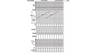
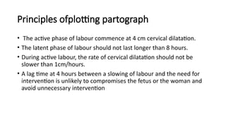
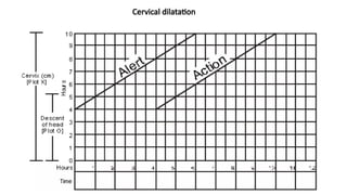
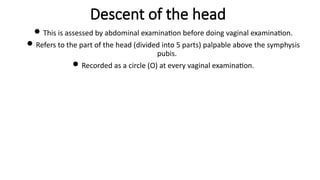
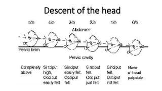
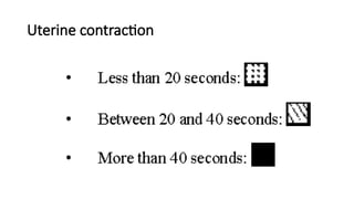
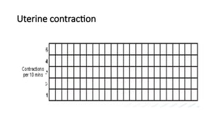
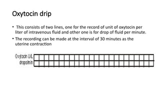
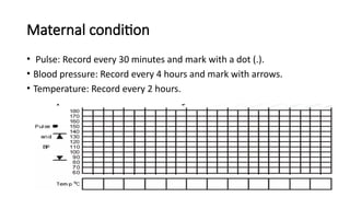
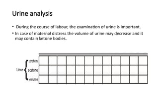
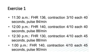
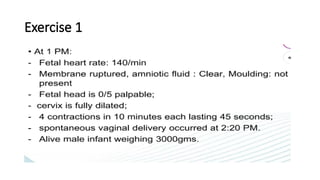
Partograph
Lecturer: Amina Abdikarim
Definition
• It is a composite graphical recording of cervical dilatation and descent
of head against duration of labour in hours. • It also gives information
about fetal and maternal condition that are all recorded on single
sheet of paper.
.
History
 E.A.freidman in 1954 provide a foundation basis for development of
partograph on the basis of observation of large number of woman in
labour.
 After that, the composite picture of labour was reported by philpott
in 1972, who combined details of progress of labour together with
information about fetal and maternal conditions
Advantage of using Partograph
1. A single sheet of paper can provide details of necessary information
at a glance.
2. No need to record labour events repeatedly
3. Gives clear picture of normality and abnormality in labour.
4. It can predict deviation from duration of labour. So appropriate steps
could be taken in time.
Advantage of using Partograph
5. It facilitates handover procedure of staffs.
6. Save working time of staff against writing labour notes in long hand.
7. Educational value for all staff.
Principles ofplotting partograph
• The active phase of labour commence at 4 cm cervical dilatation.
• The latent phase of labour should not last longer than 8 hours.
• During active labour, the rate of cervical dilatation should not be
slower than 1cm/hours.
• A lag time at 4 hours between a slowing of labour and the need for
intervention is unlikely to compromises the fetus or the woman and
avoid unnecessary intervention
Method of recording partograph
• Patient information: Fill out name, gravida, para, hospital number,
date and time of admission and time of ruptured membranes
Method of recording partograph
• Fetal heart rate: The rate of the fetal heart rate indicates the state of
the fetus inside the uterus. Record every half hour.
Method of recording partograph
• Amniotic fluid: Record the colour of amniotic fluid at every vaginal
examination:
• I: membranes intact;
• C: membranes ruptured, clear fluid;
• M: meconium-stained fluid;
• B: blood-stained fluid.
Moulding
• Moulding is a state of reduction or loss of space between skull bones.
• Presence of increased moulding of the head high in the pelvis
indicates CPD.
• Recording of degree of moulding
• 0: Bones are separated and sutures can be felt easily
• 1: sutures apposed
• 2: sutures overlapped but reducible
• 3: sutures overlapped and not reducible
Cervical dilatation
• Assessed at every vaginal examination and marked with a cross (X).
• Begin plotting on the partograph at 4 cm.
• This graph consists of homogenous squares, ten square vertically,
each square indicate one centimeter of cervical dilatation.
Cervical dilatation
Cervical dilatation
 The cross (X) in the graph are joined by a continuous line begin plotting on the
partograph at 4 cm.
 The climbing tendency of this line normally lies on the left of the middle of the graph.
 Alert line: A line starts at 4 cm of cervical dilatation to the point of expected full
dilatation at the rate of 1 cm per hour.
 Action line: Parallel and 4 hours to the right of the alert line
Descent of the head
This is assessed by abdominal examination before doing vaginal examination.
Refers to the part of the head (divided into 5 parts) palpable above the symphysis
pubis.
Recorded as a circle (O) at every vaginal examination.
Descent of the head
• Hours: Refers to the time elapsed since onset of active phase of
labour.
• Time: Record actual time
Uterine contractions
• Uterine contractions are recorded graphically on the partograph
according to their strength and frequency.
• Observation of contraction is made half hourly in the active phase.
• Palpate the number of contractions in 10 minutes and their duration
in seconds
Uterine contraction
Uterine contraction
Oxytocin drip
• This consists of two lines, one for the record of unit of oxytocin per
liter of intravenous fluid and other one is for drop of fluid per minute.
• The recording can be made at the interval of 30 minutes as the
uterine contraction
Drugs andother intravenous fluids
• Record any additional drug given and are recorded at the particular
point of time.
• This includes sedatives, antibiotics, IV fluids etc. The name of the
drugs and doses given should be written clearly in the long box.
Maternal condition
• Pulse: Record every 30 minutes and mark with a dot (.).
• Blood pressure: Record every 4 hours and mark with arrows.
• Temperature: Record every 2 hours.
Urine analysis
• During the course of labour, the examination of urine is important.
• In case of maternal distress the volume of urine may decrease and it
may contain ketone bodies.
Exercises
Exercise 1
• Mrs. Sita pokharel, 25 yrs old, Primigravida was admitted in the latent
phase of labour at 5 AM 2072/10/14:
• fetal head 4/5 palpable;
• cervix dilated 2 cm;
• 3 contractions in 10 minutes, each lasting 20 seconds;
• normal maternal and fetal condition.
Exercise 1
• At 9 AM:
• Fetal heart rate; 134/min
• Membrane: intact
• Moulding : sutures are not apposed.
• Fetal head is 3/5 palpable
• Cervix dilated 5 cm
• 4 contractions in 10 minutes, each lasting 20 seconds
• Mother’s Pulse: 80/min, BP: 110/70 mm of Hg, Temp: 98°F
Exercise1
Exercise 1
Exercise 1
partograph updated 2026 standardized visual tool.pptx

partograph updated 2026 standardized visual tool.pptx

  • 1.
  • 2.
    Definition • It isa composite graphical recording of cervical dilatation and descent of head against duration of labour in hours. • It also gives information about fetal and maternal condition that are all recorded on single sheet of paper. .
  • 4.
    History  E.A.freidman in1954 provide a foundation basis for development of partograph on the basis of observation of large number of woman in labour.  After that, the composite picture of labour was reported by philpott in 1972, who combined details of progress of labour together with information about fetal and maternal conditions
  • 5.
    Advantage of usingPartograph 1. A single sheet of paper can provide details of necessary information at a glance. 2. No need to record labour events repeatedly 3. Gives clear picture of normality and abnormality in labour. 4. It can predict deviation from duration of labour. So appropriate steps could be taken in time.
  • 6.
    Advantage of usingPartograph 5. It facilitates handover procedure of staffs. 6. Save working time of staff against writing labour notes in long hand. 7. Educational value for all staff.
  • 7.
    Principles ofplotting partograph •The active phase of labour commence at 4 cm cervical dilatation. • The latent phase of labour should not last longer than 8 hours. • During active labour, the rate of cervical dilatation should not be slower than 1cm/hours. • A lag time at 4 hours between a slowing of labour and the need for intervention is unlikely to compromises the fetus or the woman and avoid unnecessary intervention
  • 8.
    Method of recordingpartograph • Patient information: Fill out name, gravida, para, hospital number, date and time of admission and time of ruptured membranes
  • 9.
    Method of recordingpartograph • Fetal heart rate: The rate of the fetal heart rate indicates the state of the fetus inside the uterus. Record every half hour.
  • 10.
    Method of recordingpartograph • Amniotic fluid: Record the colour of amniotic fluid at every vaginal examination: • I: membranes intact; • C: membranes ruptured, clear fluid; • M: meconium-stained fluid; • B: blood-stained fluid.
  • 11.
    Moulding • Moulding isa state of reduction or loss of space between skull bones. • Presence of increased moulding of the head high in the pelvis indicates CPD. • Recording of degree of moulding • 0: Bones are separated and sutures can be felt easily • 1: sutures apposed • 2: sutures overlapped but reducible • 3: sutures overlapped and not reducible
  • 12.
    Cervical dilatation • Assessedat every vaginal examination and marked with a cross (X). • Begin plotting on the partograph at 4 cm. • This graph consists of homogenous squares, ten square vertically, each square indicate one centimeter of cervical dilatation.
  • 13.
  • 14.
    Cervical dilatation  Thecross (X) in the graph are joined by a continuous line begin plotting on the partograph at 4 cm.  The climbing tendency of this line normally lies on the left of the middle of the graph.  Alert line: A line starts at 4 cm of cervical dilatation to the point of expected full dilatation at the rate of 1 cm per hour.  Action line: Parallel and 4 hours to the right of the alert line
  • 15.
    Descent of thehead This is assessed by abdominal examination before doing vaginal examination. Refers to the part of the head (divided into 5 parts) palpable above the symphysis pubis. Recorded as a circle (O) at every vaginal examination.
  • 16.
  • 17.
    • Hours: Refersto the time elapsed since onset of active phase of labour. • Time: Record actual time
  • 18.
    Uterine contractions • Uterinecontractions are recorded graphically on the partograph according to their strength and frequency. • Observation of contraction is made half hourly in the active phase. • Palpate the number of contractions in 10 minutes and their duration in seconds
  • 19.
  • 20.
  • 21.
    Oxytocin drip • Thisconsists of two lines, one for the record of unit of oxytocin per liter of intravenous fluid and other one is for drop of fluid per minute. • The recording can be made at the interval of 30 minutes as the uterine contraction
  • 22.
    Drugs andother intravenousfluids • Record any additional drug given and are recorded at the particular point of time. • This includes sedatives, antibiotics, IV fluids etc. The name of the drugs and doses given should be written clearly in the long box.
  • 23.
    Maternal condition • Pulse:Record every 30 minutes and mark with a dot (.). • Blood pressure: Record every 4 hours and mark with arrows. • Temperature: Record every 2 hours.
  • 24.
    Urine analysis • Duringthe course of labour, the examination of urine is important. • In case of maternal distress the volume of urine may decrease and it may contain ketone bodies.
  • 25.
  • 26.
    Exercise 1 • Mrs.Sita pokharel, 25 yrs old, Primigravida was admitted in the latent phase of labour at 5 AM 2072/10/14: • fetal head 4/5 palpable; • cervix dilated 2 cm; • 3 contractions in 10 minutes, each lasting 20 seconds; • normal maternal and fetal condition.
  • 27.
    Exercise 1 • At9 AM: • Fetal heart rate; 134/min • Membrane: intact • Moulding : sutures are not apposed. • Fetal head is 3/5 palpable • Cervix dilated 5 cm • 4 contractions in 10 minutes, each lasting 20 seconds • Mother’s Pulse: 80/min, BP: 110/70 mm of Hg, Temp: 98°F
  • 28.
  • 29.
  • 30.