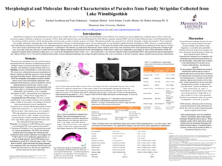
SlideShare a Scribd company logo
Morphological and Molecular Barcode Characteristics of Parasites from Family Strigeidae Collected from
Lake Winnibigoshish
Rachael Swedberg and Yuko Nakamura, Graduate Mentor: Tyler Achatz, Faculty Mentor: Dr. Robert Sorensen Ph. D
Minnesota State University, Mankato
rachael.yates-swedberg@mnsu.edu and yuko.nakamura@mnsu.edu
Introduction:
Identification of parasites can be problematic as many species have complex life cycles. To make matters for identification worse, plasticity of an organism can allow adaptations to a different species of host, which may
incorrectly suggest a different or subspecies of a parasite. In 2012, ducks and waterfowl were recovered from hunters by Holly Bloom, a graduate student of MSU, from the northern Minnesota lake, Lake Winnibigoshish. Inside
the intestines of these waterfowl, which included mallard, and ring neck, a number of similar parasites were found. The parasites initially were suspected to be of Family Strigeidae, a family of trematodes. These individuals
were initially identified as Strigeidae due to the blatant characteristic of having two distinguishable regions; the fore body usually in a cup formation and the hind body (Kostadinova, 2005). However, as mentioned before
initial identification could not be certain due to the phenotypic plasticity gained from variance in host or geographic region. In this study, the identity of the suspected individuals has been confirmed to be the species Cotylurus
brevis and Cotylurus flabelliformis and that of Apatemon . Confirmation of the identity was made from characteristics made visible by microscopy, both stained and SEM. Such characteristics included testis orientation and
size, ovary ratio, body ratio, and sizes of ventral and oral suckers. Ranges obtained and observations of sizes and morphology of the worms’ organs were comparable to past studies by Nasir (1962) and Dubois (1950). We
sequenced a portion of the cytochrome oxidase gene, cox1, to aid in the identification of these worms. Cox 1 was used because it is a universal sequence found in mitochondrial DNA and proposed for species determination
and relation (DeWalt, 2011). This will be helpful in future studies, because although morphology may change through the parasites life cycle or in relation to the host, its genetic markers should reveal an accurate identification.
Discussion
The confirmation technique that proved most
effective was internal staining. From this
structures linked to the identity of the
Stigeidae we noticeable and quantifiable.
Problems did occur in that some samples were
lost or destroyed suing the process which can
skew statistical results. However,
characteristics measured were congruent with
information presented from past research, as
seen in data tables. Such tables show evidence
of the identification of Cotylurus brevis,
Cotylurus flabelliformis, and Apatemon
gracillis.
We do recognize that variation has occurred
and that this could be due to a series of
unpredictable factors such as data of infection
of obtained birds or alteration to worms during
the staining process. It should be noted that
Apatemon species and Cotylurus brevis have
many similar characteristics but are noticeably
distinguished by number of eggs present,
ovary placement, vitellaria distribution, and
lobes of testis (Fig 1.A, and 3). C.
Flabelliformis is also identified by its small
size and characteristic c-shape (Fig 2). In
future studies we wish to more accurately
determine testis formation with cross sections
of suspected C. brevis and A. gracillis.
SEM technique, although useful in showing
external characteristics and confirmation of
family, but, was not useful in determining the
species or genus of test worms.
Gene sequencing has yet to be completed,
however, preliminary data would suggest
homology through the shared species. When
complete, results will be analyzed by genetic
software that can determine homology and
relation between species. If sequence data is
accurate, results may be posted on iBOL or
International Barcode of Life.
Sourcees
Campbell, R. A. (1973). Studies on the Biology of the Life Cycle of Cotylurus flabelliformis (Trematoda:
Strigeidae). Transactions of the American Microscopical Society, 92(4), 629-640. Retrieved from
http://www.jstor.org/discover/10.2307/3225273?
uid=3739736&uid=2&uid=4&uid=3739256&sid=21103981334153
DeWalt, R. E. (2011). DNA barcoding: a taxonomic point of view. Journal of the North American Benthological
Society, 30(1), 174–181. doi:10.1899/10-021.1
Drago, F. B., & Lunaschi, L. I. (2010). Digenea, Strigeidae, Australapatemon canadensis Dubois and Rausch, 1950:
First record in South America and a new host record. Journal of species lists and distribution, 6(3), 382-384.
Retrieved from file:///Users/Yuko/Downloads/digenea%252c%20strigeidae%252c%20australapatemon
%20canadensis.pdf
Dubois, G., & Rausch, R. (1950). A Contribution to the Study of North American Strigeids (Trematoda). American
Midland Naturalist, 43(1), 1-31. Retrieved from http://www.jstor.org/discover/10.2307/2421874?
uid=3739736&uid=2&uid=4&uid=3739256&sid=21103981334153
Kostadinova, A. (2005). Family Psilostomidae Looss, 1990. In A. Jones, R. Bray & D. Gibson (Eds.), Keys to the
Trematoda (Vol. 2, pp. 99-118). London, UK: CABI Publishing and the Natural History Museum.
McDonald, M. E. (1981). Key to Trematodes Reported in Waterfowl. Washington, D.C.: U.S. Department of the
Interior : Fish and Wildlife Service.
Nasir, P. (1962). On the Identification of the Cercaria of Cotylurus brevis Dubois and Rausch, 1950, (Trematoda:
Strigeida) and Genitalia of the Adult. HELMINTHOLOGICAL SOCIETY, 29(1), 82-87. Retrieved from
file:///Users/Yuko/Downloads/On%20the%20Identification%20of%20the%20cercaria%20of%20Cotylurus
%20brevis-%20Nasir.pdf
Results:
C. Flabelliformis Results (Literature)
Results
(Experiemental)
Total body size
0.76-1.12 mm (Campbell,
1937) 0.73- 1.21mm
Oral sucker subequal to ventral sucker
(McDonald, 1981)
oral suck is about 76%
of ventral sucker
Testie shape Bean (Campbell, 1937) bilobed or trilobed
body shape (McDonald, 1981) bent/ c- shaped x ≤ 90 °
Fig 1) The three above picture depict Cotylurus brevis. The image on the left is an illustration drawn by Nasir in 1962
which acts as a guide for identification of organ system. Image B was experimentally obtained and stained with
acetocarmine. This worms internal structure is well presented and has been labeled as VS is Ventral sucker, OS is oral
sucker, Ov is ovary, AT is Anterior testis and PT is posterior testis. An egg, E, is also seen. PT shows a trilobed structure
which identifies the specimen as C. brevis. Images C and D are SEM images showing the external structures. In D we can
see an egg emerging from the genital pore.
Fig 2) Cotylurus flabelliformis obtained from
mallard. These were distinguished by their
significantly smaller size and the characteristic
c-shape formed by the hind body and the fore
body. In the Image A noticeable adhesion
organs (AO) in the fore body is seen. Image B
is shown to show more average body shape.
Both images obtained at 100 x magnification.
Methods:
Suspected type Strigeidae were only used for analysis
and gained from the intestines of mallard and ring neck.
Sampled worms were preserved either by freezing or
formalin until staining or genetic testing could occur.
Staining was done with Semichon’s acetocarmin stain.
The worms were subjected increasing concentrations of
ethanol, totaling an eight step process to extract formalin
and water from their corpses. After two rounds in 100%
EtOH the trematodes were soaked in xylene to further
remove excess water and formalin. Specimens were
mounted in Kleermount in a series of positions to reveal
internal structures such as testis, ovaries, or any
intestinal structures. Characteristics were identified with
keys such as Key to Trematodes Reported in Waterfowl
(McDonald, 1981) and Keys to the Trematode
(Kostadinova, 2005). Measurements were obtained on
microscopy software Motic Images Plus 2.0.
Scanning Electron Microscopy was done to obtain
identification of any external structures such as holdfast
organs, ventral or oral suckers, or anything of structures
about the genital pore to aid in reproduction.
Trematodes were washed with phosphate buffer solution
(PBS) before exposed to a 1:1 ratio of 2% OsO4 and
PBS for an hour. After which worms were washed in
PBS and subjected to increasing concentrations of
acetone, from 50% to 100%. Samples were dried in a
critical point drier and then mounted to double sided
sticky tape. Before going under the SEM worms were
dust with gold and exposed to argon.
Genetics was focused on a 600-700 base pair sequence
in mitochondrial DNA, the sequence that codes for
Cytochrome O Oxidase, (Cox1), an universal gene used
for species determination .The DNA was extracted
using the DNA Easy Qiagen Kit. During the first
polymerase chain reaction replisomes were tagged with
T7-HCO and T3-LCO, artificially tagged primers, to
specify wanted sequence. After DNA presence was
determined, a second PCR with only tags T7 and T3 was
done to amplify signal and ensure the proper basepairs
were sequenced. DNA was extracted from PCR gel with
ZymoClean DNA Recovery Kit and was loaded into a
polyacrylimide gel for gene sequencing.
Table 1: A comparison of organ ranges
discovered from literature and experimental
identification.
Fig 3) The above image is of Apatemon gracillis.
This conclusion was based on the over body size ~ <
2 mm and the bilobed testis. A notable characteristic
of Apatemon is also displayed- the small patch of
vitellaria (V) in the fore body. A notable seminal
duct or uterus is also present.
Table 2) the comparison of C. flabelliformis between results
gained from experimentation and known characteristics of
literature. Averages found are placed in historic ranges and other
characteristics match literature descriptions
Cotylurus Brevis
Results (Dubois and
Rausch ,1950) mm
Results
(Experiemental)
mm
total body length 1.17-1.8 1.37 +/- 0.0330
Forebody .042-.072 0.536 +/- 0.0199
Hindbody .075-.1080 0.846 +/- 0.0261
body ratio 1.48-1.8 1.65+/- 0.0485
Diameter of ovary .091-.096/.07-.074 0.116 +/- .00614
Anterior testies .190-.2/?
0.170 +/-
0.0107/ 0.159
+/- 0.0139
Posterior testies .180-.215/?
0.213 +/-
0.0151/ 0.166
+/- 0.0122
Diameter of eggs .092-.103/.063-.07
0.0964 +/-
0.00189/0.0639
+/- 0.0013
Ovary position 0.11-0.22 0.117 +/- 0.0285
Testie shape Trilobe down Trilobe down
A. gracilis Results (Drago and Lunaschi, 2010) mm Results (Experiemental) mm
total body length upto 3.2 mm 2.108 +/- 0.405
forebody 0.510-0.96x0.37-0.770 0.766 +/- 0.141
hindbody 0.87-2.227x .420-.9 1.343 +/- 0.359
body ratio 1.2-2.8 1.72 +/- 0.610
Diameter of oral Sucker 0.12-2x 0.105-0.17 0.144 +/- 0.0548 x 0.148 +/- 0.0309
Diameter of ventral Sucker 0.14-0.245 x 0.16-0.235 0.180 +/- 0.0347 x 0.167 +/- 0.0369
Diameter of ovary 0.105-0.19 x 0.125-0.21 0.177 +/- 0.0625 x 0.230 +/- 0.0976
Anterior testies 0.25-0.47 x 0.235-0.440 0.269 +/- 0.0984 x 0.261 +/- 0.0610
Posterior testies 0.335-0.64 x 0.24-0.475 0.279 +/- 0.0617 x 0.207 +/- 0.0601
Diameter of eggs 0.095-0.125x0.065-0.08 0.0708 +/- 0.0195 X 0.0886 +/- 0.0169
egg number 20 x>35
Testie shape bilobe up bilobed
This document is available in alternative format to individuals with disabilities by calling Accessibility
Resources at 507-389-2825 (V), 800-627-3529 or 711 (MRS/TTY).
For the URC Grant: This research is supported a grant from the MSU-Mankato Undergraduate Research
Center.
Tables 3) Numbers below were published by Drago and Lunaschi (2010) comparing known A gracillis values to their
own found in in South America. We compared our averages to the same known values- supporting our identification.
A B
40x
Ov
P.T
A.T
E
V.S
O.S
A B
C D
100 x
AO
PT
V
VSSD
PT
HF

More Related Content

What's hot

Taxonomy and classification Implications for avian identification
Taxonomy and classification Implications for avian identificationTaxonomy and classification Implications for avian identification
Taxonomy and classification Implications for avian identificationNicola snow
 
UC Davis EVE161 Lecture 14 by @phylogenomics
UC Davis EVE161 Lecture 14 by @phylogenomicsUC Davis EVE161 Lecture 14 by @phylogenomics
UC Davis EVE161 Lecture 14 by @phylogenomics
Jonathan Eisen
 
RICHELLE SOPKO_resume_042215
RICHELLE SOPKO_resume_042215RICHELLE SOPKO_resume_042215
RICHELLE SOPKO_resume_042215Richelle Sopko
 
UC Davis EVE161 Lecture 15 by @phylogenomics
UC Davis EVE161 Lecture 15 by @phylogenomicsUC Davis EVE161 Lecture 15 by @phylogenomics
UC Davis EVE161 Lecture 15 by @phylogenomics
Jonathan Eisen
 
Microbial Phylogenomics (EVE161) Class 14: Metagenomics
Microbial Phylogenomics (EVE161) Class 14: MetagenomicsMicrobial Phylogenomics (EVE161) Class 14: Metagenomics
Microbial Phylogenomics (EVE161) Class 14: Metagenomics
Jonathan Eisen
 
EVE 161 Winter 2018 Class 10
EVE 161 Winter 2018 Class 10EVE 161 Winter 2018 Class 10
EVE 161 Winter 2018 Class 10
Jonathan Eisen
 
Microbial Phylogenomics (EVE161) Class 3: Woese and the Tree of Life
Microbial Phylogenomics (EVE161) Class 3: Woese and the Tree of LifeMicrobial Phylogenomics (EVE161) Class 3: Woese and the Tree of Life
Microbial Phylogenomics (EVE161) Class 3: Woese and the Tree of Life
Jonathan Eisen
 
Microbial Phylogenomics (EVE161) Class 10-11: Genome Sequencing
Microbial Phylogenomics (EVE161) Class 10-11: Genome SequencingMicrobial Phylogenomics (EVE161) Class 10-11: Genome Sequencing
Microbial Phylogenomics (EVE161) Class 10-11: Genome Sequencing
Jonathan Eisen
 
Evolutionary genomics
Evolutionary genomicsEvolutionary genomics
Evolutionary genomics
boussau
 
Marine Host-Microbiome Interactions: Challenges and Opportunities
Marine Host-Microbiome Interactions: Challenges and OpportunitiesMarine Host-Microbiome Interactions: Challenges and Opportunities
Marine Host-Microbiome Interactions: Challenges and Opportunities
Jonathan Eisen
 
Dave Lunt presentation to Nottingham UKNGS 2013
Dave Lunt presentation to Nottingham UKNGS 2013Dave Lunt presentation to Nottingham UKNGS 2013
Dave Lunt presentation to Nottingham UKNGS 2013davelunt
 
EVE 161 Winter 2018 Class 17
EVE 161 Winter 2018 Class 17EVE 161 Winter 2018 Class 17
EVE 161 Winter 2018 Class 17
Jonathan Eisen
 
1 history of genetics
1 history of genetics1 history of genetics
1 history of genetics
AshokD25
 
EveMicrobial Phylogenomics (EVE161) Class 9
EveMicrobial Phylogenomics (EVE161) Class 9EveMicrobial Phylogenomics (EVE161) Class 9
EveMicrobial Phylogenomics (EVE161) Class 9
Jonathan Eisen
 
EVE 161 Winter 2018 Class 18
EVE 161 Winter 2018 Class 18EVE 161 Winter 2018 Class 18
EVE 161 Winter 2018 Class 18
Jonathan Eisen
 
Microbial Phylogenomics (EVE161) Class 15: Shotgun Metagenomics
Microbial Phylogenomics (EVE161) Class 15: Shotgun Metagenomics Microbial Phylogenomics (EVE161) Class 15: Shotgun Metagenomics
Microbial Phylogenomics (EVE161) Class 15: Shotgun Metagenomics
Jonathan Eisen
 

What's hot (16)

Taxonomy and classification Implications for avian identification
Taxonomy and classification Implications for avian identificationTaxonomy and classification Implications for avian identification
Taxonomy and classification Implications for avian identification
 
UC Davis EVE161 Lecture 14 by @phylogenomics
UC Davis EVE161 Lecture 14 by @phylogenomicsUC Davis EVE161 Lecture 14 by @phylogenomics
UC Davis EVE161 Lecture 14 by @phylogenomics
 
RICHELLE SOPKO_resume_042215
RICHELLE SOPKO_resume_042215RICHELLE SOPKO_resume_042215
RICHELLE SOPKO_resume_042215
 
UC Davis EVE161 Lecture 15 by @phylogenomics
UC Davis EVE161 Lecture 15 by @phylogenomicsUC Davis EVE161 Lecture 15 by @phylogenomics
UC Davis EVE161 Lecture 15 by @phylogenomics
 
Microbial Phylogenomics (EVE161) Class 14: Metagenomics
Microbial Phylogenomics (EVE161) Class 14: MetagenomicsMicrobial Phylogenomics (EVE161) Class 14: Metagenomics
Microbial Phylogenomics (EVE161) Class 14: Metagenomics
 
EVE 161 Winter 2018 Class 10
EVE 161 Winter 2018 Class 10EVE 161 Winter 2018 Class 10
EVE 161 Winter 2018 Class 10
 
Microbial Phylogenomics (EVE161) Class 3: Woese and the Tree of Life
Microbial Phylogenomics (EVE161) Class 3: Woese and the Tree of LifeMicrobial Phylogenomics (EVE161) Class 3: Woese and the Tree of Life
Microbial Phylogenomics (EVE161) Class 3: Woese and the Tree of Life
 
Microbial Phylogenomics (EVE161) Class 10-11: Genome Sequencing
Microbial Phylogenomics (EVE161) Class 10-11: Genome SequencingMicrobial Phylogenomics (EVE161) Class 10-11: Genome Sequencing
Microbial Phylogenomics (EVE161) Class 10-11: Genome Sequencing
 
Evolutionary genomics
Evolutionary genomicsEvolutionary genomics
Evolutionary genomics
 
Marine Host-Microbiome Interactions: Challenges and Opportunities
Marine Host-Microbiome Interactions: Challenges and OpportunitiesMarine Host-Microbiome Interactions: Challenges and Opportunities
Marine Host-Microbiome Interactions: Challenges and Opportunities
 
Dave Lunt presentation to Nottingham UKNGS 2013
Dave Lunt presentation to Nottingham UKNGS 2013Dave Lunt presentation to Nottingham UKNGS 2013
Dave Lunt presentation to Nottingham UKNGS 2013
 
EVE 161 Winter 2018 Class 17
EVE 161 Winter 2018 Class 17EVE 161 Winter 2018 Class 17
EVE 161 Winter 2018 Class 17
 
1 history of genetics
1 history of genetics1 history of genetics
1 history of genetics
 
EveMicrobial Phylogenomics (EVE161) Class 9
EveMicrobial Phylogenomics (EVE161) Class 9EveMicrobial Phylogenomics (EVE161) Class 9
EveMicrobial Phylogenomics (EVE161) Class 9
 
EVE 161 Winter 2018 Class 18
EVE 161 Winter 2018 Class 18EVE 161 Winter 2018 Class 18
EVE 161 Winter 2018 Class 18
 
Microbial Phylogenomics (EVE161) Class 15: Shotgun Metagenomics
Microbial Phylogenomics (EVE161) Class 15: Shotgun Metagenomics Microbial Phylogenomics (EVE161) Class 15: Shotgun Metagenomics
Microbial Phylogenomics (EVE161) Class 15: Shotgun Metagenomics
 

Similar to parasite poster for linkin

Gutell 088.mpe.2003.29.0258
Gutell 088.mpe.2003.29.0258Gutell 088.mpe.2003.29.0258
Gutell 088.mpe.2003.29.0258Robin Gutell
 
2012 -- Alderslade_McFadden -- Jasonisis
2012 -- Alderslade_McFadden -- Jasonisis2012 -- Alderslade_McFadden -- Jasonisis
2012 -- Alderslade_McFadden -- JasonisisPhil Alderslade
 
Bioinformatica 24-11-2011-t6-phylogenetics
Bioinformatica 24-11-2011-t6-phylogeneticsBioinformatica 24-11-2011-t6-phylogenetics
Bioinformatica 24-11-2011-t6-phylogenetics
Prof. Wim Van Criekinge
 
Paper to Upload, MOLECULAR PHYLOGENY OF CATFISHES.pdf
Paper to Upload, MOLECULAR PHYLOGENY OF CATFISHES.pdfPaper to Upload, MOLECULAR PHYLOGENY OF CATFISHES.pdf
Paper to Upload, MOLECULAR PHYLOGENY OF CATFISHES.pdf
OanhTrng13
 
Genetic analysis of cavefish reveals molecular convergencein.docx
Genetic analysis of cavefish reveals molecular convergencein.docxGenetic analysis of cavefish reveals molecular convergencein.docx
Genetic analysis of cavefish reveals molecular convergencein.docx
budbarber38650
 
D_leucolena_poster_MRB
D_leucolena_poster_MRBD_leucolena_poster_MRB
D_leucolena_poster_MRBFreddy Castro
 
Eight Primate Research
Eight Primate ResearchEight Primate Research
Eight Primate Research
Jan Champagne
 
Reproductive traits in dicyemids
Reproductive traits in dicyemidsReproductive traits in dicyemids
Reproductive traits in dicyemidsdreicash
 
Age Related Histomorphological and Transmission Electron Microscopic Studies ...
Age Related Histomorphological and Transmission Electron Microscopic Studies ...Age Related Histomorphological and Transmission Electron Microscopic Studies ...
Age Related Histomorphological and Transmission Electron Microscopic Studies ...
iosrjce
 
Evolution of North American Micruracarus
Evolution of North American MicruracarusEvolution of North American Micruracarus
Evolution of North American Micruracarus
Rachel Shoop
 
Goldy ABRCMS Poster Final
Goldy ABRCMS Poster FinalGoldy ABRCMS Poster Final
Goldy ABRCMS Poster FinalGoldy Landau
 
Query Sequence And Analysis Of Verify3D
Query Sequence And Analysis Of Verify3DQuery Sequence And Analysis Of Verify3D
Query Sequence And Analysis Of Verify3D
Amber Wheeler
 
5.4 cladistics student copy
5.4 cladistics student copy5.4 cladistics student copy
5.4 cladistics student copy
lucascw
 
Conventional & newer aspects in taxonomy
Conventional & newer aspects in taxonomyConventional & newer aspects in taxonomy
Conventional & newer aspects in taxonomy
Manideep Raj
 
The need for a phylogeny driven genomic encyclopedia of eukaryotes #SMBEEuks
The need for a phylogeny driven genomic encyclopedia of eukaryotes #SMBEEuksThe need for a phylogeny driven genomic encyclopedia of eukaryotes #SMBEEuks
The need for a phylogeny driven genomic encyclopedia of eukaryotes #SMBEEuks
Jonathan Eisen
 
485 lec2 history and review (i)
485 lec2 history and review (i)485 lec2 history and review (i)
485 lec2 history and review (i)
hhalhaddad
 

Similar to parasite poster for linkin (20)

Gutell 088.mpe.2003.29.0258
Gutell 088.mpe.2003.29.0258Gutell 088.mpe.2003.29.0258
Gutell 088.mpe.2003.29.0258
 
2000 JME (51)278-285
2000 JME (51)278-2852000 JME (51)278-285
2000 JME (51)278-285
 
2012 -- Alderslade_McFadden -- Jasonisis
2012 -- Alderslade_McFadden -- Jasonisis2012 -- Alderslade_McFadden -- Jasonisis
2012 -- Alderslade_McFadden -- Jasonisis
 
Bioinformatica 24-11-2011-t6-phylogenetics
Bioinformatica 24-11-2011-t6-phylogeneticsBioinformatica 24-11-2011-t6-phylogenetics
Bioinformatica 24-11-2011-t6-phylogenetics
 
Paper to Upload, MOLECULAR PHYLOGENY OF CATFISHES.pdf
Paper to Upload, MOLECULAR PHYLOGENY OF CATFISHES.pdfPaper to Upload, MOLECULAR PHYLOGENY OF CATFISHES.pdf
Paper to Upload, MOLECULAR PHYLOGENY OF CATFISHES.pdf
 
Genetic analysis of cavefish reveals molecular convergencein.docx
Genetic analysis of cavefish reveals molecular convergencein.docxGenetic analysis of cavefish reveals molecular convergencein.docx
Genetic analysis of cavefish reveals molecular convergencein.docx
 
D_leucolena_poster_MRB
D_leucolena_poster_MRBD_leucolena_poster_MRB
D_leucolena_poster_MRB
 
Eight Primate Research
Eight Primate ResearchEight Primate Research
Eight Primate Research
 
Reproductive traits in dicyemids
Reproductive traits in dicyemidsReproductive traits in dicyemids
Reproductive traits in dicyemids
 
Age Related Histomorphological and Transmission Electron Microscopic Studies ...
Age Related Histomorphological and Transmission Electron Microscopic Studies ...Age Related Histomorphological and Transmission Electron Microscopic Studies ...
Age Related Histomorphological and Transmission Electron Microscopic Studies ...
 
Brayner,2005
Brayner,2005Brayner,2005
Brayner,2005
 
Evolution of North American Micruracarus
Evolution of North American MicruracarusEvolution of North American Micruracarus
Evolution of North American Micruracarus
 
Goldy ABRCMS Poster Final
Goldy ABRCMS Poster FinalGoldy ABRCMS Poster Final
Goldy ABRCMS Poster Final
 
Query Sequence And Analysis Of Verify3D
Query Sequence And Analysis Of Verify3DQuery Sequence And Analysis Of Verify3D
Query Sequence And Analysis Of Verify3D
 
5.4 cladistics student copy
5.4 cladistics student copy5.4 cladistics student copy
5.4 cladistics student copy
 
Conventional & newer aspects in taxonomy
Conventional & newer aspects in taxonomyConventional & newer aspects in taxonomy
Conventional & newer aspects in taxonomy
 
The need for a phylogeny driven genomic encyclopedia of eukaryotes #SMBEEuks
The need for a phylogeny driven genomic encyclopedia of eukaryotes #SMBEEuksThe need for a phylogeny driven genomic encyclopedia of eukaryotes #SMBEEuks
The need for a phylogeny driven genomic encyclopedia of eukaryotes #SMBEEuks
 
485 lec2 history and review (i)
485 lec2 history and review (i)485 lec2 history and review (i)
485 lec2 history and review (i)
 
SICB Poster
SICB PosterSICB Poster
SICB Poster
 
Pielak_DevBiol2004
Pielak_DevBiol2004Pielak_DevBiol2004
Pielak_DevBiol2004
 

parasite poster for linkin

  • 1. Morphological and Molecular Barcode Characteristics of Parasites from Family Strigeidae Collected from Lake Winnibigoshish Rachael Swedberg and Yuko Nakamura, Graduate Mentor: Tyler Achatz, Faculty Mentor: Dr. Robert Sorensen Ph. D Minnesota State University, Mankato rachael.yates-swedberg@mnsu.edu and yuko.nakamura@mnsu.edu Introduction: Identification of parasites can be problematic as many species have complex life cycles. To make matters for identification worse, plasticity of an organism can allow adaptations to a different species of host, which may incorrectly suggest a different or subspecies of a parasite. In 2012, ducks and waterfowl were recovered from hunters by Holly Bloom, a graduate student of MSU, from the northern Minnesota lake, Lake Winnibigoshish. Inside the intestines of these waterfowl, which included mallard, and ring neck, a number of similar parasites were found. The parasites initially were suspected to be of Family Strigeidae, a family of trematodes. These individuals were initially identified as Strigeidae due to the blatant characteristic of having two distinguishable regions; the fore body usually in a cup formation and the hind body (Kostadinova, 2005). However, as mentioned before initial identification could not be certain due to the phenotypic plasticity gained from variance in host or geographic region. In this study, the identity of the suspected individuals has been confirmed to be the species Cotylurus brevis and Cotylurus flabelliformis and that of Apatemon . Confirmation of the identity was made from characteristics made visible by microscopy, both stained and SEM. Such characteristics included testis orientation and size, ovary ratio, body ratio, and sizes of ventral and oral suckers. Ranges obtained and observations of sizes and morphology of the worms’ organs were comparable to past studies by Nasir (1962) and Dubois (1950). We sequenced a portion of the cytochrome oxidase gene, cox1, to aid in the identification of these worms. Cox 1 was used because it is a universal sequence found in mitochondrial DNA and proposed for species determination and relation (DeWalt, 2011). This will be helpful in future studies, because although morphology may change through the parasites life cycle or in relation to the host, its genetic markers should reveal an accurate identification. Discussion The confirmation technique that proved most effective was internal staining. From this structures linked to the identity of the Stigeidae we noticeable and quantifiable. Problems did occur in that some samples were lost or destroyed suing the process which can skew statistical results. However, characteristics measured were congruent with information presented from past research, as seen in data tables. Such tables show evidence of the identification of Cotylurus brevis, Cotylurus flabelliformis, and Apatemon gracillis. We do recognize that variation has occurred and that this could be due to a series of unpredictable factors such as data of infection of obtained birds or alteration to worms during the staining process. It should be noted that Apatemon species and Cotylurus brevis have many similar characteristics but are noticeably distinguished by number of eggs present, ovary placement, vitellaria distribution, and lobes of testis (Fig 1.A, and 3). C. Flabelliformis is also identified by its small size and characteristic c-shape (Fig 2). In future studies we wish to more accurately determine testis formation with cross sections of suspected C. brevis and A. gracillis. SEM technique, although useful in showing external characteristics and confirmation of family, but, was not useful in determining the species or genus of test worms. Gene sequencing has yet to be completed, however, preliminary data would suggest homology through the shared species. When complete, results will be analyzed by genetic software that can determine homology and relation between species. If sequence data is accurate, results may be posted on iBOL or International Barcode of Life. Sourcees Campbell, R. A. (1973). Studies on the Biology of the Life Cycle of Cotylurus flabelliformis (Trematoda: Strigeidae). Transactions of the American Microscopical Society, 92(4), 629-640. Retrieved from http://www.jstor.org/discover/10.2307/3225273? uid=3739736&uid=2&uid=4&uid=3739256&sid=21103981334153 DeWalt, R. E. (2011). DNA barcoding: a taxonomic point of view. Journal of the North American Benthological Society, 30(1), 174–181. doi:10.1899/10-021.1 Drago, F. B., & Lunaschi, L. I. (2010). Digenea, Strigeidae, Australapatemon canadensis Dubois and Rausch, 1950: First record in South America and a new host record. Journal of species lists and distribution, 6(3), 382-384. Retrieved from file:///Users/Yuko/Downloads/digenea%252c%20strigeidae%252c%20australapatemon %20canadensis.pdf Dubois, G., & Rausch, R. (1950). A Contribution to the Study of North American Strigeids (Trematoda). American Midland Naturalist, 43(1), 1-31. Retrieved from http://www.jstor.org/discover/10.2307/2421874? uid=3739736&uid=2&uid=4&uid=3739256&sid=21103981334153 Kostadinova, A. (2005). Family Psilostomidae Looss, 1990. In A. Jones, R. Bray & D. Gibson (Eds.), Keys to the Trematoda (Vol. 2, pp. 99-118). London, UK: CABI Publishing and the Natural History Museum. McDonald, M. E. (1981). Key to Trematodes Reported in Waterfowl. Washington, D.C.: U.S. Department of the Interior : Fish and Wildlife Service. Nasir, P. (1962). On the Identification of the Cercaria of Cotylurus brevis Dubois and Rausch, 1950, (Trematoda: Strigeida) and Genitalia of the Adult. HELMINTHOLOGICAL SOCIETY, 29(1), 82-87. Retrieved from file:///Users/Yuko/Downloads/On%20the%20Identification%20of%20the%20cercaria%20of%20Cotylurus %20brevis-%20Nasir.pdf Results: C. Flabelliformis Results (Literature) Results (Experiemental) Total body size 0.76-1.12 mm (Campbell, 1937) 0.73- 1.21mm Oral sucker subequal to ventral sucker (McDonald, 1981) oral suck is about 76% of ventral sucker Testie shape Bean (Campbell, 1937) bilobed or trilobed body shape (McDonald, 1981) bent/ c- shaped x ≤ 90 ° Fig 1) The three above picture depict Cotylurus brevis. The image on the left is an illustration drawn by Nasir in 1962 which acts as a guide for identification of organ system. Image B was experimentally obtained and stained with acetocarmine. This worms internal structure is well presented and has been labeled as VS is Ventral sucker, OS is oral sucker, Ov is ovary, AT is Anterior testis and PT is posterior testis. An egg, E, is also seen. PT shows a trilobed structure which identifies the specimen as C. brevis. Images C and D are SEM images showing the external structures. In D we can see an egg emerging from the genital pore. Fig 2) Cotylurus flabelliformis obtained from mallard. These were distinguished by their significantly smaller size and the characteristic c-shape formed by the hind body and the fore body. In the Image A noticeable adhesion organs (AO) in the fore body is seen. Image B is shown to show more average body shape. Both images obtained at 100 x magnification. Methods: Suspected type Strigeidae were only used for analysis and gained from the intestines of mallard and ring neck. Sampled worms were preserved either by freezing or formalin until staining or genetic testing could occur. Staining was done with Semichon’s acetocarmin stain. The worms were subjected increasing concentrations of ethanol, totaling an eight step process to extract formalin and water from their corpses. After two rounds in 100% EtOH the trematodes were soaked in xylene to further remove excess water and formalin. Specimens were mounted in Kleermount in a series of positions to reveal internal structures such as testis, ovaries, or any intestinal structures. Characteristics were identified with keys such as Key to Trematodes Reported in Waterfowl (McDonald, 1981) and Keys to the Trematode (Kostadinova, 2005). Measurements were obtained on microscopy software Motic Images Plus 2.0. Scanning Electron Microscopy was done to obtain identification of any external structures such as holdfast organs, ventral or oral suckers, or anything of structures about the genital pore to aid in reproduction. Trematodes were washed with phosphate buffer solution (PBS) before exposed to a 1:1 ratio of 2% OsO4 and PBS for an hour. After which worms were washed in PBS and subjected to increasing concentrations of acetone, from 50% to 100%. Samples were dried in a critical point drier and then mounted to double sided sticky tape. Before going under the SEM worms were dust with gold and exposed to argon. Genetics was focused on a 600-700 base pair sequence in mitochondrial DNA, the sequence that codes for Cytochrome O Oxidase, (Cox1), an universal gene used for species determination .The DNA was extracted using the DNA Easy Qiagen Kit. During the first polymerase chain reaction replisomes were tagged with T7-HCO and T3-LCO, artificially tagged primers, to specify wanted sequence. After DNA presence was determined, a second PCR with only tags T7 and T3 was done to amplify signal and ensure the proper basepairs were sequenced. DNA was extracted from PCR gel with ZymoClean DNA Recovery Kit and was loaded into a polyacrylimide gel for gene sequencing. Table 1: A comparison of organ ranges discovered from literature and experimental identification. Fig 3) The above image is of Apatemon gracillis. This conclusion was based on the over body size ~ < 2 mm and the bilobed testis. A notable characteristic of Apatemon is also displayed- the small patch of vitellaria (V) in the fore body. A notable seminal duct or uterus is also present. Table 2) the comparison of C. flabelliformis between results gained from experimentation and known characteristics of literature. Averages found are placed in historic ranges and other characteristics match literature descriptions Cotylurus Brevis Results (Dubois and Rausch ,1950) mm Results (Experiemental) mm total body length 1.17-1.8 1.37 +/- 0.0330 Forebody .042-.072 0.536 +/- 0.0199 Hindbody .075-.1080 0.846 +/- 0.0261 body ratio 1.48-1.8 1.65+/- 0.0485 Diameter of ovary .091-.096/.07-.074 0.116 +/- .00614 Anterior testies .190-.2/? 0.170 +/- 0.0107/ 0.159 +/- 0.0139 Posterior testies .180-.215/? 0.213 +/- 0.0151/ 0.166 +/- 0.0122 Diameter of eggs .092-.103/.063-.07 0.0964 +/- 0.00189/0.0639 +/- 0.0013 Ovary position 0.11-0.22 0.117 +/- 0.0285 Testie shape Trilobe down Trilobe down A. gracilis Results (Drago and Lunaschi, 2010) mm Results (Experiemental) mm total body length upto 3.2 mm 2.108 +/- 0.405 forebody 0.510-0.96x0.37-0.770 0.766 +/- 0.141 hindbody 0.87-2.227x .420-.9 1.343 +/- 0.359 body ratio 1.2-2.8 1.72 +/- 0.610 Diameter of oral Sucker 0.12-2x 0.105-0.17 0.144 +/- 0.0548 x 0.148 +/- 0.0309 Diameter of ventral Sucker 0.14-0.245 x 0.16-0.235 0.180 +/- 0.0347 x 0.167 +/- 0.0369 Diameter of ovary 0.105-0.19 x 0.125-0.21 0.177 +/- 0.0625 x 0.230 +/- 0.0976 Anterior testies 0.25-0.47 x 0.235-0.440 0.269 +/- 0.0984 x 0.261 +/- 0.0610 Posterior testies 0.335-0.64 x 0.24-0.475 0.279 +/- 0.0617 x 0.207 +/- 0.0601 Diameter of eggs 0.095-0.125x0.065-0.08 0.0708 +/- 0.0195 X 0.0886 +/- 0.0169 egg number 20 x>35 Testie shape bilobe up bilobed This document is available in alternative format to individuals with disabilities by calling Accessibility Resources at 507-389-2825 (V), 800-627-3529 or 711 (MRS/TTY). For the URC Grant: This research is supported a grant from the MSU-Mankato Undergraduate Research Center. Tables 3) Numbers below were published by Drago and Lunaschi (2010) comparing known A gracillis values to their own found in in South America. We compared our averages to the same known values- supporting our identification. A B 40x Ov P.T A.T E V.S O.S A B C D 100 x AO PT V VSSD PT HF

Editor's Notes

  1. + redo stats to reflect averages and S.E Disscussion: describe data to support conclusions any questions about interperptation