
Recommended
Recommended
More Related Content
What's hot
What's hot (20)
Viewers also liked
Viewers also liked (20)
Similar to Paramedic update
Similar to Paramedic update (20)
Recently uploaded
Recently uploaded (20)
Paramedic update
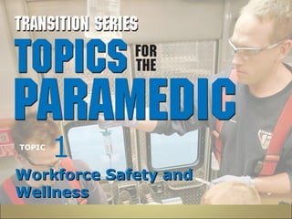
- 1. 1 TOPIC WWoorrkkffoorrccee SSaaffeettyy aanndd WWeellllnneessss
- 2. Introduction • Now more than ever, paramedics must employ multiple strategies to ensure their safety: – Disease transmission – Recognition of a dangerous scene – Personal safety – Health and wellness
- 3. Actual Safety Threats • The leading cause of death to EMS providers is being involved in a motor vehicle crash. • EMS providers are as likely to die from a heart attack as to be murdered. • Leading causes of injury in the EMS workplace include back injuries and exposures to bloodborne pathogens.
- 4. RReessppoonnddiinngg ttoo tthhee AAccttuuaall TThhrreeaattss –– WWeellllnneessss aanndd IInnjjuurryy PPrreevveennttiioonn • Motor Vehicle Crashes – Account for nearly 80 percent of EMS line-of-duty deaths – It is imperative to safely operate the ambulance. – Seatbelts save lives. – Seatbelts worn inside the ambulance can protect the EMS providers.
- 5. RReessppoonnddiinngg ttoo tthhee AAccttuuaall TThhrreeaattss –– WWeellllnneessss aanndd IInnjjuurryy PPrreevveennttiioonn • Back injuries – Most common cause of lost work and long-term disability among EMS providers – Proper lifting and moving techniques should be used in order to prevent injury
- 6. RReessppoonnddiinngg ttoo tthhee AAccttuuaall TThhrreeaattss –– WWeellllnneessss aanndd IInnjjuurryy PPrreevveennttiioonn • Key Elements of Proper Lifting – Anticipate a career of lifting – Know your limitations and request assistance when needed – Lift using the proper power-lift technique – Pay attention to minor injuries
- 7. RReessppoonnddiinngg ttoo tthhee AAccttuuaall TThhrreeaattss –– WWeellllnneessss aanndd IInnjjuurryy PPrreevveennttiioonn • Key Elements of Proper Lifting – You must set the example and help build a culture in which lift assistance is the norm, rather than the exception. – Know when your capabilities are outmatched by the weight of your patient. – Attempting a lift without proper capabilities is unsafe to both you and to your patient.
- 8. Infection Control • Prevent high-risk exposures by using appropriate personal protective equipment and using simple strategies such as: – Washing your hands – Handling sharps safely – Using Standard Precautions
- 9. Standard Precautions • Decide what precautions are needed as you consider the circumstances. – Gloves and hand washing are a minimum. – Face, gowns, and respiratory precautions as needed.
- 10. Standard Precautions • Decide what precautions are needed as you consider the circumstances. – Modalities such as IV catheterization, advanced airway placement, or medication administration require additional attention to Standard Precautions.
- 11. Standard Precautions • Re-evaluate and choose the appropriate level of personal protective equipment accordingly. • As a paramedic, your decisions will be setting the example for others. • It is necessary for a paramedic to handle sharps safely.
- 12. Wellness • Leading a healthy lifestyle can benefit paramedics. • Concepts to incorporate into a wellness plan include: – Regular exercise – Healthy diet – Rest – Routine and regular medical care – Stress management
- 13. Stress Management • Stress can damage your health and well-being. • Types of stress reactions include: – Acute stress reaction – Delayed stress reaction – Cumulative stress reaction • Employ strategies to minimize stress.
- 14. Summary • Self-protection is an imperative part of safely going home at the end of the day. • The paramedic must remain vigilant to all threats to their well-being. • Paramedics should take steps to prevent injury and stay safe and well.
- 15. TOPIC 2 PPaattiieenntt SSaaffeettyy
- 16. Introduction • Many patients die every year as a result of preventable medical errors. • As paramedics, you are entrusted to treat your patients and do no harm. • Your responsibilities include preventing medical errors and ensuring the safety of your patient. • Improper actions or treatments can result in harm or death to your patient.
- 17. Recognizing Risks • Scene assessment and situational awareness can help identify and avoid problems. • Patient transfer and handoffs account for the single largest situation associated with patient errors.
- 18. Patient Transfer and Handoff You arrive at a busy ED at a time when your shift has three priority 1 calls holding. Your suspected stroke patient seems stable enough, but you are obviously concerned about the overall outcome. En route you give a radio report; on arrival, you recognize the triage nurse as the person with the voice you spoke to on the radio.
- 19. Patient Transfer and Handoff She says, “Go ahead and put him in the hall bed; we will be right there.” In the meantime, dispatch radios you for the fourth time and asks if you are available. Having been acknowledged by the nurse, you and your partner transfer the patient and leave for the next call.
- 20. Patient Transfer and Handoff • What risks have you exposed the patient to? • What consequences can occur because of your actions? • How could this have been avoided?
- 21. Communication Difficulties • Miscommunication or communication difficulties can lead to patient errors. • Communication difficulties may put the patient at risk. • As a paramedic, it is imperative that you communicate well with others.
- 22. Medication Issues • Incorrect medication administration can potentially result in disastrous consequences. • Ever-changing medication lists, packaging, and dosage calculations can all pose potential problems. • Use the “five Rights” to help reduce medication errors.
- 23. Airway Issues • Mishandled airways have proven to be both prevalent and disastrous. • Misplaced endotracheal intubations continue to be a serious problem in the world of EMS. • Paramedics must incorporate good airway decision-making skills into the assessment and management of each patient.
- 24. Patient Movement • Patients are at risk whenever they are moved. • Dropping a patient can lead to injury and possible legal and civil liabilities. • Utilize the appropriate resources and/or technology for safely moving patients.
- 25. Ambulance Crashes • Ambulance crashes remain the largest cause of lawsuits against EMS providers. • They account for the majority of injuries to patients by providers. • Safe ambulance operation is a responsibility of the paramedic.
- 26. Spinal Immobilization • Proper spinal immobilization is designed to prevent secondary injuries. • When performed inappropriately or not applied when necessary, it can present a disastrous risk to the patient.
- 27. How Errors Happen • Types of errors – Skill-Based errors – Knowledge errors – Rule-based failure • Each category is potentially dangerous and can be prevented.
- 28. Preventing Errors • The two main approaches to preventing errors are systemic strategies and individual tactics. • Know your own limitations and capabilities. • Seek help when needed. • Learn from your mistakes. • Embrace quality improvement and continuing education.
- 29. TOPIC 3 LLeeggaall IIssssuueess iinn EEMMSS
- 30. Introduction • Legal issues impact every patient contact. • Laws are designed to protect both the patient and the care provider. • If paramedics do not adhere to the legislation that they must operate within, severe legal punishments may result.
- 31. Legal Terms • Scope of practice • Negligence • Intentional torts • Duty to act • Ethical behavior • Medical direction • Good Samaritan laws • Sovereign immunity • Statute of limitations • Standard of care
- 32. Figure 3–1 A paramedic may be required to testify in court in a variety of legal settings.
- 33. Ethics • Branch of philosophy directed toward the study of morals or concepts such as right or wrong. • NAEMT has issued a Code of Ethics. • Ethical decision making should guide the choices paramedics make everyday.
- 34. PPaattiieennttss’’ RRiigghhttss • Every patient that summons EMS has certain “rights.” These include: – Privacy and confidentiality – Access to emergency care – Consent – Ability to refuse care
- 35. PPaattiieennttss’’ RRiigghhttss • Every patient that summons EMS has certain “rights.” These include: – Advance directives – Organ donation – Transport – Privacy – Refusal
- 36. Special Reporting Situations • EMS providers are legally bound to report certain types of emergencies. • These mandatory reporting points may vary from state to state. • Paramedics should remain abreast of what their state requires and learn the reporting system used.
- 37. Summary • So long as there is EMS, there will be laws governing EMS. • The paramedic is solely responsible for staying abreast of laws that apply in his state. • The paramedic should always behave ethically and act in the best interest of the patient.
- 38. Summary • The best defense for preventing a lawsuit is to provide conscientious care to the patient, maintain the standard of care, follow state guidelines, and provide quality documentation on the patient care report.
- 39. 6 TOPIC CCeelllluullaarr EEnnvviirroonnmmeenntt aanndd MMeettaabboolliissmm
- 40. Introduction • Understand how changes in the patient are due to changes in cellular integrity. • The basic intention of emergency medical care is to keep the cells alive. • Cellular integrity must be the core of a paramedic’s assessment and treatment.
- 41. Figure 6–1 The cell.
- 42. Physiology • Metabolism – Metabolism refers to the sum total of chemical reactions taking place in the body. – Many metabolic activities build upon each other. – Disturbances can lead to cellular death, which in turn ultimately leads to death of the organism.
- 43. Physiology • Anabolism – Creation of larger structures from smaller molecules – Requires energy • Catabolism – Process that breaks down large molecules into smaller ones – Requires enzymes and water, and produces energy in the process
- 44. Physiology • Cellular Respiration – Process of transferring energy from a glucose molecule to a cell. – Oxidation is necessary for energy production and heat. – Glucose is the building block of cellular energy. – ATP is the primary energy-carrying molecule.
- 45. Physiology • Aerobic Cellular Metabolism – Glycolysis – Citric acid cycle (Krebs cycle) – Electron transport chain
- 46. Figure 6–2 Aerobic metabolism. Glucose broken down in the presence of oxygen produces a large amount of energy (ATP).
- 47. Physiology • Anaerobic Cellular Metabolism – Without oxygen, cellular production of ATP is very low. – Glycolysis still occurs. – Hydrogen molecules build up, increasing lactic acidosis. – The cell fails and dies.
- 48. Figure 6–3 Anaerobic metabolism. Glucose broken down without the presence of oxygen produces pyruvic acid, which converts to lactic acid and only a small amount of energy (ATP). A lack of glucose and oxygen will create a disturbance to cellular metabolism and may lead to dysfunction and eventual cell death. Cell dysfunction and death lead to organ dysfunction. When a critical mass of cells dies within an organ, the organ itself then dies
- 49. Physiology • Sodium/Potassium Pump – Maintains normal levels of Na+ and K+ on either side of the cellular wall. – Exchanges three sodium molecules for two potassium molecules. – The pump requires ATP to operate. – If ATP is lacking (anaerobic metabolism), the pump fails and the cell ruptures.
- 50. Summary • Understanding the need for normal cellular function underlies all branches of medicine. • Although we tend to treat the obvious (airway, breathing, circulation), doing so ultimately treats the ability to maintain cellular integrity.
- 51. Summary • Once cells start dying, the syndrome progresses rapidly and may be irreversible. • The paramedic should always consider how their treatment will impact cellular activity.
- 52. 7 TOPIC AAnnaattoommyy aanndd PPhhyyssiioollooggyy:: TThhee BBlloooodd
- 53. Introduction • The blood is the body’s transport mechanism. • Understanding the composition and role of the blood can help the paramedic understand perfusion, shock, and the circulatory system in general.
- 54. Composition of the Blood • Formed elements (45%) – RBC – WBC – Platelets • Plasma (55%) – 91 percent water – Albumin, antibodies, clotting factors
- 55. Blood Plasma • Plasma is the yellow-colored liquid medium of the blood – 91 percent water – -9 percent plasma proteins • Albumin (maintains the fluid balance in the blood) • Antibodies (defence against infectious) • Clotting factors (key in coagulation)
- 56. Erythrocytes • Created during erythropoiesis. • Eliminated during eryptosis. • Cytoplasm contains hemoglobin. • Genesis and elimination of RBCs provide for maintaining adequate oxygen-carrying capabilities.
- 57. Leukocytes • Protect the body against infection and eliminate dead and injured cells and debris. • Types of leukocytes – Neutrophils destruction and removal of bacterial – Eosinophils deal’swith invaders to the body & inflamation – Basophile releases histamine – Lymphocytes respond to and destroy foreign invaders – Monocytes assist antibodies with identifying unwanted invaders
- 58. Thrombocytes • Platelets are fragments that play a major role in hemostasis. • Adhere to each other to form clots and stop bleeding.
- 59. Hemostasis • Hemostasis is the process of protecting the circulatory system from blood loss. • Phases of hemostasis: – Vasoconstriction – Platelet plugging – Coagulation
- 60. Coagulation Cascade • During coagulation, fibrin is introduced. • Fibrin is regulated by chemical factors and proteins, • Factor X is activated and initiates a series of events which cause coagulation.
- 61. Coagulation Cascade • Prothrombin is converted to thrombin. • Thrombin converts fibrinogen to fibrin fibers which envelope platelet plug and stabilize the clot.
- 62. The Complete Blood Count • The complete blood count is a test performed on a sample of blood • Used to determine the presence of key elements of blood composition.
- 63. Table 7–1 Complete Blood Count Normal Values
- 64. Blood Types and Rh Factor • ABO system categorizes blood based on the presence or lack of antigens on red blood cells and antibodies in plasma. • Blood types: A, B, AB, and O. • The Rh factor looks for a specific third antigen and is represented as positive or negative
- 65. Summary • Understanding the composition and role of the blood can help the paramedic identify, treat, and manage patients. • The paramedic should understand how hemostasis is accomplished.
- 66. BREAK
- 67. TOPIC 8 TThhee NNeerrvvoouuss SSyysstteemm
- 68. Objectives • Identify the major components of the nervous system. • Differentiate between the central and peripheral nervous system and their roles in maintaining homeostasis. • Discuss the clinical application of how the nervous system can affect a patient’s physiological presentation.
- 69. Introduction • The nervous system allows the body to: – Receive information from the environment – Transport that information to the brain – Process and react to the information
- 70. Introduction • Categorized into the central and peripheral nervous systems. • Thoughts, movements, senses, and reflexes are all results of the actions of the nervous system.
- 71. Neurons • The building blocks of the nervous system. • The three types of neurons include sensory, motor, and interneurons. • Nerves transmit impulses to convey information. • Damage to the nerves can be detrimental to the body’s natural function.
- 72. Figure 8–1 The neuron.
- 73. Central Nervous System • Composed of two components. – The brain – The spinal cord • Damage can result in the ability to perform even basic functions. • Sensory pathways of the spinal cord: – Posterior column – Spinothalamic pathway – Spinocerebellar pathway
- 74. Figure 8–2 The divisions of the brain.
- 75. Peripheral Nervous System • Composed of structures not covered by the central nervous system. • The PNS is divided into two main sections: – Somatic division – Autonomic division • Sympathetic branch • Parasympathetic branch
- 76. Table 8–1 The Cranial Nerves
- 77. Figure 8–3 Spinal nerves.
- 78. The Senses • Allows the body to relay information about the environment to the nervous system. • Helps prevent the body from sustaining injuries.
- 79. The Senses • The general senses are: – Pain – Temperature – Touch/pressure/position – Chemical detection
- 80. Special Senses • The special senses have specialized organs which relay information. • The special senses include: – Sight – Smell – Hearing – Taste
- 81. Reflexes • Reflexes are physiologic responses from the body to a stimulus. • Categories of reflexes include: – Spinal reflexes – Cranial reflexes – Somatic – Autonomic
- 82. Summary • The nervous system is the collector, transporter, and interpreter for the world around us. • A paramedic should understand that it is vital for maintaining homeostasis and the ability to move, breathe, think, and understand the environment we live in.
- 83. TOPIC 9 MMeeddiiccaall TTeerrmmiinnoollooggyy
- 84. Objectives • Review the components of a medical term. • Review a list of common medical terms.
- 85. Introduction • Medical terminology is the language of health care. • By understanding terms, components, even complex words, can be broken down. • Understanding and utilizing proper terminology can improve communication between members of the healthcare team.
- 86. Medical Terms Origin • Terms are often derived from Greek and Latin sources. • Common parts compose the terms. – Prefixes – Suffixes – Combining forms • Some memorization will be required to get a basic grasp of the language.
- 87. Structure of Medical Terms • Three basic components – Combining form • Root • Combining vowel – Suffix – Prefix
- 88. HHooww ttoo DDeeffiinnee MMeeddiiccaall TTeerrmmss • Terms can easily be defined by determining the meaning of their parts. • Read left to right, but define by interpreting the suffix, then the prefix, then the combining form. prefix combining form suffix hyper- glyc/o -emia (above or excessive) (sugar) (blood condition)
- 89. Use proper medical terminology to communicate with other health care professionals.
- 90. Figure 9–1 Sometimes it will be more convenient to use an accepted medical abbreviation or symbol in your report instead of writing the entire term.
- 91. Table 9–2 Common Prefixes in Medical Terms
- 92. Table 9–2 (continued) Common Prefixes in Medical Terms
- 93. Table 9–3 Common Suffixes in Medical Terms
- 94. Table 9–3 (continued) Common Suffixes in Medical Terms
- 95. Table 9–4 Common Combining Forms in Medical Terms
- 96. Table 9–4 (continued) Common Combining Forms in Medical Terms
- 97. Table 9–4 (continued) Common Combining Forms in Medical Terms
- 98. Table 9–4 (continued) Common Combining Forms in Medical Terms
- 99. Table 9–4 (continued) Common Combining Forms in Medical Terms
- 100. Table 9–4 (continued) Common Combining Forms in Medical Terms
- 101. Table 9–4 (continued) Common Combining Forms in Medical Terms
- 102. Table 9–4 (continued) Common Combining Forms in Medical Terms
- 103. Table 9–4 (continued) Common Combining Forms in Medical Terms
- 104. Table 9–4 (continued) Common Combining Forms in Medical Terms
- 105. Table 9–4 (continued) Common Combining Forms in Medical Terms
- 106. Table 9–4 (continued) Common Combining Forms in Medical Terms
- 107. Table 9–4 (continued) Common Combining Forms in Medical Terms
- 108. Table 9–4 (continued) Common Combining Forms in Medical Terms
- 109. SSuummmmaarryy • The proper use of medical terminology will help ensure clarity in the sharing of information regarding the patient. • The paramedic should keep abreast of medical terms and abbreviations as they pertain to the practice. • A paramedic is expected to use proper medical terminology.
- 110. 11 TOPIC SSeellff--DDeeffeennssee MMeecchhaanniissmmss aanndd IInnffllaammmmaattiioonn
- 111. Objectives • Review the inherent mechanisms of cellular self-defense and the inflammatory process. • Discuss the first-line and second-line defenses of the inflammatory response. • Understand the local and systemic manifestations of inflammation.
- 112. Introduction • The immune system provides a defense against the challenges faced by the body. • Native immunity includes natural barriers and inflammation. • Protective physical, mechanical, and biochemical barriers provide protection against infection.
- 113. Figure 11–1 The defense mechanisms of the body.
- 114. Figure 11–2 White blood cells form the basis for the phagocytic response.
- 115. The Inflammatory Response • The inflammatory response is a complex sequence of events designed to prevent damage and repair existing damage to cells. • It is stimulated by any process that can kill cells or damage connective tissue.
- 116. Figure 11–3 The process of inflammation.
- 117. Manifestations of Inflammation • Local manifestations of inflammation include: – Heat – Redness – Swelling – Pain
- 118. Manifestations of Inflammation • Systemic manifestations of acute inflammation include: – Fever – Leukocytosis – Plasma protein synthesis
- 119. Manifestations of Inflammation • Acute – Short time of activation • Chronic – Over two weeks of activation – Common pathways include: • Persistent accute inflammation • Neutrophil degranualation and death • Lymphocyte activation • Fibroblast activation
- 120. Summary • The immune system provides a defense against the challenges faced by the body. • It is important to understand how the body responds to theses challenges, especially at the cellular level. • Paramedics should understand how inflammation impacts the body.
- 121. 12 TOPIC TThhee CCaarrddiioovvaassccuullaarr SSyysstteemm
- 122. Objectives • Distribution of blood within the vascular compartment and the physiologic determinants that affect movement of fluid into and out of the vascular compartment: – Hydrostatic pressure. – Plasma oncotic pressure.
- 123. Objectives • Normal cardiac output, and how certain variables can alter it from normal: – Changes in heart rate. – Changes in stroke volume. • Systemic vascular resistance, and the effects should it become deranged: – Tissue perfusion. – Systolic and diastolic blood pressure. – Pulse pressure.
- 124. Objectives • Microcirculation, and how changes of the aforementioned principles have a positive or negative effect on it. • Blood pressure, and how it becomes deranged from disturbances in the aforementioned principles.
- 125. Objectives • How the autonomic nervous system (sympathetic and parasympathetic) can alter cellular perfusion through manipulation of the aforementioned principles.
- 126. Introduction • The heart, the blood, and the blood vessels each play an essential role in maintaining adequate tissue perfusion and homeostasis. • Understanding how the cardiovascular system functions will help the paramedic to recognize critical situations and anticipate further patient deterioration.
- 127. Blood Volume • Blood volume is one of the determinants of adequate blood pressure and perfusion. • Blood is distributed throughout the cardiovascular system. • Hydrostatic pressure and plasma oncotic pressure play important roles in maintaining the fluid balance.
- 128. Blood Volume • Hydrostatic pressure—is the “push” force inside the vessel or capillary bed generated by the contraction of the heart and blood pressure • Plasma oncotic pressure,colloid oncotic pressure, or oncotic pressure—is the “pull” force responsible for keeping fluid inside the vessels
- 129. Table 12–1 Distribution of Blood in the Cardiovascular System
- 130. Figure 12–1 Hydrostatic pressure pushes water out of the capillary. Plasma oncotic pressure pulls water into the capillary.
- 131. Pump Function of the Myocardium • The heart must pump effectively to maintain adequate blood pressure and perfusion. • Cardiac output is the amount of blood ejected by the left ventricle in 1 minute.
- 132. Pump Function of the Myocardium • Systolic blood pressure is a relative indicator of cardiac output. • Cardiac output = Heart rate × Stroke volume
- 133. Systemic Vascular Resistance • The resistance that is offered to blood flow through a vessel – Vasodilation typically decreases the pressure. – Vasoconstriction typically increases the pressure.
- 134. Systemic Vascular Resistance • Diastolic pressure is the basic measure of SVR. • Pulse pressure is the difference between the systolic and diastolic blood pressure readings. • Vasoconstriction decreases vessel diameter, increases resistance, and increases blood pressure. • Vasodilation increases vessel diameter, decreases resistance, and decreases blood pressure
- 135. Microcirculation • Microcirculation is the flow of blood through the arterioles, capillaries, and venules. • True capillaries are the sites of exchange between the blood and the cells.
- 136. Microcirculation • Capillary blood flow is influenced by: – Local factors – Neural factors – Hormonal factors
- 137. Microcirculation • In a resting state, the local factors predominantly control blood flow through the capillaries. • When adaptation is necessary, the neural factors will change the capillary blood flow. • Hormones are usually responsible for a sustained effect on the arterioles and capillaries.
- 138. Microcirculation is the flow of blood through the smallest blood vessels: arterioles, capillaries, and venules. Precapillary sphincters control the flow of blood through the capillaries.
- 139. Blood Pressure • Blood pressure (BP) is derived by multiplying two major factors: cardiac output (CO) and systemic vascular resistance (SVR). • Blood pressure is monitored and regulated by: – Baroreceptors – Chemoreceptors
- 140. Summary • Maintaining adequate metabolism and perfusion is essential for the survival of the cells, organs, and the patient. • Understanding the ways in which the cardiovascular system compensates will help the paramedic not only recognize critical situations, but also anticipate further patient deterioration.
- 141. 15 TOPIC MMeeddiiccaattiioonn AAddmmiinniissttrraattiioonn
- 142. Objectives • Discuss patient safety strategies associated with medication administration. • Understand the responsibilities of paramedic-level pharmacology. • Discuss ways to prevent medication errors. • Review nontraditional medication routes.
- 143. Introduction • Paramedics have access to and provide a wide array of medications to benefit patients. • With this ability, comes great responsibility.
- 144. Introduction • Paramedics must keep the patient’s safety at the center of care and treatment. • Paramedics must maintain, improve, and enhance their capabilities to utilize medications.
- 145. Patient Safety • Patient safety is imperative. • Medication errors can result in fatal consequences to the patient. • Some medication errors encountered in EMS include those involving: – Dose – Route – Rate of administration – Allergies
- 146. Figure 15–1 Check the medication.
- 147. The Five Rights • The five rights of medication administration include: – Right medicine – Right dose – Right time – Right route – Right patient
- 148. Figure 15–2 Double-check the concentration and expiration date.
- 149. Maintaining Competency • Paramedics must ensure that their knowledge base meets and exceeds the standard of care. • It is imperative that the paramedic is familiar with the regulations and protocols that guide their practice.
- 150. Advances in Medication Administration • Paramedics have adopted a number of changes associated with the delivery of medications. – Intraosseous administration for adult and pediatric patients. – Intranasal administration can allow for rapid medication absorption and a safer needle-free environment.
- 151. The EZ-IO (Vida-Care Corporation).
- 152. Summary • Medication administration is an important responsibility of a paramedic and should always be taken seriously. • The paramedic should always consider the patient’s safety and the “five rights” before administering any medication.
- 153. TOPIC 16 PPaarraammeeddiicc MMeeddiiccaattiioonnss
- 154. Objectives • Review the paramedic formulary. • Discuss new approaches with traditional prehospital medications. • Understand some of the issues surrounding specific prehospital medications.
- 155. Introduction • New research has influenced the medications being administered by paramedics. • Paramedics should be aware of various debates pertaining to the administration of some medications. • Paramedics should understand how these debates may impact their protocols.
- 156. Oxygen Reconsidered • Hypoxic patients should still receive oxygen. • Hyperoxia may be harmful and lead to systemic vasoconstriction and the release of free radicals in the body. • Oxygen therapy should be titrated based on the monitoring of the oxyhemoglobin saturation to ≥94 percent.
- 157. Figure 16–1 Use of supplemental oxygen is being reconsidered.
- 158. Acute Pulmonary Edema Medications • Morphine Sulfate – Morphine has been found to not possess the vasodilatory property once believed. – Cardiac toxicity and reduced cardiac output may occur with administration. – Low-dose benzodiazepines may provide the same anxiolytic effects without the negative side effects.
- 159. Acute Pulmonary Edema Medications • Furosemide (Lasix) – Once believed that the diuresis would benefit the patient’s hypervolemic state and was often administered in high doses. – Research studies have found that many patients in APE are not hypervolemic. – The diuresis in a normovolemic patient can lead to hypovolemia, which must be corrected.
- 160. Cardiac Arrest Medications • The following medications have been recently reviewed by the AHA and have remained a cause of much debate and research with respect to appropriate care of cardiac arrest: – Atropine (no therapeutic benefit) – Vasopressin (no better than standard EPI) – Sodium bicarbonate (no benefit)
- 161. Other Controversial Medications • Thiamine – Thiamine deficiency is rare and for thiamine to be effective, it should be administered over days. • Procainamide – Antidysrhythmic used in the treatment of wide complex tachycardia. But avoid in pt. with a prolonged QT or CHF
- 162. Summary (cont'd) • Paramedics must stay abreast of the changes and understand how they can impact their practice.
- 163. 17 TOPIC AAiirrwwaayy AAsssseessssmmeenntt aanndd DDeecciissiioonn MMaakkiinngg
- 164. Objectives • Delineate between respiratory distress and respiratory failure. • Review the signs and symptoms that illustrate ventilatory adequacy of inadequacy.
- 165. Objectives • Determine when or when not to ventilate a patient. • Review and integrate the airway treatment options for a patient suffering from a disturbance to the airway. • Review core treatment interventions for a patient suffering from disturbance to the airway.
- 166. Introduction • Paramedics must be able to properly assess and recognize airway dysfunction. • Airway management is a process that should be guided by the assessment findings and should be goal oriented.
- 167. Introduction • The paramedic must utilize critical thinking and good decision-making skills in order to provide the best treatment for the patient.
- 168. Anatomy of the upper airway.
- 169. Pathophysiology • Upper airway dysfunction – Obstruction can result from foreign bodies or more commonly as a result of poor muscle tone. – Structural changes can also impede airflow.
- 170. Loss of control of the upper airway may occur, when the muscles of the upperairway relax too much and the epiglottis is allowed to fall back and cover the glottic opening.
- 171. Pathophysiology • Lower airway dysfunction – Bronchoconstriction is the most common cause. – Other disorders can structurally change how gas is exchanged in the alveoli.
- 172. Airway Assessment • The paramedic must ensure and secure the airway. • Consider the following: – Mental status, speech, and voice – Pathophysiology or other findings that may threaten airway • Ensure breathing is adequate to meet the needs of the body
- 173. Patient suffering respiratory distress, indicated by his tripod position.
- 174. Respiratory Distress • Compensation to a respiratory challenge – Respiratory rate increases – Accessory muscles are engaged – Heart beats faster and stronger • The compensatory efforts are sustaining normal function despite the problem.
- 175. Respiratory Failure • Compensatory mechanisms fail. – Oxygen may not be distributed – Carbon dioxide is retained – Muscles of respiration tire
- 176. Respiratory Failure • The patient will require ventilatory assistance. • Altered mental status, hypoxia, cyanosis, and irregular respiratory patterns are key findings that indicate respiratory failure.
- 177. The continuum of breathing ranges from normal, adequate breathing to no breathing at all. It is essential to recognize the need for assisted ventilations even before severe respiratory distress develops.
- 178. Using Assessment to Guide Treatment • Quality assessment allows for recognition of a problem and provides valuable information. • Critical thinking is a must for using the correct tools in the right circumstance. • Cost and benefits must be considered. • Consider the pathophysiology.
- 179. Goals of Airway Management • Assess the ability to move air and exchange oxygen and carbon dioxide. • Determine weather the patient is in respiratory distress or respiratory failure.
- 180. Goals of Airway Management • Goals of airway management should include: – Securing and protecting the airway – Oxygenating the patient – Ventilating the patient
- 181. Outcome-Based Management • Depends on critical thinking. • Links assessment findings to desired outcome in order to form a treatment plan. • Allows for the most appropriate tools for the best patient outcome.
- 182. Opening/Securing the Airway • Basic airway interventions are frequently the most appropriate to open and secure the airway. • Consider both short-term and long-term airway management. • Utilize a cost–benefit analysis. • Consider the nature of the disorder.
- 183. Oxygenating and Ventilating • Ensure adequate oxygenation and ventilation. • Patients in respiratory failure require positive pressure ventilation. – Consider the ability to secure the airway – Consider minute volume – Consider pharmacologic treatments including oxygen
- 184. Oxygenating and Ventilating • Support compensatory efforts and reverse the challenge for patients in respiratory distress. – Oxygen therapy – Pharmacologic treatments
- 185. Summary • The paramedic must be able to assess and promptly treat respiratory failure. • Airway management should be guided by the assessment findings and should be goal oriented. • Critical thinking is necessary for the paramedic to choose what is the most appropriate treatment for their patient.
- 186. 18 TOPIC NNoonniinnvvaassiivvee AAiirrwwaayy IInntteerrvveennttiioonn
- 187. Objectives • Discuss the core interventions for a patient suffering from a disturbance to the airway. • Review the concepts of oxygen therapy and positive pressure ventilation. • Discuss the use of continuous positive airway pressure during the management of a patient in respiratory distress.
- 188. Introduction • Paramedics must use assessment and critical thinking to decide which tool is right for a specific patient. • A wide range of tools are available for managing patients with airway problems. • It is the responsibility of the paramedic to determine the most appropriate intervention.
- 189. DDoonn’’tt FFoorrggeett tthhee BBaassiiccss • A paramedic must weigh the costs and benefits to determine the best treatment for the patient. • In many cases, basic interventions are the most valuable and/or appropriate. • Advanced procedures are important in the right circumstances.
- 190. Supplemental Oxygen Revisited • Oxygen is a drug that must be used correctly. • Never withhold oxygen from a hypoxic patient. • Continued high-flow oxygen beyond normal oxygen saturations may cause hyperoxia.
- 191. Supplemental Oxygen Revisited • Oxygen should be titrated to maintain a normal saturation levels of 94 percent to 95 percent
- 192. Positive Pressure Revisited • Positive pressure ventilation is needed to correct respiratory failure. • Minimize the effect of positive pressure on the heart and cardiac output. • Keep gastric insufflation in mind. • Ventilate at age-appropriate rates to avoid hyperventilation.
- 193. Bag-Mask Device and Cardiac Arrest • Intubation interrupts compressions and may negatively affect resuscitation. • Bag-mask ventilations alone may not be an effective way to move air. • Blind airway insertion devices should be considered. • The costs and benefits of moving to a more aggressive airway must be weighed by the paramedic.
- 194. Continuous Positive Airway Pressure • CPAP creates a constant slight flow of air against which the patient will breathe. • CPAP is most commonly used to treat acute pulmonary edema, but can be used to treat other forms of respiratory distress.
- 195. Continuous positive airway pressure (CPAP) is used for the awake and spontaneously breathing patient who needs ventilatory support.
- 196. Summary • Paramedics must use assessment findings and critical thinking to determine the most appropriate way to manage a patient suffering from an airway disturbance.
- 197. Summary • Many options are available for the paramedic to manage the airway. • Utilizing noninvasive airways may be the most beneficial for the short-and long-term outcomes for some patients.
- 198. 19 TOPIC IInnvvaassiivvee AAiirrwwaayy MMaannaaggeemmeenntt
- 199. Objectives • Discuss the decision-making process when utilizing an advanced airway. • Review blind insertion airway devices. • Understand the current endotracheal intubation dilemma. • Discuss how to help preserve endotracheal intubation in the paramedic scope of practice.
- 200. Introduction • Paramedics can utilize advanced airway skills within their scope of practice. • Paramedics should select the most appropriate intervention for each situation after weighing the costs and benefits.
- 201. Introduction • The responsibility to make good airway management decisions is especially true with the recent controversy surrounding endotracheal intubations.
- 202. Progressing to Invasive Airway Management • Airway management decision should consider: – Assessment findings – Pathophysiology – Other circumstances to create best treatment plan • Invasive procedures should be utilized when their benefits clearly outweigh their risks.
- 203. Progressing to Invasive Airway Management • Consider the following indications for invasive airways: – More basic maneuvers have failed – Invasive airways are indicated by the pathophysiology of the situation – Invasive airways represent the better choice given an analysis of the circumstances – The clinical course of the patient indicates invasive maneuvers.
- 204. Benefits and Risks of Advanced Airway Procedures
- 205. The Endotracheal Intubation Dilemma • Endotracheal intubation is the most secure airway and when performed correctly. • Risks and complications can include hypoxia, increased intracranial pressure, trauma, and death. • Success rates are reported to be low. • Training and ongoing education are challenging.
- 206. Preserving Intubation • Preserving intubation should be a priority for all paramedics and proactive steps must be taken. – Recognize the problem – Select appropriate patients – Improving confirmation is an essential step
- 207. Intubation Confirmation • Confirmation of proper placement is essential. • Positive confirmation recognizes and corrects errors that happen. • The gold standard for confirmation is waveform capnography.
- 208. Intubation Confirmation • Other confirmation devices can be used. • Multiple methods should be used to achieve a definitive confirmation.
- 209. Blind Insertion Airway Devices • Blind airway devices do not require specialized equipment to insert. • They offer an alternative to ETI, but do not definitively protect the airway. • Various types of BIADs exist. – Esophageal obturation devices – Supraglottic devices
- 210. Case Study • You are working a shift at the fire department and you are toned to a house fire. You throw your gear into the ambulance and follow the fire engine to the scene. Upon arrival, you find a crowd standing around a man who is down in the grass. There are flames shooting out of the windows of the house.
- 211. Summary • The paramedic must use good decision making in order to select and utilize the most appropriate interventions for maintaining the airway of a patient. • Controversy surrounds the use of prehospital endotracheal intubation and other advanced airway skills.
- 212. Summary (cont'd) • Paramedics may help preserve endotracheal intubation intervention by recognizing the issues, selecting appropriate situations to use the skill, and improving their ability to confirm proper placement.
- 213. TOPIC 32 NNeeuurroollooggyy:: SSttrrookkee
- 214. Objectives • Review the frequency with which strokes occur. • Discuss the common types of occlusive strokes to include pathophysiology and findings. • Review "mini-strokes" such as TIA and RIND. • Discuss strokes caused by hypoperfusion.
- 215. Objectives • Relate the stroke location with cerebral arteries. • Review the stroke scale assessment tools. • Review current treatment standards for patients suffering from a stroke.
- 216. Introduction • Stroke is an acute emergency resulting in disruption of blood flow to a region of the brain. • Can result in temporary or permanent abnormalities of cerebral functioning. • EMS must rapidly identify and transport the potential stroke patient.
- 217. Epidemiology • 700,000 strokes occur per year. – About one every 45 seconds • Strokes are the third leading cause of death in the United States – One stroke-related death every 3 minutes • Higher risk to women, African Americans, and Hispanics/Latinos. • Major cause of permanent disability.
- 218. Pathophysiology • Types of strokes – Ischemic • Thrombotic • Embolic • Transient ischemic attack • Reversible neurologic deficit • Hypoperfusion – Most common • 80 percent to 85 percent
- 219. Pathophysiology • Types of strokes – Hemorrhagic • Intracerebral hemorrhage • Subarachnoid hemorrhage – Etiology • Arteriovenous malformations • Aneurysm – Frequency • 10 percent to 15 percent
- 220. Causes of stroke. Blood is carried from the heart to the brain via the carotid and vertebral arteries, which form a ring and branches within the brain. An ischemic stroke occurs when a thrombus is formed on the wall of an artery or when an embolus travels from another area until it lodges in and blocks an arterial branch. A hemorrhagic stroke occurs when a cerebral artery ruptures and bleeds into the brain (examples shown: subarachnoid bleeding on the surface of the brain and intracerebral bleeding within the brain).
- 221. Pathophysiology • Progression of neurologic dysfunction and damage in stroke – Loss/diminishment of blood flow. – Cells become electrically “silent.” – Na+/K+ pump failure, cells swell and rupture. • “Cytotoxic edema”
- 222. Pathophysiology • Progression of neurologic dysfunction and damage in stroke – Ischemic penumbra receives diminished flow. • It may also become electrically silent.
- 223. Clinical Findings • Assessment of the stroke patient – Time is paramount. – Narrow window for thrombolytic drugs. – Careful assessment for baseline findings and changes is important. • Always try to determine onset time for symptoms.
- 224. Clinical Findings • Signs and symptoms of stroke – Facial droop and/or slurred speech – Dysphasia and aphasia – Unilateral numbness – Headache/dizziness (severe in ICH/SAH)
- 225. Clinical Findings • Signs and symptoms of stroke – Weakness/Paralysis – Mental status changes – Vision changes – Cognitive changes – Incontinence
- 226. (a) The face of a nonstroke patient has normal symmetry. (b) The face of a stroke patient often has an abnormal, drooped appearance on one side. abnormal, drooped appearance on one side. normal symmetry
- 227. A patient who has not suffered a stroke can generally hold the arms in an extended position with eyes closed. (b) A stroke patient will often display “arm drift” or “pronator drift”—one arm will remain extended when held outward with eyes closed, but the other arm will drift or drop downward and pronate (palm turned downward). arms in an extended position with “arm drift” eyes closed
- 228. Cincinnati Prehospital Stroke Scale (CPSS)
- 229. Los Angeles Prehospital Stroke Screen (LAPSS)
- 230. Emergency Medical Care • Consider spinal precautions, determine onset of symptoms. • Support lost function. – Airway, breathing, circulation • Initiate intravenous therapy and titrate as necessary. – Normal saline to keep open rate – Increase if systolic blood pressure drops below 90 mmHg
- 231. Emergency Medical Care • Assess blood glucose level level. – Hypoglycemia may mimic stroke. – Treat hypoglycemia as indicated. • Protect paralyzed limbs. – Be sure to properly secure paralyzed limbs to prevent accidental trauma during patient movement. • Transport.
- 232. Summary • A stroke occurs when there is interruption of blood flow to a region of the brain. • Although symptoms may present as mild initially, it is often not known early on how severely the patient may deteriorate.
- 233. Summary • Prehospital identification and treatment are integral to the successful overall management of stroke patients.
- 234. 34 TOPIC IImmmmuunnoollooggyy:: AAnnaapphhyyllaaccttiicc aanndd AAnnaapphhyyllaaccttooiidd RReeaaccttiioonnss
- 235. Objectives • Review the frequency with which immunologic emergencies occur. • Understand the pathology of immunologic emergencies. • Discuss chemical mediators and their reactions. • Illustrate the relationship between pathology and symptomatology.
- 236. Objectives • Differentiate between a mild and severe reactions. • Discuss treatment strategies such as epinephrine.
- 237. Introduction • Allergic reactions may present from mild to severe. • Manifestations can be related to the body system failing due to the reaction. • Although an allergic reaction is designed to be beneficial to the body, when the response is severe it can be fatal.
- 238. Epidemiology • Anaphylaxis is not a reportable disease. • An estimated 20,000 to 50,000 persons suffer an anaphylactic reaction each year in the United States • Most common triggers include penicillin, insect stings, radiocontrast media, and food.
- 239. Pathophysiology • Anaphylactic reaction – Patient must be sensitized – Chemical mediators released with subsequent exposure – Effects of mediators causes organ and system failure – Characteristic presentation
- 240. Table 34–1 Common Causes of Anaphylactic Reactions
- 241. Pathophysiology • Anaphylactoid reaction – Not the typical immunologic antigen-antibody reaction – Anaphylactoid trigger “directly” causes the breakdown of mast cells and basophils – Chemical mediators released – Characteristic presentation similar to anaphylactic reaction
- 242. Table 34–2 Common Causes of Anaphylactoid Reactions
- 243. Pathophysiology • Effects of chemical mediator release – Increased capillary permeability – Decreased vascular smooth muscle tone – Increased bronchial smooth muscle tone – Increased mucus secretions in the tracheobronchial tract
- 244. responses in anaphylactic reaction: bronchoconstriction, capillary permeability, vasodilation, and an increase in mucus production.
- 245. Pathophysiology • General considerations – Fatal episodes related to airway occlusion, respiratory failure, severe hypoxia, and circulatory collapse
- 246. Figure 34–2 Localized angioedema to the tongue from an anaphylactic reaction. (© Edward T. Dickinson, MD)
- 247. Table 34–3 Common Signs and Symptoms of Anaphylactic Reactions.
- 248. Table 34–3 (continued) Common Signs and Symptoms of Anaphylactic Reactions.
- 249. Table 34–3 (continued) Common Signs and Symptoms of Anaphylactic Reactions.
- 250. Figure 34–3 Urticaria (hives) from an allergic reaction to a penicillin-derivative drug.
- 251. Assessment Findings • Other notable assessment characteristics – Parenteral injections produce the severest reactions. – The faster the onset, the worse the reaction. – Signs and symptoms peak in 15–30 minutes.
- 252. Assessment Findings • Other notable assessment characteristics – Skin and respiratory reactions are the earliest to present. – Mild reactions could suddenly turn severe. – Most fatalities occur within 30 minutes. – The patient may have a biphasic or multiphasic reaction following treatment.
- 253. Table 34–4 Differentiating Between a Mild and a Moderate to Severe Reaction
- 254. EEmmeerrggeennccyy MMeeddiiccaall CCaarree • Keep airway patent. • Suction secretions. • Administer oxygen and ventilate the patient if needed. – Maintain SpO2 above 94 percent • Initiate intravenous infusion – Large bore catheter – Maintain systolic BP of 90 mmHg
- 255. Emergency Medical Care • Administer epinephrine if patient presents with systemic symptoms. – Preferred routes: auto-injector or IM – Adult dose: • 0.2 to 0.5mg of 1:1,000 IM • 0.3 mg auto-injector
- 256. Emergency Medical Care • Administer epinephrine if patient presents with systemic symptoms. – Pediatric dose: • 0.1 mg/kg not to exceed adult dose • 0.15 mg auto-injector • If patient weighs more than 66 lbs. Use adult injector – Repeat every 3 to 5 minutes if severe symptoms persist
- 257. Emergency Medical Care • Administer epinephrine if patient presents with systemic symptoms. – Consider concurrent glucagon with the epinephrine if the patient is taking beta blockers. • Administer diphenhydramine to negate the effects of the histamine.
- 258. Emergency Medical Care • Administer corticosteroids to help stabilize capillary permeability and prevent swelling. • Initiate rapid transport.
- 259. Emergency Medical Care • If an extremity is involved consider application of a loose tourniquet. • Treat wheezing with beta2 agonist. • Treat hypotension with IV fluid bolus. • Treat hypotension secondary to beta blockers with glucagon.
- 260. Summary • An allergic reaction may range from mild to severe. • Anaphylactic and anaphylactoid reactions can rapidly cause death to the patient. • The paramedic must recognize the acute allergic reaction and provide appropriate care based on findings.
- 261. 35 TOPIC EEnnddooccrriinnee EEmmeerrggeenncciieess:: HHyyppooggllyycceemmiiaa
- 262. Objectives • Review the frequency with which diabetic emergencies occur. • Discuss the etiologies of diabetes mellitus (type 1 and type 2). • Review the roles of insulin and glucagon. • Discuss the causes of hypoglycemia.
- 263. Objectives • Review the symptoms of hypoglycemia and relate to hyperadrenergic or neuroglycopenic pathophysiology. • Review the role of oral glucose in patient management.
- 264. Introduction • Diabetes mellitus (DM) is a condition in which the body no longer metabolizes glucose correctly. • This inability can lead to seriously high or low levels of blood sugar. • The paramedic must quickly identify the problem and support lost function to reduce morbidity and mortality.
- 265. Epidemiology • Most common endocrine disorder. • 6 percent of the population is afflicted with the disease. • Whites are more likely to have the disease than non whites. • Type 1 DM accounts for 5 percent to 10 percent • Type 2 DM accounts for 90 percent to 95 pecent
- 266. Epidemiology • Type 1 diabetes mellitus – Autoimmune disease process – Characteristic to younger patients – Requires supplemental insulin – Prone to hypoglycemia and diabetic ketoacidosis
- 267. Epidemiology • Type 2 diabetes mellitus – Impaired insulin production – Impaired insulin effects – Commonly an adult onset – Associated with a higher body mass index – Controlled through diet and oral pills – Prone to HHNS
- 268. Pathophysiology • Role of hormones in glucose regulation – Insulin and glucagon – Cellular metabolism of glucose
- 269. Glucose movement into the cell with insulin and the inability of glucose to get into the cell without insulin.
- 271. Pathophysiology • Hypoglycemia – Precipitating causes – Patients become symptomatic when the blood glucose level falls to 40–50 mg/dL – Brain most sensitive to low levels of glucose – Body then releases additional hormones aimed at trying to raise glucose back up
- 272. Assessment Findings • General considerations – Findings can be broadly categorized • Hyperadrenergic—increases sympathetic tone • Neuroglucopenic—brain dysfunction from lack of glucose
- 273. Signs and Symptoms of Hypoglycemia
- 274. Assessment Findings • Other notable assessment characteristics – Hypoglycemia may occur suddenly. – Hypoglycemia may present like a stroke. – Once referred to as “insulin shock” as many presentation findings mirrored hypovolemic shock.
- 275. Emergency Medical Care • Keep airway patent; be alert for vomiting. • Place patient in lateral recumbent position. • Administer oxygen based on ventilatory needs. – Keep SpO2 >95 percent.
- 276. Emergency Medical Care • Deliver glucose to the cells. – Administer oral glucose if criteria is met – Administer 50% dextrose if criteria is met via IV or IO – Administer glucagon IM if criteria is met
- 277. Emergency Medical Care • Reassess the patient after medication administration. • Use good clinical judgment when considering refusal requests.
- 278. Summary • Diabetic patients are a fairly common type of patient seen by the paramedic. • Based on the type of diabetes they have, the resulting emergency may cause high or low levels of glucose to develop.
- 279. Summary • The paramedic's goal is to recognize the type of diabetic reaction and provide appropriate care.
- 280. 36 TOPIC EEnnddooccrriinnee EEmmeerrggeenncciieess:: HHyyppeerrggllyycceemmiicc DDiissoorrddeerrss
- 281. Objectives • Review the frequency and demographic of hyperglycemic emergencies. • Discuss the pathophysiologic changes associated with hyperglycemia. • Review the symptomatology of diabetic ketoacidosis (DKA). • Discuss pathophysiology in hyperglycemic patients.
- 282. Objectives • Diabetic ketoacidosis and hyperglycemic hyperosmolar nonketotic syndrome • Review appropriate emergency care steps.
- 283. Introduction • Hyperglycemic episodes are at the opposite end of diabetic emergencies. • DKA or HHNS must be considered in all patients with altered consciousness. • History of onset and monitored BGL levels are the best way to differentiate hyperglycemic episodes from other problems.
- 284. Epidemiology • DKA is more common in type 1 DM. • HHNS is more common in type 2 DM. • HHNS occurs with higher frequency than DKA does, and is more prevalent in females. • Mortality rates can be 10 percent to 20 percent in hyperglycemic emergencies. • 20 percent to 33 percent of patients with HHNS have no history of DM.
- 285. Pathophysiology • Diabetic ketoacidosis (DKA) – Relative or absolute insulin deficiency. – BGL rises greater than 300 mg/dL. – The brain has plenty of glucose, but the body cannot use it without insulin. – Progression produces: • Metabolic acidosis • Osmotic diuresis • Electrolyte disturbance
- 286. Assessment Findings • Diabetic ketoacidosis – Slow change in mental status – Signs of severe dehydration – Polyuria and polydipsia – Nausea and vomiting, abdominal pain – Fatigue, weakness, lethargy, confusion – Kussmaul respirations – Fruity or acetone odor on breath – ECG changes, dysrhythmias
- 288. Pathophysiology • Hyperglycemic hyperosmolar nonketotic syndrome (HHNS) – Severe elevations in BGL (>600 mg/dL) – Some insulin still present • Not enough or not effective – Changes in physiology • Osmotic diuresis • Electrolyte disturbance – No ketogenesis
- 289. Assessment Findings • HHNS – Slow progression of symptoms – Dehydration findings – Polyuria early, oliguria late – Changes in mental status – Possible seizure activity – Findings of volume depletion
- 290. Signs and Symptoms of Diabetic Emergency Conditions
- 291. Treatment Considerations • General considerations for the prehospital emergency care – Focus of hypoglycemia is the administration of glucose. – Focus of DKA and HHNS is rehydration of the patient.
- 292. EEmmeerrggeennccyy MMeeddiiccaall CCaarree • Establish and maintain a patent airway. • Establish and maintain adequate ventilation. • Establish and maintain oxygenation – Titrate oxygen to keep SpO2 >95 percent.
- 293. Emergency Medical Care • Assess blood glucose level. • Initiate intravenous therapy. – Fluid administration based on patient presentation
- 294. Case Study • You are called one afternoon to evaluate an elderly female patient at home. Upon arrival PD is on scene and has forced entry into the home based on the neighbor saying that the elderly occupant has not been seen for days. You find the patient lying on the couch, dried vomit on the face, with loud sonorous respirations.
- 295. Case Study (cont'd) • Scene Size-Up – Standard Precautions taken. – Scene is safe, no entry or egress problems. – One patient, elderly female, looks unresponsive on the couch. – Nature of illness is unknown mental status change. – No signs of struggle or trauma.
- 296. Case Study (cont'd) • What are some concerns you have based on the scene size-up? • What are possible conditions you suspect at this time?
- 297. Case Study (cont'd) • Primary Assessment Findings – Patient does not respond to painful stimuli. – Sonorous respirations. – Breathing is tachypneic with alveolar breath sounds. – Peripheral perfusion absent; skin dry, carotid pulse present. – No indication of significant trauma.
- 298. Case Study (cont'd) • Is this patient a high or low priority? Why? • What are the life threats to this patient? • What emergency care should you provide based on the primary assessment findings?
- 299. Case Study (cont'd) • Medical History – Unknown • Medications – Unknown • Allergies – Unknown
- 300. Case Study (cont'd) • Pertinent Secondary Assessment Findings – Pupils midsize and midposition. – Airway now maintained with OPA. – Breathing still adequate, regular and the rate is fast. – No abnormal odors noted on the patient’s breath.
- 301. Case Study (cont'd) • Pertinent Secondary Assessment Findings – Carotid pulse present, peripheral perfusion absent. – Skin cool and dry, tongue furrowed, membranes pale.
- 302. Case Study (cont'd) • Pertinent Secondary Assessment Findings – B/P 84/64, heart rate 128, respirations 30/min. – Finger prick test of BGL reveals 860 mg/dL. – Pulse oximeter intermittently reading 94 percent.
- 303. Case Study (cont'd) • Pertinent Secondary Assessment Findings – Dried urine stains on patient's clothing and couch. – No other findings contributory to presentation.
- 304. Case Study (cont'd) • With this information, has your field impression changed at all? • What do you suspect is the underlying pathophysiology? • What would be the next steps in management you would provide to the patient?
- 305. Case Study (cont'd) • Care provided: – Patient placed in lateral recumbent position. – Oxygen applied to maintain SpO2 of 95 percent – OPA kept in place, airway remained patent.
- 306. Case Study (cont'd) • Care provided: – Intravenous therapy and fluid resuscitation. – Patient packaged and prepared for transport to hospital.
- 307. Case Study (cont'd) • In a patient with this field impression, discuss why the following findings were present: – Decrease in mental status – Tachycardia – Dry skin and furrowed tongue – Low blood pressure – High glucose level
- 308. Summary • Hyperglycemia can be recognized by its onset and elements of dehydration and confirmed by BGL. • Although the patient needs insulin, immediate initiation of intravenous therapy by the paramedic can allow for rehydration to begin during transport to the hospital.
- 309. 59 TOPIC PPaattiieennttss wwiitthh SSppeecciiaall CChhaalllleennggeess
- 310. Objectives • Discuss the complexity of problems when people are living at home with medical technology or are victims of abuse. • Review the pathophysiology of certain special challenges. • Review current treatment strategies for the special challenged or technology-assisted patient.
- 311. Introduction • Advances in medical care and technology allow people with certain deficits to live at home. • When the patients special challenges worsen or their medical devices fail, EMS is the first called to intervene. • Paramedics must be able to assess, intervene, treat, and transport these individuals.
- 312. Epidemiology • Determining the number of “specially challenged” patients is next to impossible. • More than 3 million children are victims of abuse annually. • More than 560,000 cases of elder abuse are reported each year in the United States
- 313. Epidemiology • 3 to 4 million people are victims of spousal or partner abuse. • More than 8 million disabled patients receive health care from professional providers.
- 314. Pathophysiology • A person may be receiving care at home for any of multiple reasons. • When the patient deteriorates or the technology being used fails, EMS is usually called to assist the primary care provider.
- 315. Pathophysiology • Abuse – Child abuse • Physical abuse (which can include neglect) • Emotional abuse • Sexual abuse
- 316. Pathophysiology • Abuse – Elder abuse • Neglect (active and passive) • Physical abuse • Sexual abuse • Financial abuse • Emotional/mental abuse
- 317. Pathophysiology • Mental or emotional illness – May range from mild to severe – Can make assessment challenging – Mental retardation encompasses disabilities that affect the nervous system and have a negative impact on intelligence and learning.
- 318. Physical abuse of an elderly person can have dire consequences because of the patient’s frailty.
- 319. Pathophysiology • Disabilities – Can be caused by disease, trauma, inheritance, or other factors that necessitate sustained medical care for the individual. – Commonly disabilities encountered by EMS include paralysis, obesity, neuromuscular diseases, those susceptible to multiple organ problems.
- 320. Effects of Excess Weight on Organ Systems
- 321. Pathophysiology • Traumatized patients – Head or brain trauma can present with a multitude of residual disabilities. – Can occur at any age. – May result in permanent damage, as evidenced by changes in cognition, learning abilities, emotional abilities, and/or muscle weakness or paralysis.
- 322. Pathophysiology • Technology assistance/dependency – Apnea monitors – CPAP/BiPAP – Tracheosotmy – Ventilators – Vascular access devices – Dialysis – Feeding tubes – Intraventricular shunts
- 323. CPAP and BiPAP • Continuous positive airway pressure (CPAP) and bi-level positive airway pressure (BiPAP) machines – Keep airways open during exhalation; improves both oxygenation and ventilation
- 324. CPAP and BiPAP • CPAP provides a constant positive pressure during the entire ventilatory cycle • BiPAP provides higher pressure during inhalation and lower pressure during exhalation. • Some CPAP and BiPAP machines also allow the administration of oxygen during use.
- 325. A tracheostomy tube for older children and adults has an outer cannula and an inner cannula.
- 326. Ventilators • Home mechanical ventilators are designed to assist a patient who cannot breathe adequately on his own. • Two types of ventilators – Negative pressure ventilators – Positive pressure ventilators.
- 327. Ventilators • Negative pressure ventilators encircle the patient’s chest and generate a negative pressure around the thoracic cage. • Positive pressure ventilators push air into the airway. Exhalation ensues when the positive pressure stops, and the chest wall and lungs recoil.
- 328. Ventilators • Controls on a ventilator – Ventilatory rate – Adjust size of each breath – Adjusts amount of oxygen provided during ventilation
- 329. Ventilators • Alarms: – High-pressure alarm – Low-pressure alarm – Apnea alarm – Low FiO2 alarm
- 330. Vascular access devices include central IV catheters such as a PICC line, central venous lines such as the Broviac catheter, and implants ports such as the MediPort system.
- 331. Pathophysiology • Dialysis – Hemodialysis – Peritoneal dialysis • Feeding tubes • Intraventricular shunts
- 332. Assessment • Consider the challenge. • Relate it to the pathophysiology. • You may need to rely on the care provider to obtain the patient’s medical history and information about any care that has been provided thus far relative to the current emergency.
- 333. Emergency Medical Care • Ensure scene safety. • Consider spinal immobilization. • Assess the airway and maintain a patent airway. • Assess the breathing adequacy. – Ventilate with O2 if inadequate. – Provide oxygen therapy based on patient need.
- 334. Emergency Medical Care • Assess central and peripheral circulation. – Treat hemorrhage as you normal would – Treat for shock if necessary • Complete the secondary assessment. • Transport to appropriate facility.
- 335. Emergency Medical Care • The care you render for specially challenged patients will depend on the condition(s) for which you were summoned.
- 336. Case Study • You are called to a local residence for a 2-year-old male patient for uncontrollable crying and vomiting. Upon your arrival, the mother meets you at the door and states that her son has been crying for the past half hour and has vomited twice.
- 337. Case Study (cont'd) • What possible differentials are you considering at this time? • What Standard Precautions would you take based on what you have been told?
- 338. Case Study (cont'd) • Scene Size-up – One patient – 2-year-old boy, approximately 25 lbs. – Patient lying on bathroom floor crying and holding his bald head. – He runs to his mother when she enters the room. – No entry or egress problems – No signs of trauma or external bleeding
- 339. Case Study (cont'd) • Primary assessment – Patient is alert and anxious. – Airway is patent and maintained by the patient. – Breathing is fast and, patient is crying vigorously.
- 340. Case Study (cont'd) • Primary assessment – Circulation is intact. Peripheral and central pulses are a little slow and bounding. – No obvious signs of trauma noted.
- 341. Case Study (cont'd) • The mother begins to tell you that her son has “water on his brain” and had surgery three weeks ago. She says they implanted a shunt in his head. She asks you if that could be the problem. – How would respond?
- 342. Case Study (cont'd) • What would be your first priority? • What condition do you suspect his mother is referring to? • Explain what an intraventricular shunt does.
- 343. Case Study (cont'd) • If the problem is the shunt, what signs and symptoms would you expect to find? • What challenges will you face in assessing this patient?
- 344. Case Study (cont'd) • Medical History – Hydrocephalus, heart murmur • Medications – None at this time • Allergies – None
- 345. Case Study (cont'd) • Secondary assessment findings – Pupils are slightly dilated – Projectile vomiting – Respirations are still masked by the crying – Slight murmur heard on auscultation
- 346. Case Study (cont'd) • Secondary assessment findings – Systolic blood pressure 96 mmHg, HR 78 bpm, RR 35 – SpO2 96 percent on room air – No other significant pertinent findings
- 347. Case Study (cont'd) • What effects could hypoxia and hypercapnia have on this patient? • Why is this patient bradycardic? • What emergency care would you provide to this patient? • What transport considerations might you have?
- 348. Case Study (cont'd) • Care provided: – Maintain an open airway. Suction if needed. – Administer oxygen and provide ventilations if necessary. – Transport to appropriate facility.
- 349. Case Study (cont'd) • Care provided: – Initiate IV en route and reassess. Limit fluid administration. – Provide supportive care to both patient and family.
- 350. Summary • Paramedics should be familiar with a wide variety of conditions that require special needs such as technology to sustain their vital functioning. • Ultimately, the care rendered will be based on the condition; however, the paramedic must always maintain the airway, breathing, and perfusion first.
- 352. Objectives • Discuss statistics relating to the aging geriatric imperative. • Discuss pathophysiologic changes that occur to the body due to aging. • Integrate assessment findings with related pathophysiology in geriatric patients. • Review current treatment strategies for geriatric patients.
- 353. Introduction • People over the age of 65 make up the fastest-growing segment of the population. • Changes in physiology due to aging and lifestyle have an effect on pathophysiology as compared to younger adults.
- 354. Epidemiology • Almost 40 million in 2008, or 12.8 of the population. • Cardiovascular disease is the leading cause of death, followed by cancer, strokes, and COPD. • They use one-third of all prescriptions. • The average geriatric patient takes 4.5 medications per day.
- 355. Pathophysiology • Human body changes with age: cellular, organ, and system functions. • Changes in normal physiology start around age 30. • Process can be slowed with diet and exercise, but it cannot be stopped entirely.
- 356. Pathophysiology • Cardiovascular system – Degenerative process to the myocardium – Damage to valves – Thickening of the walls – Loss of artery elasticity – Decrease in baroreceptor activity
- 357. Pathophysiology • Respiratory system – Size and strength of respiratory muscles decrease. – Alveolar surfaces degrade, impairing gas exchange. – Chemoreceptors begin to fail. – More turbulent airflow through the bronchioles.
- 358. Pathophysiology • Nervous system – Nerve cells degenerate and die as early as in the mid-20s. – Reflexes slow, proprioception falters. – Brain atrophies with a resultant increase in cerebrospinal fluid. – Regulation of basal bodily functions becomes less sensitive.
- 359. Pathophysiology • Gastrointestinal system – Sense of taste and smell is diminished. – Cardiac sphincter becomes weaker. – Hepatic function decreases. – Lining of GI system degenerates, resulting in lesser absorption of nutrients.
- 360. Pathophysiology • Endocrine system – Hormones that elevate blood pressure and those that regulate fluid balance become deranged. – Stimulation of adrenergic sites diminishes due to failure of sensitivity of receptor cells.
- 361. Pathophysiology • Musculoskeletal system – Loss of minerals from the bones. – Vertebral disks narrow. – Joints lose flexibility. – Synovial fluid thickens.
- 362. Pathophysiology • Renal system – Decrease in nephrons, kidneys shrink – Diminished ability to filter blood – Fluid and electrolyte disturbances
- 363. Pathophysiology • Integumentary system – Skin becomes thinner from a loss of subcutaneous layer. – Replacement cells generate more slowly. – Sense of touch is dulled, less perspiration. – Less effectiveness as an external barrier.
- 364. Changes in the body systems of the elderly.
- 365. Clues to Illness Found in the Scene Size-Up
- 366. Special Considerations in the Primary Assessment of the Geriatric Patient
- 367. Special Considerations in the Primary Assessment of the Geriatric Patient
- 368. Special Considerations in the Primary Assessment of the Geriatric Patient
- 369. Potential Differential Diagnoses Based on Clinical Findings in Geriatric Patients
- 370. Potential Differential Diagnoses Based on Clinical Findings in Geriatric Patients
- 371. Potential Differential Diagnoses Based on Clinical Findings in Geriatric Patients
- 372. Potential Differential Diagnoses Based on Clinical Findings in Geriatric Patients
- 373. Potential Differential Diagnoses Based on Clinical Findings in Geriatric Patients
- 374. Emergency Medical Care • Manual cervical spine considerations • Assess and maintain the airway. • Determine breathing adequacy. – Provide positive pressure ventilations with supplemental oxygen if breathing is inadequate. – Titrate to maintain saturation >95 percent of breathing adequately.
- 375. Emergency Medical Care • Assess circulatory components. – Check pulse, skin characteristics. – Control major bleeds.
- 376. Emergency Medical Care • Position the patient appropriately. • Obtain intravenous access. • Consider history and medications before initiating any treatment. • Transport and reassess.
- 377. Case Study • Your EMS unit is dispatched for a “possible cardiac arrest” in the low-income housing district. Upon arrival, police escort you into a single-bedroom dwelling where an unresponsive elderly male is found in bed. The report is that the neighbor has not seen him in a few days so he asked the building manager to gain access.
- 378. Case Study (cont'd) • Scene Size-Up – Standard Precautions taken. – Scene is safe, no entry or egress problems. – 70–75-year-old male, about 200 pounds.
- 379. Case Study (cont'd) • Scene Size-Up – Patient dressed in pajamas, time is 1430 hrs. – Nature of illness, is unknown/unresponsive, possible arrest. – Friend is on scene, but is not much help regarding history.
- 380. Case Study (cont'd) • Describe possible ways to learn about the patient's medical history. • For each body system, name at least one differential that could cause unresponsiveness. – Nervous – Respiratory – Cardiac – Endocrine
- 381. Case Study (cont'd) • Primary Assessment Findings – Patient unresponsive. – Pupils reactive, membranes dry, tongue furrowed. – Some vomitus in airway, gurgling with breathing.
- 382. Case Study (cont'd) • Primary Assessment Findings – Respirations rapid and deep. – Carotid pulse 120/min, peripheral pulse absent. – Peripheral skin warm and dry. – No major bleeding noted.
- 383. Case Study (cont'd) • How would you prioritize this patient? • What are the patient's life threats, if any? • What care should be administered immediately?
- 384. Case Study (cont'd) • Medical History – Unknown • Medications – Glucophage found in bathroom – Aspirin and other over-the-counter medications found in cabinet • Allergies – Unknown
- 385. Case Study (cont'd) • Pertinent Secondary Assessment Findings – Pupils reactive to light, membranes dry. – Airway patent, patient breathing fast and deep. – Central pulse present, peripheral absent. – Skin is dry, delayed capillary refill.
- 386. Case Study (cont'd) • Pertinent Secondary Assessment Findings – No bruising, guarding, nor rigidity to abdomen. – Blood glucose level 710 mg/dL, SpO2 96 percent on high flow. – BP 82/62, HR 112, RR 28 and deep. – No other findings contributory to this report.
- 387. Case Study (cont'd) • Is this a structural or metabolic cause of unresponsiveness? • What is the likely underlying cause for the emergency? • Explain the pathology for the following: – Unresponsiveness – Rapid heart rate, dehydration findings
- 388. Case Study (cont'd) • Care provided: – Patient immobilized as a precaution. – High-flow oxygen via nonrebreather mask. – Patient loaded on wheeled cot and taken to ambulance.
- 389. Case Study (cont'd) • Care provided: – Initiated intravenous access. • Fluid administration to rehydrate and maintain systolic blood pressure of 90 mmHg. – Emergent transport to the hospital.
- 390. Summary • Geriatric patients, like pediatric patients, have an altered physiology that needs to be considered given illness and injuries. • The normal decline in the body systems renders the geriatric patient susceptible to a multitude of emergencies.
- 391. Summary • Carefully manage and closely watch elderly patients, as they may deteriorate suddenly.
- 393. Objectives • Identify personal, EMS, and health care system resources for managing pediatric patients. • Discuss how to approach the pediatric patient. • Review the Pediatric Assessment Triangle and how to implement it with pediatrics.
- 394. Objectives • Discuss common pediatric pathologies and their corresponding management. • Discuss current treatment standards for a patient with a pediatric emergency.
- 395. Introduction • Managing pediatrics requires: – Personal preparation – EMS system preparation – Hospital network system preparation
- 396. Approach: First Impression • First impressions matter more to children. – They do not have the experiences to make correct judgments. – Get down to their level with the caregiver present. – Assessment starts as soon as you arrive.
- 397. Approach a young child on the child’s level, with the caregiver present.
- 398. Parents and Caretakers • Parents and caretakers know you are there to help. – It does not mean they trust you. – Gaining parent's trust will help in gaining the child's trust. – Take time to listen and address the parent’s fears and concerns honestly.
- 399. Assessment • Assessment of the pediatric patient differs from that of the adult patient. • Rapid changes in anatomy, physiology, and cognitive ability. • Vitals change during development. • Pediatric Assessment Triangle – Allows for objective and reproducible evaluation of sick pediatrics patients.
- 400. The Pediatric Assessment Triangle (PAT).
- 401. Assessment • Appearance – Often the first clues to a problem are found in the appearance. – TICLS mnemonic can help. • Tone • Interactiveness • Consolability • Look/Gaze • Speech/Cry
- 402. Assessment • Breathing – Ventilation needed for respiration. – Respiration needed for energy and cellular activity. – Pediatric respiratory is system ill-equipped to handle significant disturbances.
- 403. Assessment • Circulation – Relationship of pump, pipes, and fluid. – When one fails, the other two have to cover. – Causes • Volume loss • Pump failure • Low vascular tone – IV versus IO access.
- 404. Treatment Guidelines • Have the appropriate tools • Provide the appropriate care • If needed, fluid challenges are based on age – 20 mL/kg in children – 10 mL/kg is infants • Education, quality improvement, and cooperation can help improve care.
- 405. Case Study • You are called to the home of a 5- year-old child who reportedly fell off a trampoline in his backyard, and now has left leg pain. The parents are gone and the child is in the care of the babysitter.
- 406. Case Study (cont'd) • Scene Size-Up – Standard Precautions taken. – Scene is safe, no entry or egress problems. – 5-year-old male, about 35 pounds.
- 407. Case Study (cont'd) • Scene Size-Up – Patient found sitting under tree in back yard. – Mechanism of injury is fall from a jungle gym (fall <5 feet). – Parents on way home, per babysitter.
- 408. Case Study (cont'd) • Primary Assessment Findings – Patient is responsive. – Airway is clear. – Breathing adequate, patient crying, calms with babysitter.
- 409. Case Study (cont'd) • Primary Assessment Findings – Carotid pulse 120/min, peripheral pulse present. – Peripheral skin warm and slightly diaphoretic. – Good muscle tone.
- 410. Case Study (cont'd) • How would you characterize this patient according to PAT? • What are the patient's life threats, if any? • What care should be administered immediately?
- 411. Case Study (cont'd) • Medical History – None per babysitter • Medications – None per babysitter • Allergies – None per babysitter • Parents arrive home and consent to treatment and tansport.
- 412. Case Study (cont'd) • Pertinent Secondary Assessment Findings – Pupils reactive to light, membranes hydrated. – Airway patent, patient breathing at 24/min. – Central and peripheral pulses present, 90/minute. – Skin is still warm, not as diaphoretic.
- 413. Case Study (cont'd) • Pertinent Secondary Assessment Findings – Pulse oximeter reads 100 percent with low-flow oxygen. – Patient markedly calmer, interacting appropriately.
- 414. Case Study (cont'd) • Pertinent Secondary Assessment Findings – Abdomen is non-tender, no bruising, guarding, nor rigidity. – Left lower leg is painful, tender to touch, contusions, swelling, deformity noted with good distal circulation, motor, and sensory findings.
- 415. Case Study (cont'd) • Is the child improving or deteriorating? • What is the likely underlying cause for the emergency? • Is there any additional treatment or change in treatment required?
- 416. Case Study (cont'd) • Care provided: – Patient immobilized supine, secured for transport. – Low-flow oxygen. – Fracture immobilized and splinted.
- 417. Case Study (cont'd) • Care provided: – Transported with parent in front of ambulance. – IV initiated en route. – Consider medication for pain based on protocol.
- 418. Summary • Pediatric emergencies can be stressful for the provider, the parent, and the child. • Approach to treatment of the pediatric patient should follow the PAT assessment triangle. • Interventions should be provided based upon need, and in concert with the patient and/or parents if possible.
- 420. Objectives • Identify incidence and morbidity/mortality in neonatal complications. • Review leading causes of death in the <1 year bracket. • Discuss the assessment format and interventions for a newborn child. • Review a mnemonic to assist the paramedic in remembering steps and interventions on a neonate.
- 421. Introduction • In utero, the fetus is totally dependent on the mother for survival. • Once born, the neonate now needs to rely on his own body processes for survival. • Many times, there are congenital or acquired anomalies that disturb the body's processes.
- 422. Epidemiology • 2 percent to 5 percent of all live births have some type of congenital anomalies. • 20 percent to 30 percent of perinatal deaths are the result of congenital anomalies.
- 423. Epidemiology • 10 percent of births will need some medical help at birth to begin life. • 1 percent will need aggressive resuscitation to survive the neonatal period.
- 424. Terminology • Review of terminology related to newborns – Fetal or in utero – Gestational period – Premature – Term – Late term – Perinatal – Infancy
- 425. Transitioning • Review the anatomic and physiologic changes from in utero to the extrauterine environment. • The lungs must open and allow gas to be exchanged for the first time. • Discuss how meconium aspiration, structural defects, and infection can affect the neonate.
- 426. AABBCCss ““IInn TThhaatt OOrrddeerr,, EEvveerryy TTiimmee”” • Airway – Anatomical differences make positive pressure ventilation challenging. – Do not place pressure on the trachea. – The insertion of an oropharyngeal airway or a nasopharyngeal airway may help control the airway. – The use of a bag-valve-mask does not require much force or strength.
- 427. To provide positive pressure ventilation, use a bag-valve mask. Maintain a good mask seal. Ventilate with just enough force to raise the infant’s chest. Ventilate at a rate of 40–60 per minute for 30 seconds, then reassess
- 428. Pathophysiology • Breathing – Rate of 40–60 per minute. • 30–40 for older neonate.
- 429. Pathophysiology • Breathing – Tidal volumes • 15–25 mL for a newborn. • 25–50 for a neonate up to 1 month of age. • “Just enough to move the chest.” – Use a manometer to keep airway pressure <30 cmH2O.
- 430. Pathophysiology • Breathing – If adequate: • Rapid improvement in color and perfusion will occur. • Heart rate will normalize. • Spontaneous respirations may return. • Use a blended mix of oxygen to achieve a desired pulse oximetry level.
- 431. Pathophysiology • Careful and efficient basic airway management is preferred over advanced techniques. • Meconium aspiration should only be performed to distressed babies. • Review the 2010 AHA Guidelines for achieving the desired SpO2 levels.
- 432. Pathophysiology • Circulation – If persistently bradycardic (<60 bpm), signs of poor perfusion after 1 minute of BVM with oxygen, start compressions. – “Thumb technique” is recommended. – Compression: Breath ratio 3:1
- 433. To provide chest compressions, circle the torso with the fingers and place both thumbs on the lower third of the infant’s sternum. If the infant is very small, you may need to overlap the thumbs. If the infant is very large, compress the sternum with the ring and middle fingers placed one finger’s depth below the nipple line. In the newborn, compress the chest one-third the depth of the chest at the rate of 120 per minute and a ratio of 3:1 compressions to ventilations.
Editor's Notes
- Paramedics are being exposed to more and more diseases as patient volume and the prevalence of certain communicable diseases continue to rise. Discuss how defensive reactive steps against violence are often addressed. Stress the need for preventive measures that can potentially stop the paramedic’s career from ending. The Paramedic must remain abreast of changes in science or procedures that contribute to the safe delivery of prehospital care. Stress that the paramedic should be leader and protect the EMS team.
- According to the National Highway Traffic Safety Administration Fatality Analysis Reporting System: EMS providers are six times more likely to be killed in a vehicle crash than to be murdered EMS providers are as likely to die from a heart attack as to be murdered The American Journal of Industrial Medicine and several other related smaller studies show that back injuries and exposures to bloodborne pathogens are the leading causes of missed work among EMS professionals. Discuss other threats that can result in loss of work days to EMS providers.
- Review the statistics for in the line-of-duty deaths involving motor vehicle crashes. Discuss the importance of wearing seatbelts in the ambulance. Ask student to identify perceived barriers to seat belt use in the back of an ambulance. Discuss strategies and reasons to overcome these perceived barriers.
- Discuss the prevalence of back injuries in EMS. Stress that proper lifting and moving techniques can prevent injuries which could potentially end a career on the ambulance. Explain why it is in the paramedic’s best interest to prevent rather than to treat and recover from a back injury.
- Explain why a paramedic must consider the long-term effects of a career involving lifting. Stress that a paramedic should know their limitations when lifting and that requesting additional assistance can prevent disasters. Discuss why it is necessary to develop a safe strategy before moving any patient. Consider the following when moving a patient: The location and obstacles present The weight of the patient The capabilities of the EMTs The device or devices needed The patient&apos;s condition The equipment that will be transported with the patient Review the proper techniques of lifting and moving. Explain why medical attention should be sought for minor injuries.
- Discuss with the class acceptable guidelines for lifting techniques. Discuss how advances in technology are helping paramedics perform lifting and moving. Discuss the importance of proper body mechanics.
- Discuss ways to minimize your risk to all infectious exposures. Explain how to properly wash your hands. Ask students to demonstrate the technique and time them. Explain the uses and limitations of alcohol-based hand gels. Review how to properly use and dispose of sharps. Stress that the paramedic sets examples that can protect others from exposure.
- Too often the paramedic will don gloves while exiting the ambulance and then when they get to the patient, more precautions may be needed but not be readily available.
- Too often the paramedic will don gloves while exiting the ambulance and then when they get to the patient, more precautions may be needed but not be readily available.
- Discuss how to maintain a safe environment and prevent exposure by disposing of sharps appropriately. Discuss why it is necessary to have a post-exposure plan in place prior to having an exposure.
- Although leading a healthy lifestyle may seem like a simple concept, the life of a paramedic often runs counter to this goal. Discuss why it is important to establish a cultural shift away from the unhealthy patterns of yesterday and toward a future culture of fitness and health. Identify ways the paramedic can incorporate these activities into their daily routines. Regular exercise Healthy diet Rest Relaxation Routine and regular medical care Stress management Explain how the wellness of the paramedic effects more than just the individual.
- Explain how stress can damage the health and well-being of a paramedic. Differentiate between the different types of stress reactions. Acute stress reaction – Most commonly occur immediately in response to an unusual or extra ordinary event and can result in physiologic and behavioral changes. Delayed stress reaction – Most commonly is caused by a stressor that triggers a response days, months, or even years later. Post traumatic stress disorder is an example of a delayed reaction. Cumulative stress reaction – Develops over time and results in what is known as burnout. Explain the importance of obtaining professional mental health assistance if it is needed. Discuss strategies to minimize stress such as exercising regularly, relaxing, sleeping, eating right, and seeking medical attention if needed. Remind students that their health impacts others.
- Review and expand on the topics as needed.
- Discuss the responsibility of the paramedic to provide for the patient’s safety. Stress that paramedics have the ability to perform advanced invasive skills that increase the risks to their patients.
- Discuss how high risk safety situations should be dealt with in a similar fashion as you would conduct a scene size up. Discuss how situational awareness can help you recognize and avoid problems. Explain why patient handoff situations are associated with the most patient errors. Review ways to eliminate patient errors during patient transfer.
- Discuss the following: You arrive at a busy ED at a time when your shift has three priority 1 calls holding. Your suspected stroke patient seems stable enough, but you are obviously concerned about the overall outcome. En route you give a radio report; on arrival, you recognize the triage nurse as the person with the voice you spoke to on the radio. She says, “Go ahead and put him in the hall bed; we will be right there.” In the meantime, dispatch radios you for the fourth time and asks if you are available. Having been acknowledged by the nurse, you and your partner transfer the patient and leave for the next call.
- Discuss the following: You arrive at a busy ED at a time when your shift has three priority 1 calls holding. Your suspected stroke patient seems stable enough, but you are obviously concerned about the overall outcome. En route you give a radio report; on arrival, you recognize the triage nurse as the person with the voice you spoke to on the radio. She says, “Go ahead and put him in the hall bed; we will be right there.” In the meantime, dispatch radios you for the fourth time and asks if you are available. Having been acknowledged by the nurse, you and your partner transfer the patient and leave for the next call.
- What risks have you exposed the patient to? What consequences can occur because of your actions? How could this have been avoided?
- Discuss why communications must be treated with the utmost seriousness. Review ways to reduce communication difficulties. Stress that the paramedic must be able to communicate effectively with all persons throughout an emergency call. Discuss how impaired communication can negatively affect the patient’s outcome.
- Review the five rights of medication administration : right medication right dose right time right route right patient. Discuss how asking these questions can help reduce patient errors. Discuss ways to prevent other medication errors. Discuss why orders should be repeated back or written down.
- Discuss why mishandled airways are extremely dangerous to our patients. Discuss how airway issues—particularly misplaced endotracheal intubations—continue to be a serious problem in the world of EMS. Discuss how intubation should be properly performed and should always be verified with technology such as waveform capnography and end tidal carbon dioxide detectors. Discuss ways to reduce the stress that can lead to poor decision making.
- Explain why patients are at risk whenever they are moved. Discuss how dropping a patient can lead to injury and possible legal and civil liabilities. Discuss why it is necessary to utilize the appropriate resources and/or technology for safely moving patients. Review safe lifting practices and the importance of communication in moving a patient.
- Discuss the prevalence of ambulance crashes in EMS. Discuss how ambulance crashes can put both the patient and others at risk. Discuss how to safely operate an ambulance.
- Discuss the purpose of spinal immobilization. Discuss the role spinal immobilization plays in the prevention of secondary injuries. Explain when performed inappropriately or not applied when necessary, spinal immobilization can present a disastrous risk to the patient. Review the proper procedure for applying spinal immobilization.
- Errors involving patients can generally be categorized into one of three types: Skill-based errors: technology fails or a technical skill is completed incorrectly Knowledge errors: wrong decisions are made as a result of either incomplete or incorrect information Rule-based failures: when a provider fails to follow prescribed rules, regulations, or protocols Identify ways that each type of error can be avoided.
- Discuss the two main approaches to preventing errors: systemic strategies: engineer safety into rules, regulations, and procedures individual tactics: include situational awareness, reflection, and an understanding of personal limitations Discuss the importance of constantly evaluating your own performance and learning from every mistake. Review the importance of quality improvement and continuing education in helping prevent, reduce, and eliminate errors.
- Legal issues are integrated into every part of each call to which a paramedic responds. Laws provide a basis for emergency care and serve to protect those who provide and receive the care. Thus, it is important to follow your state&apos;s legal framework for EMS. Your risk of being named in a lawsuit may be reduced if you: Behave ethically Maintain the standard of care Properly complete your documentation
- Review common legal terms and concepts that are important to EMS practices. Review the four elements of negligence: duty to act, breach of duty, damages, and direct cause. Review examples of intentional torts. Discuss the importance of medical practice acts, which vary from state to state.
- Explain that a paramedic may be required to testify in a variety of legal settings. Discuss how often these testimonies occur years after the incident. Stress that the paramedic should always act in the best interest of the patient and should always obey the law.
- Discuss the importance of ethics in providing care to patients and functioning as a member of the healthcare team. Review the ethical responsibilities of all EMS professionals. Discuss the importance of incorporating ethics into the decision-making process. Discuss consequences of not behaving ethically.
- Every patient that summons EMS has “rights”. Explain that there are many other rights. Review each concept as it relates to rendering patient care. Discuss the importance of obtaining true consent from a patient by educating the patient in regard to the benefits, risks, complications, and consequences of accepting and refusing the treatment in question. Discuss the need to document refusal of care thoroughly. Review possible consequences for violating the rights of a patient.
- Review the “rights” of the patient. Discuss how the rights of a patient may conflict with the feelings and beliefs of family members. Discuss ways to approach end-of-life issues and decisions with family members. Explain the importance of having written and signed documentation reflecting the end-of-life decisions.
- Paramedics and other health care professionals are required to report certain types of incidents. Review some special reporting situations: Suspected abuse or neglect Potential crime scenes Suspected infectious disease exposure Treatment or transport of incapacitated patients Dog bites Discuss the purpose of Safe haven laws.
- Discuss as needed.
- Discuss as needed. Compassionate, professional care in the best interest of our patients should always be the guiding principle of paramedic-level treatment. Being familiar with the legal concepts that are important to EMS will help you ensure that you are protected from liability while doing the right thing for your patient.
- Discuss how almost every aspect of emergency care provided is geared to keeping cells alive. Review the way organs and organ systems are created through the combination and interaction of cells. Discuss how to recognize exterior signs and symptoms as they relate to cellular dysfunction. Paramedics should understand that actions such as establishing and maintaining an airway, ventilating, oxygenating, and maintaining adequate circulation are designed to meet and sustain cellular needs.
- The human cell is the smallest unit of life. Review the structures of the cell. Cell membrane Cytoplasm Nucleus Endoplasmic reticulum Ribosomes Golgi apparatus Lysosomes Mitochondria Explain why the function of each of these individual subunits is critical to the overall life of the cell. Discuss how these organelles allow the cell to accomplish its individual function and help the body maintain homeostasis.
- Discuss how a cell must maintain its metabolism, which requires a constant supply of fuel and oxygen and a normal cellular environment (milieu), in order to stay alive. Explain how many of thousands of chemical reactions are linked, where the product of one metabolic reaction is the impetus to start another set of reactions. Explain how cellular death can lead to tissue death which in turn contributes to organ death and system failure, which ultimately causes organism death.
- Discuss the two types of metabolic processes: Anabolism: the process in which larger molecules are made from smaller ones. Anabolism uses energy and forms water in the process. The material provided is needed for continuous cellular growth and repair. Catabolism: the process that breaks down large molecules into smaller ones. Catabolism requires specific enzymes to break down large molecules into smaller ones. The enzymes use water to split the molecules, and energy is released during the process. Thus, dehydration can impact the effectiveness of catabolism. Explain why the rate of catabolism must occur similar to the rate of anabolism in order to prevent cell damage or death.
- Cellular respiration is the set of the chemical reactions that take place in the cell to convert nutrients into energy in the form of adenosine triphosphate (ATP). Oxidation is the process of breaking down the glucose molecules in the cell. Discuss how cellular respiration occurs and releases energy and heat. Discuss why a constant source of energy in the form of ATP is necessary for normal cellular function. Discuss how an inadequate rate of oxidation will lead to hypothermia.
- Aerobic refers to the fact that oxygen is available during the later part of the reaction Review how the creation of sufficient cellular energy (ATP) is dependent upon three reactions: Glycolysis: Takes a glucose molecule that crosses the cell membrane and breaks it down into two pyruvic acid molecules releasing two ATP (energy) molecules and high-energy electrons. (This process is anaerobic.) Citric acid cycle: The pyruvic acid that was produced enters the mitochondria, where carbon dioxide, more high-energy electrons, and more ATP are produced. The electron transport chain: The high-energy electrons are passed along the chain and energy is transferred to form even more ATP. The final electron carrier is oxygen. With oxygen available, the final byproduct of aerobic cellular metabolism is: Water (H2O) Carbon dioxide (CO2) A large amount of energy (32 to 34 molecules of ATP) Heat
- Discuss how the final byproducts of aerobic cellular metabolism (ATP, water, and heat) are necessary for normal cell function. Explain how the carbon dioxide is passed to the blood and transported to the lungs, where it is eliminated during exhalation.
- Anaerobic cellular metabolism refers to cellular respiration that occurs without the availability of oxygen. Discuss how without oxygen available, the hydrogen molecules and the electrons are given back to the pyruvic acid, which then forms lactic acid. Discuss how the cells become acidotic which leads to a loss of the cell membrane integrity and cellular death.
- Discuss how the lactic acid produced will also diffuse out of the cell and enter the blood, making it acidotic as well.
- Sodium is normally found outside the cell and potassium is found inside the cell. Review how the Na+/K+ pump exchanges three sodium molecules from inside the cell for two potassium molecules located outside the cell. Discuss how the exchange is dependent on ATP and maintains a normal balance of sodium and potassium which prevents the cell from swelling and rupturing.
- Discuss as needed.
- Discuss as needed.
- The blood is the body’s transport mechanism. It carries nutrients, oxygen, and water to the cells to support the vital functions of the body. Discuss how understanding the composition and role of blood is important to understanding perfusion, shock, and the circulatory system in general.
- Discuss/review the basic components and function of the blood.
- Discuss the components and purpose of blood plasma. Albumin plays a major role in maintaining the fluid balance in the blood. Antibodies are responsible for the defence against infectious organisms. Clotting factors include prothrombin and fibrinogen and are key in coagulation of blood from damaged vessels.
- Red blood cells make up approximately 48 percent of the blood cell volume in men and 42 percent in women. Explain the process of erythropoiesis. Discuss how eryptosis is accomplished. Hemoglobin is a molecule that contains iron and is primarily responsible for carrying oxygen and delivering it to cells for metabolism and gives blood red color.
- Discuss the five different types of leukocytes. Neutrophils are responsible primarily for the destruction and removal of bacterial and fungal invaders of the body. Eosinophils are used to deal with invaders to the body and play a large role in the inflammation associated with hypersensitivity reactions. Basophils help the body respond to foreign invaders by releasing histamine. Lymphocytes are the key cells of immune response. B cell lymphocytes produce antibodies that help the body recognize invaders. T cell and natural killer cell lymphocytes respond to and destroy foreign invaders. Monocytes assist antibodies with identifying unwanted invaders, they destroy and remove unwanted materials, and they produce cytokines.
- Platelets play a major role in hemostasis. Explain what happens when platelets are activated. Discuss the role of platelets in the clotting cascade.
- Define hemostasis. Discuss what happens when the integrity of the container is challenged. Discuss the phases of hemostasis: Vasoconstriction: shunts blood away from the damage and thereby minimizes loss. Platelet plugging: rapidly creates a plug for the hole and can limit blood loss. Coagulation: produces a more stable, longer lasting fix than plugging. Chemical triggers from the damaged area activate self-defense mechanisms to begin a sequence of events called the coagulation or clotting cascade.
- Explain the coagulation cascade. Discuss how the accumulation of fibrin actually inhibits the production of thrombin. Discuss the use of fibrinolytic drugs in pathologic clotting situations.
- Explain the coagulation cascade. Discuss how the accumulation of fibrin actually inhibits the production of thrombin. Discuss the use of fibrinolytic drugs in pathologic clotting situations.
- The complete blood count is a test used to determine the presence of key elements of blood composition. Normal values for the complete blood count can be found in Table 7-1. Discuss how to read and interpret values for a complete blood count.
- Review the normal values of a complete blood count.
- Typing used to assess compatibility in the event of transfusion. Review the four major blood types: A: only the A antigen is present on red blood cells (and B antibody in the plasma) B: only the B antigen is present on blood red cells (and A antibody in the plasma) AB: has both A and B antigens on red blood cells (but neither A nor B antibody in the plasma) O: has neither A nor B antigens on red blood cells (but both A and B antibody are in the plasma) The Rh, or rhesus, factor looks for the presence of a specific third antigen and is usually represented as positive or negative.
- Discuss as needed.
- Review the objectives.
- This topic examines the major components, functionality, and building blocks of the nervous system, and then applies that knowledge to the physiology and operation of the senses and reflexes in the body. The nervous system allows the body to: receive information from the environment transport that information to the brain process and react to the information gathered Review actions of the nervous system. Explain that the nervous system is quite large and has many different cmplex components.
- This topic examines the major components, functionality, and building blocks of the nervous system, and then applies that knowledge to the physiology and operation of the senses and reflexes in the body. The nervous system allows the body to: receive information from the environment transport that information to the brain process and react to the information gathered Review actions of the nervous system. Explain that the nervous system is quite large and has many different cmplex components.
- Review the importance of the anatomy of a neuron in understanding how it allows for communication. Review the three types of neurons: Sensory neurons—bring information from the body back to the central nervous system (CNS). Motor neurons—bring messages from the CNS out to the receiving part of the body. Interneurons—conduct messages within the CNS and work to take in the information from the sensory neurons, process that information, and then send out the appropriate response through motor neurons. Discuss how nerves transmit impulses by changing the charges inside and outside of a cell. Explain why damaged extremities are more likely to regain function and sensation than damage sustained to the spinal cord.
- Review the anatomy and physiology of the neuron. Discuss the purpose of the myelin sheath and its concentration in forming white matter. Review the purpose of neuroglia in maintaining homeostasis.
- The central nervous system (CNS) includes two parts—the brain and the spinal cord—which work in conjunction to maintain homeostasis in the body. Explain how the meninges and blood–brain barrier are protective structures for the brain. Review the three sensory pathways of the spinal cord: The posterior column pathway forms the white matter on the posterior side of the spinal cord and brings sensory information from the periphery to the cerebral cortex. The spinothalamic pathway brings information from and to the same places, but is the gray matter of the spinal cord. The spinocerebellar pathway is responsible for helping to maintain gait and balance. Differentiate between the pyramidal and extrapyramidal systems. Explain why an injury on one side of the brain can cause neurogenic problems associated with the other side of the body.
- Review the anatomy and physiology of the brain. The divisions of the brain can be based on anatomic landmarks or on the functions of the body controlled by the specific part of the brain. The brain itself is divided into four lobes: temporal, parietal, occipital, and frontal lobes. Review other parts of the brain including the midbrain, pons, diencephalon, cerebellum, medulla oblongata, basal ganglia, thalamus, and hypothalamus.
- The peripheral nervous system (PNS) composes the second set of structures not covered by the central nervous system. Differentiate between the somatic and autonomic divisions. Somatic division—is generally related to conscious thoughts and movements. Consists of cranial and spinal nerves. Autonomic division—encompasses nonconscious actions. Composed of the sympathetic and parasympathetic branches. Differentiate between and discuss the sympathetic and parasympathetic branches. Discuss how some medications, such as beta-blockers, affect the way the body’s sympathetic and parasympathetic divisions work.
- Review the names, numbers, and functions of the cranial nerves. Discuss how the 12 cranial nerves affect most movements of the face. They are used when people speak, chew food, and simply look from left to right.
- Spinal nerves exit the spine from cervical to coccyx. Review the distribution of the spinal nerves: eight cervical pairs twelve thoracic pairs five lumbar pairs five sacral pairs one coccygeal pair
- Review that the purpose of sensations in the body is to be able to relay information about the environment to the nervous system. Discuss how sensation can help to prevent the body from sustaining burn injuries. Identify and review the basic components of a sense include: Sensory receptors Sensory neurons Sensory tracts Sensory areas Review the general senses: pain, temperature, touch/pressure/position, and chemical detection. Discuss how sensation is perceived differently throughout the body. Review how to assess for and differentiate between different types of pain.
- Review that the purpose of sensations in the body is to be able to relay information about the environment to the nervous system. Discuss how sensation can help to prevent the body from sustaining burn injuries. Identify and review the basic components of a sense include: Receptors Sensory neurons Sensory tracts Sensory areas Review the general senses: pain, temperature, touch/pressure/position, and chemical detection. Discuss how sensation is perceived differently throughout the body. Review how to assess for and differentiate between different types of pain.
- Discuss the importance of the special senses. Review the anatomy and physiology of the eye and ear. Discuss the special senses.
- Reflexes are physiologic responses from the body in response to a stimulus. Review and discuss the different categories of reflexes: Spinal reflexes process information in the spinal cord gray matter. Cranial reflexes integrate stimuli in the gray matter of the brainstem. Autonomic reflexes describe the actions and responses of autonomic muscle and glands, whereas somatic reflexes affect skeletal muscles. Somatic reflexes can include the subcategories of stretch and flexor reflexes. Differentiate between a reflex and a purposeful movement. Discuss the importance of discerning a reflex from a purposeful movement when assessing a patient.
- Discuss and review as needed.
- Discuss the objectives.
- As a paramedic, it is important that you are able to utilize and understand medical terminology. Explain that we adjust our communication styles for our patients, but we should use proper medical terminology when addressing others on the health care team and when documenting patient care reports. Discuss how a paramedic’s ability to effectively communicate with other professionals on the health care team can improve patient care and help prevent errors.
- Many medical terms may appear difficult to read, understand, or pronounce. Most medical terms are derived from Greek and Latin origins. Many medical terms get their meaning from: Anatomical structures Organs Systems with which they are associated Discuss how knowing the common parts that compose the term, the words can become easier to understand and interpret.
- Review the three basic components of medical terms. The combining form is the subject or foundation of the word that gives the word its essential meaning. It is composed of a root and a combining vowel. The suffix is the term located at the end of the word. It modifies the root and gives it an additional meaning. A prefix is a term that begins the word. It is also used to modify the root. Discuss how words may contain different amounts of the basic parts (e.g. cardiovascular does not have a prefix, but has two combining forms and a suffix).
- In this example of hyperglycemia, the meaning is derived from the suffix -emia (meaning blood condition), then the prefix hyper- (meaning above or excessive), followed by the combining form glyc/o (meaning glucose or sugar). So the meaning of the term hyperglycemia would be a blood condition that has an excessive amount of glucose (sugar) in it.
- Discuss how using complex medical terms may actually cause confusion, as opposed to clarity. Discuss the necessity of considering your audience when using medical terms. Explain that professional communication in healthcare is dependent on proper medical terminology. Explain that if you are unsure of what term to use, it is acceptable to fall back on plain language.
- Review why only approved medical abbreviations should be used. Discuss how abbreviations can have more than one meaning and can lead to unclear communication and medical errors. Review abbreviations that are on the Joint Commision’s “do not use list” (http://www.jointcommission.org/assets/1/18/Do_Not_Use_List.pdf).
- Review the prefixes. Ask students to use the prefixes to form terms and then define them.
- Review the prefixes. Ask students to use the prefixes to form terms and then define them.
- Review the common suffixes. Ask students to use them to form terms and then define them.
- Discuss as needed.
- Review the objectives.
- Discuss the role of the immune system in maintaining homeostasis.
- Review the defense mechanisms of the body. Discuss how the physical and mechanical barriers are self-defense mechanisms that consist of special cells or secretions. Discuss how these special adaptations prevent infection by providing an environment too extreme for rapid cell growth and division by pathogens. Explain that the end result is a network of interconnected cells that resists intrusion by outside invaders that wish to damage it. Discuss how biochemical barriers often work with physical barriers to trap bacteria or other pathogens.
- Discuss how the microphages and macrophages ingest and destroy cellular debris and mircoorganisms. Differentiate between the different cells. Microphages are the circulating neutrophils (target bacteria and debris) and eosinophils (target antibody-rich foreign material) in the bloodstream. They work to actively ingest foreign material; they may be fixed to a particular cell or tissue or may roam in the blood and lymphatic system. Natural killer cells are specialized lymphocytes that constantly monitor tissues for invaders by detecting the presence of antigens. When activated by antigens, the natural killer cells create special substances called perforins, which destroy the invaders by creating large holes in the cell membrane. Interferons are special cytokines that slow viral infections and stimulate the activity of macrophages and natural killer cells. Discuss the importance of the complement system in improving antibody function.
- Discuss how the inflammatory response is designed to prevent further damage and repair existing damage to cells of the body, when possible. Discuss the two phases that occur in inflammation: the vascular response the cellular response. Explain the inflammatory response. Discuss that control of the inflammatory process occurs through the different biochemical mediators that act locally and do not spread to healthy tissue. Review the three different plasma protein systems: the complement system, the clotting system, and the kinin system.
- Discuss the inflammatory response. Explain the process of inflammation. Discuss how the end result is an enhanced inflammatory response due to the actions of these chemical messengers.
- Local manifestations of inflammation accompany all types of cellular injury. All the typical manifestations of infection are present: Heat Redness Swelling Pain Exudate is a collection of fluid and cellular debris that occur as cells die. Exudate initially is watery but can progress to becoming more thick and clotted. Pus is the local collection of purulent exudates from cysts or abscesses.
- Systemic manifestations of acute inflammation include: Fever Leukocytosis Plasma protein synthesis Fever is induced by response to specific cytokines such as endogenous pyrogens. Leukocytosis is a proliferation of leukocytes, primarily neutrophils. Plasma protein synthesis involves release of either pro- or antiinflammatory proteins in the early phases of the immune response that help activate additional biochemical mediators of infection. Explain that these biochemical mediators, in turn, activate additional biochemical pathways in a stepwise fashion, leading to additional responses by the body.
- Differentiate between acute and chronic inflammation. Explain the common pathways associated with chronic inflammation: persistent acute inflammation neutrophil degranualation and death lymphocyte activation fibroblast activation. Explain that resolution and repair of the chronic inflammatory state occurs when tissue repair leads to a scar or when lymphocyte and monocyte/macrophage infiltration leads to pus that must be reabsorbed.
- Discuss as needed.
- Review the function of the cardiovascular system. Explain that the blood volume and composition, cardiac function, and vascular resistance all contribute to the movement of oxygenated blood out of the alveolar capillaries and to the cells throughout the body. Discuss how an understanding of the cardiovascular system is extremely relevant to the paramedic’s assessment and care.
- Explain that blood volume correlates with body mass. An adult has approximately 70 mL of blood for every kilogram of body weight. Review the distribution of the blood throughout the heart and body. Differentiate between hydrostatic and plasma oncotic pressure. Hydrostatic pressure—is the “push” force inside the vessel or capillary bed generated by the contraction of the heart and blood pressure. Plasma oncotic pressure,colloid oncotic pressure, or oncotic pressure—is the “pull” force responsible for keeping fluid inside the vessels.
- Review how the blood is distributed throughout the cardiovascular system. Discuss how any type of hemorrhage reduces the venous volume available to supply the heart and to the body.
- Differentiate between hydrostatic pressure and plasma oncotic pressure. Stress that a balance between the pressures must be maintained for equilibrium of fluid balance. Explain how a high hydrostatic pressure would force more fluid out of the vessel or capillary and promote edema. Discuss that when this occurs in the lungs it results in pulmonary edema. Explain how a high oncotic pressure would pull fluid from outside the vessel, through the vessel wall, and into the vessel and can lead to fluid overload.
- Stress that the cardiac output has a major influence on blood pressure. Review that the normal cardiac output for an adult is 5 to 7 liters per minute. Discuss how cardiac output is affected by changes to the heart rate and stroke volume. Review factors that can influence the heart rate. Automaticity Autonomic nervous system Hormonal factors Review the definition of stroke volume—the volume of blood ejected by the left ventricle with each contraction. Discuss the components of stroke volume and how they affect the cardiac output: Preload Myocardial contractility Afterload Explain how Frank-Starling law of the heart impacts the myocardial contractility. Explain that if the heart rate is too fast, it can reduce preload and decrease cardiac output.
- Stress that the cardiac output has a major influence on blood pressure. Review that the normal cardiac output for an adult is 5 to 7 liters per minute. Discuss how cardiac output is effected by changes to the heart rate and stroke volume. Review factors that can influence the heart rate. Automaticity Autonomic nervous system Hormonal factors Review the definition of stroke volume- the volume of blood ejected by the left ventricle with each contraction. Discuss the components of stroke volume and how they effect the cardiac output: Preload Myocardial contractility Afterload Explain how Frank-Starling law of the heart impacts the myocardial contractility. Explain that if the heart rate is too fast, it can reduce preload and decrease cardiac output.
- Discuss how vessel size influences blood pressure. Vasoconstriction decreases vessel diameter, increases resistance, and increases blood pressure. Vasodilation increases vessel diameter, decreases resistance, and decreases blood pressure. Explain that pressure within the vessels is greatest during systole and least during diastole. Discuss how a higher diastolic blood pressure can cause the heart to fail. It is all related to resistance of flow and harder workloads. Differentiate between a narrow and wide pulse pressure. Stress that the paramedic must consider the patient presentation when considering the pulse pressure.
- Discuss how vessel size influences blood pressure. Vasoconstriction decreases vessel diameter, increases resistance, and increases blood pressure. Vasodilation increases vessel diameter, decreases resistance, and decreases blood pressure. Explain that pressure within the vessels is greatest during systole and least during diastole. Discuss how a higher diastolic blood pressure can cause the heart to fail. It is all related to resistance of flow and harder workloads. Differentiate between a narrow and wide pulse pressure. Stress that the paramedic must consider the patient presentation when considering the pulse pressure.
- Microcirculation is the flow of blood through the smallest blood vessels: arterioles, capillaries, and venules. Review the functions of each of the vessels. Explain that true capillaries branch from metarterioles and are the sites of exchange between the blood and the cells. Precapillary sphincters control the movement of blood through the capillary and help maintain arterial pressure. Discuss the three regulatory influences control blood flow through the capillaries: local factors neural factors hormonal factors
- Microcirculation is the flow of blood through the smallest blood vessels: arterioles, capillaries, and venules. Review the functions of each of the vessels. Explain that true capillaries branch from metarterioles and are the sites of exchange between the blood and the cells. Precapillary sphincters control the movement of blood through the capillary and help maintain arterial pressure. Discuss the three regulatory influences control blood flow through the capillaries: local factors neural factors hormonal factors
- Discuss microcirculation is the flow of blood through the smallest blood vessels: arterioles, capillaries, and venules. Precapillary sphincters control the flow of blood through the capillaries. Explain that in a resting state, the local factors predominantly control blood flow through the capillaries. When adaptation is necessary, the neural factors will change the capillary blood flow. Hormones are usually responsible for a sustained effect on the arterioles and capillaries.
- To maintain adequate perfusion, the blood must be pushed with enough force to constantly deliver oxygen and glucose to the cells and remove carbon dioxide and other waste products. Discuss how both the cardiac output and systemic vascular resistance have a direct effect on blood pressure. Review the general effect blood pressure has on cellular perfusion. Discuss how baroreceptors regulate blood pressure. Review the role of the chemorecpetors in monitoring the and regulating blood pressure. Oxygen Carbon dioxide Hydrogen ions pH of blood
- Review and discuss as necessary.
- Review the objectives.
- Stress that as prehospital pharmacology evolves, the professional responsibility associated with handling and administering medication increases. Identify ways to maintain medication administration skills and knowledge. Discuss the importance of patient safety as the corner stone of every medication administration. Explain that advances in pharmacology require that the paramedic maintain their education.
- Stress that as prehospital pharmacology evolves, the professional responsibility associated with handling and administering medication increases. Identify ways to maintain medication administration skills and knowledge. Discuss the importance of patient safety as the corner stone of every medication administration. Explain that advances in pharmacology require that the paramedic maintain their education.
- Review that medication errors are preventable. Identify complications that can occur as a result of a medication error.
- Identify and discuss ways to help reduce medication errors: Know the medications you carry and your protocols. Utilize reference resources. Many medication containers look similar. Be sure you have obtained the medication you intend to administer and double-check before administering it. Verify and write down all medication orders. Calm down and concentrate. Double-check drug math and be accurate.
- It is essential that the paramedic review all five rights before administering any medication. Review and discuss the five rights of medication administration. Right medication Right dose Right time Right route Right patient Discuss other considerations the paramedic should have including the “right evaluation, documentation, reassessment” and the “right to refuse.”
- Review the importance of calculating drug dosages correctly. Explain that some medications look similar and that it is imperative to check the concentration. Give the example of epinephrine 1:1000 and epinephrine 1:10,000.
- Identify ways a paramedic can continue to maintain competency. Discuss the frequency of protocol changes and the inclusion of new medications.
- Explain that even 10 years ago, the intraosseous (IO) route was thought to be limited to pediatric emergencies only because it was once thought that the bones of an adult were too tough to penetrate and that circulation was limited in the intermedullary space. Discuss how research and experience have proven these facts to be resoundingly false. Review how to administer medication by intraosseous route. Discuss the benefits of using intranasal medications. Discuss when and how to administer an intranasal medication.
- Discuss how modern technology, such as powered devices, have allowed paramedics quick access to the marrow space within the bone. Stress that the paramedic should always follow the manufacturer’s recommendations when using a specific device. Review when an IO should be considered. Explain that in most cases, any prehospital medication can be administered through the IO route. Discuss complications that may be associated with an IO infusion.
- Discuss as needed.
- Discuss the objectives.
- Discuss how the application and indications of a particular medication may have evolved through the emergence of new and better research. Discuss how local protocols are often affected by new research. Stress that the paramedic must always adhere to their local protocols.
- Review and discuss the AHA 2010 guidelines which concluded that “no benefit and potential harm” from administering continued high-flow oxygen to patients with saturations above 94 percent. Discuss the effects of hyperoxia and free radicals on the body. Discuss how these findings has influenced the provision of oxygen to the: uncomplicated acute coronary syndrome patients dyspneic patients hypoxemic patients stroke patients heart failure patients Neonates Stress that research suggests that providers should titrate therapy, based on monitoring of oxyhemoglobin saturation, to ≥94 percent.
- Review how oxygen is a drug that should be monitored and that unmitigated administration can be harmful to the patient. Stress that no research has ever proposed withholding oxygen from a patient with low saturation. Patients in need of oxygen, should always be given it.
- Explain that the old cocktail of morphine, nitroglycerin, and furosemide is now a more measure approach. Explain that it was falsely believed that morphine possessed properties similar to those of nitroglycerin and would decrease preload. Discuss how there is growing concern also over the potentially cardiac toxic properties of morphine and that it may actually decrease cardiac output. Explain how low-dose benzodiazepines can provide the calming effect without the negative side effects of morphine.
- Explain that furosemide was administered with the idea that diuresis would benefit their hypervolemic state. Discuss how many acute pulmonary patients (in some studies, as many as 50 percent to 60 percent) are not, in fact, hypervolemic at all, but rather normovolemic; therefore, the removal of fluid secondary to diuresis leads to hypovolemia that must be corrected. Explain that hypervolemia may be difficult if not impossible to determine in the field.
- Atropine: Atropine sulfate has been removed from the asystole and pulseless electrical activity treatment algorithm because “available evidence suggests that the routine use of atropine during PEA or asystole is unlikely to have a therapeutic benefit.” Vasopressin: Vasopressin is a nonadrenergic peripheral vasoconstrictor that can be used interchangeably with epinephrine in either the first or second dose in adult cardiac arrest. Numerous controlled trials have been conducted to assess the efficancy of this drug, but none thus far has shown it to be significantly better or different than standard epinephrine in the treatment of cardiac arrest. Sodium bicarbonate: The scientific review conducted by the AHA concluded that “the majority of studies showed no benefit or found a relationship with poor outcome.” Although the evidence is still incomplete, there seems to be growing research pointing against its use in cardiac arrest.
- Explain why thiamine is now being described by some as a waste of resources. Discuss how the incidence of thiamine deficiency (especially Wernicke encephalopathy) seems to be rather rare; furthermore, to effectively correct this syndrome, thiamine would need to be administered over days. Explain that Procainamide is an antidysrhythmic used for the treatment of wide complex tachycardias. In the AHA science review, at least “one randomized comparison found procainamide to be superior to lidocaine (1.5 mg/kg) for termination of hemodynamically stable monomorphic VT.” It is important, however, to remember that procainamide should be avoided in patients with prolonged QT and congestive heart failure.
- Discuss as needed.
- Discuss the objectives.
- Discuss the objectives.
- Discuss the importance of airway management. Explain that paramedics have many tools available to treat airway dysfunctions and must be able to progress rapidly from basic obstructed airway procedures if necessary. Explain airway management should be based on the outcomes you wish to achieve. Stress that critical thinking and decision making must be used.
- Discuss the importance of airway management. Explain that paramedics have many tools available to treat airway dysfunctions and must be able to progress rapidly from basic obstructed airway procedures if necessary. Explain airway management should be based on the outcomes you wish to achieve. Stress that critical thinking and decision making must be used.
- Review the anatomy of the upper airway.
- Review how the classic upper airway problem is the obstructed airway. Identify the most common causes of upper airway obstruction. Discuss how an altered mental status can result in a compromised airway and the inability to keep it open. Discuss how the upper airway can be affected by structural changes. Review that conditions such as burns, infection, anaphylaxis, and even direct trauma can cause laryngeal edema and inflammation and result in a rapid decrease in the size of the glottic opening, significantly obstructing airflow.
- Upper airway issues affect the airway structures above the glottic opening, and lower airway disorders affect the structures found from the trachea to the alveoli. Explain that when lying supine, a patient with an altered mental status may relax the muscles of the upper airway too much and allow the epiglottis to fall back and cover the glottic opening.
- Discuss that the most common cause of lower airway dysfunction is bronchoconstriction. Identify diseases that cause bronchoconstriction. Identify other disorders that can structurally change how gas is exchanged in the alveoli. congestive heart failure near drowning altitude sickness pulmonary edema infections
- Stress that recognizing and treating respiratory failure is more important than identifying the exact nature of the disorder. Every patient needs a clear path for air to move. If this path is obstructed or threatened, steps must be taken to secure it. Discuss the role of speech in assessing the airway. Consider the pathophysiology and other findings that might point to a threatened airway. When assessing breathing, you also must: ensure that the patient actually is breathing—look, listen, and feel ensure that the patient’s breathing is adequate to meet the needs of his body always keep oxygenation and ventilation in mind
- Identify signs of hypoxia and signs of poor ventilation. Always keep minute ventilation and alveolar ventilation in mind when assessing breathing. In the primary assessment, you need to look at the adequacy of breathing. How fast or slow is the patient breathing? Quickly listen to both sides of the patient’s chest to assure that air is moving in and out on both sides. Stress that advanced modalities such as medications and intravenous lines must be postponed until the primary assessment is complete and all immediate life threats have been addressed.
- Discuss how the body compensates to a respiratory challenge. When the brain senses increasing carbon dioxide and low oxygen: the respiratory center in the medulla increases the respiratory rate Additional muscles in the neck, chest, and abdomen are engaged to assist with breathing. The sympathetic nervous system tells the heart to beat faster and stronger. Review signs and symptoms of respiratory distress. Stress that the key to differentiating respiratory distress from respiratory failure is identifying normal function.
- Unfortunately, the body’s compensation is limited. Some respiratory challenges exceed the body’s ability to compensate. Other times, compensation simply fails over time. At this point, the challenge continues and the body may be attempting to compensate, but function has been affected. Oxygen may not be getting distributed, carbon dioxide is being retained, and the muscles of respiration tire. As a paramedic, you must be ever vigilant to recognize respiratory failure because it demonstrates that what the patient is doing on his own is not enough. Discuss the signs and symptoms that would indicate compensation has failed. Stress that altered mental status is a key indicator.
- Unfortunately, the body’s compensation is limited. Some respiratory challenges exceed the body’s ability to compensate. Other times, compensation simply fails over time. At this point, the challenge continues and the body may be attempting to compensate, but function has been affected. Oxygen may not be getting distributed, carbon dioxide is being retained, and the muscles of respiration tire. As a paramedic, you must be ever vigilant to recognize respiratory failure because it demonstrates that what the patient is doing on his own is not enough. Discuss the signs and symptoms that would indicate compensation has failed. Stress that altered mental status is a key indicator.
- Discuss how the continuum of breathing ranges from normal, adequate breathing to no breathing at all. Explain that it is essential to recognize the need for assisted ventilations even before severe respiratory distress develops. Stress that management should be goal oriented.
- Quality airway assessment not only allows for recognition of a problem, but also feeds information to a critical decision-making process. The paramedic must weigh costs and benefits and consider the usefulness of a treatment in the context of the assessment findings. Discuss why the paramedic should always consider the underlying pathophysiology before making a decision.
- In the primary assessment you assess the basic, most vital functions of the respiratory system and determine whether these functions are being achieved. The goals of airway management should be linked to these key functions. Given this idea, three key goals should be kept in mind: secure/protect the airway oxygenate the patient ventilate the patient. Discuss how these three key goals have many subcategories, but all these outcomes should provide the basis for any treatment strategy.
- In the primary assessment you assess the basic, most vital functions of the respiratory system and determine whether these functions are being achieved. The goals of airway management should be linked to these key functions. Given this idea, three key goals should be kept in mind: secure/protect the airway oxygenate the patient ventilate the patient. Discuss how these three key goals have many subcategories, but all these outcomes should provide the basis for any treatment strategy.
- Discuss the importance of outcome-based management.
- Securing the airway is a complex decision-making pathway with both short-term and long-term considerations. Discuss how frequently, basic airway interventions are most appropriate to open and even secure an airway. Discuss how both short-term versus long-term management must be considered. All airway maneuvers must be reviewed in a cost–benefit analysis. Discuss why the paramedic must consider the pathophysiology against which you are trying to defend.
- Ensuring oxygenation and ventilation are essential goals of any airway management intervention. If the patient is in respiratory failure, you must rapidly move to positive pressure ventilation. Sometimes this will be important, but in other situations basic bag-mask ventilation may suffice. Explain that in many cases, positive pressure ventilation can reverse the effects of poor oxygenation and ventilation, but in some situations, you may also have to address the root cause of the disorder. Additional pharmacologic treatments may be necessary.
- The treatment goals when dealing with a patient in respiratory distress are to support the compensatory efforts of the patient and work on reversing the challenge. Supplemental oxygen to normalize saturation is important. As a paramedic, your approach now will be to focus on a treatment plan that helps reverse the pathology of the oxygenation and ventilation imbalance.
- Discuss if needed.
- Discuss the objectives.
- Discuss as needed. Stress that just because you can perform a skill does not mean that you should. Stress that judgement and critical thinking are essential for the proper management of the patient.
- As a paramedic, you must weigh the costs and benefits to determine the best treatment plan for your patient. Cost–benefit analysis must weigh: the condition of the patient crew capabilities equipment transport time In many cases, basic noninvasive maneuvers are the most appropriate interventions. Discuss the use of basic tools in the following situations: Cardiac arrest Traumatic brain injuries Pediatric patients Advanced procedures such as endotracheal intubation certainly play an important role in the right circumstances, but that time and place should be carefully examined in the airway management decision-making process. Remember also that basic interventions often must precede advanced techniques and that frequently, advanced skills can be avoided simply by performing quality basic interventions.
- Explain that oxygen is a drug and it must be used correctly to avoid complications associated with its administration. Although hypoxic patients certainly need oxygen, continued high-flow oxygen beyond normal oxygen saturations may cause a condition called hyperoxia. Hyperoxia is theorized to cause a systemic vasoconstriction that limits essential blood flow and release free radicals into the bloodstream that many consider to be cardiac-toxic. Discuss why many experts currently recommend that oxygen be titrated to normal saturation levels. Discuss that the paramedic must evaluate the patient and adjust treatments based on improvement and current needs.
- Explain that oxygen is a drug and it must be used correctly to avoid complications associated with its administration. Although hypoxic patients certainly need oxygen, continued high-flow oxygen beyond normal oxygen saturations may cause a condition called hyperoxia. Hyperoxia is theorized to cause a systemic vasoconstriction that limits essential blood flow and release free radicals into the bloodstream that many consider to be cardiac toxic. Discuss why many experts currently recommend that oxygen be titrated to normal saturation levels. Discuss that the paramedic must evaluate the patient and adjust treatments based on improvement and current needs.
- Positive pressure ventilation is an incredibly important skill used to correct respiratory failure, and paramedics must be aggressive with its application. Discuss how with mechanical ventilation, positive pressure is applied externally to force air in and can sometimes disrupts normal body functions and decrease cardiac output. Discuss how gastric insufflation can lead to pressure on the diaphragm and decreased lung capacity. Keeping the following side effects in mind will help you improve your positive pressure ventilation technique. Minimize the effect of positive pressure. Keep gastric insufflation in mind. Hyperventilation kills. Explain that far too often, respiratory failure is identified but allowed to worsen because of indecision.
- Is there a need for intubation in cardiac arrest? The answer is a resounding “maybe.” Discuss how intubation interrupts compressions and, as a result, may negatively affect the resuscitation effort. Explain that for some patients, bag-mask ventilation may not be an effective means to move air. The better alternative would most commonly be a blind insertion airway device, such as a King Airway or a laryngeal mask airway, but in some cases bag-mask ventilation will suffice. Review some of the potential risks of BVM ventilations. The airway management decision-making process must assess: the efficacy of current interventions the success or failure of initial steps
- CPAP is a technology that uses positive pressure in a different manner from a bag-mask system. The positive pressure created by a CPAP system does not force air in but rather creates a constant, slight flow of air against which the patient will breathe. This “wall of resistance” will often make the work of breathing easier, keep alveoli open, and make breathing more effective. Explain that CPAP keeps the alveoli open and makes breathing easier. CPAP is also used to treat other forms of respiratory distress including bronchospasm and pneumonia.
- Describe how to apply CPAP. A variety of different CPAP systems are available. In general, CPAP systems create a higher flow of air by mixing oxygen with room air. Stress that CPAP is not artificial ventilation and if the patient cannot maintain an airway or breathe on his own, he is not a candidate for CPAP. Explain that the positive pressure of CPAP can also drop cardiac output by counteracting the negative filling pressure of the heart, so it should never be applied to a hypotensive patient. Discuss how CPAP can also be psychologically difficult for a patient and that a patient may not be able to tolerate this treatment. Discuss why reassessment is critical.
- Discuss as needed.
- Discuss as needed.
- Discuss the objectives.
- Discuss how skills such as endotracheal intubation and surgical airways represent definitive management and are used to secure the most difficult and threatened airways. Explain that the decision to move to invasive procedures should be made after careful consideration of the costs and benefits of the intervention. Discuss how the use of prehospital ETI has been a point of controversy in recent years. Explain that paramedics should be familiar with the ongoing debate and incorporate these valid concerns into their airway management decision-making process.
- Discuss how skills such as endotracheal intubation and surgical airways represent definitive management and are used to secure the most difficult and threatened airways. Explain that the decision to move to invasive procedures should be made after careful consideration of the costs and benefits of the intervention. Discuss how the use of prehospital ETI has been a point of controversy in recent years. Explain that paramedics should be familiar with the ongoing debate and incorporate these valid concerns into their airway management decision-making process.
- The primary assessment is used to identify airway and breathing issues and typically will be the point at which airway interventions take place. Explain how pathophysiology should be used to determine the most appropriate method to resolve the issue. Consider other circumstances to create the best treatment plan for the patient: costs and benefits of the procedure risks crew capabilities equipment on hand other situational data
- Discuss he following indications for invasive airways: More basic maneuvers have failed. Invasive airways are indicated by the pathophysiology of the situation. Invasive airways represent the better choice given an analysis of the circumstances. The clinical course of the patient indicates invasive maneuvers. Discuss how no airway should be approached in a predetermined manner. Stress that each circumstance must be evaluated individually and a plan tailor-made to suit its own particulars.
- Discuss the table. Stress that all invasive airway interventions should take place only after a careful cost–benefit analysis. Invasive procedures may represent the highest level of airway management, but in most cases they also represent the highest level of risk. Definitive airway management can be critically important. In many cases, the benefits of invasive procedures will outweigh the risks. Providers must recognize those circumstances but at the same time resist temptation to apply invasive procedures to situations for which basic maneuvers would be equally effective.
- When performed correctly, ETI represents the highest level of prehospital airway management. Discuss the many risks of failed or unrecognized esophageal intubation. Explain why any success rate less than 100 percent is too low. Discuss some of the causes for low success rates including: Poor initial training Infrequent use of the procedure Nonexistent continuing education Difficulty to improve these circumstances Explain how operating rooms are more frequently using blind insertion devices instead of ETI for short-term anesthesia and how liability questions and competition from other professions have also limited the OR experience. Discuss how intubation is a technical skill and just as with any other technique, lack of use decreases skill. When one considers the educational challenges in context with the difficult situations in which paramedics are charged with performing the skill, it is no wonder that success rates are as low as they are.
- Intubation can be preserved by undertaking three major philosophical shifts: First, we need to recognize and admit that we have a problem. Second, we need to better select the situations in which intubation is used. Third, we must concentrate on improving endotracheal confirmation. Discuss other steps, such as selecting who intubates and providing higher quality initial education that should be considered.
- Discuss the importance of intubation confirmation. Explain that the worst outcomes result from unrecognized incorrect placements. Discuss the importance of waveform capnography. Review the limitations of capnography. Discuss other confirmation devices. Explain why multiple methods should be used to achieve a definitive confirmation.
- Discuss the importance of intubation confirmation. Explain that the worst outcomes result from unrecognized incorrect placements. Discuss the importance of waveform capnography. Review the limitations of capnography. Discuss other confirmation devices. Explain why multiple methods should be used to achieve a definitive confirmation.
- Blind insertion airway devices (BIADs) are a general category of airway adjuncts so named because they do not require specialized equipment, such as a laryngoscope, to insert. Discuss how they are designed to offer a simple alternative to ETI and provide a level of protection from aspiration by (at least in theory) isolating the glottic opening. Discuss the benefits and limitations of BIADs. Stress that these devices should not be substituted for endotracheal intubation when definitive airway management is necessary. Identify complications associated with their use such as eliciting a gag reflex, stimulating the vagus nerve, and causing bradycardia. Discuss the two main categories of BIADs: Esophageal obturation devices Suprglottic devices
- Discuss as needed.
- Discuss the objectives.
- Discuss the objectives.
- Stroke, or acute cerebrovascular syndrome, is an emergency involving the disruption of blood flow through a cerebral vessel within the brain. It may result in: Significant motor (movement) abnormality Sensory abnormality Cognitive (thought or perception) dysfunction Death It is also commonly referred to as a “brain attack,” as immediate recognition and management can reduce the amount of disability or death associated with stroke. Most recently, stroke is being referred to as an acute cerebrovascular syndrome.
- Discuss the epidemiological findings relating to strokes.
- Ischemic strokes occur most frequently, and are caused by an obstruction of blood flow to a region of the brain. Often caused by atherosclerosis. Thrombotic stroke—stationary clot that develops in a cerebral blood vessel. Symptoms are often progressive over time from continued occlusion of blood flow. Embolic stroke—a clot or piece of intravascular material breaks off and travels through blood vessels until it lodges in a cerebral hemisphere. Typically symptoms present very suddenly as blockage is a sudden event. TIA—transient ischemic attack occurs when there is a temporary disruption of blood flow from either an embolism or disruption of an atherosclerotic blood vessel in the brain. Symptoms last commonly for minutes or hours—usually resolves in 24 hours. RIND—reversible ischemic neurologic deficit is similar to a TIA in etiology. RIND resolves in 24–72 hours after onset. Hypoperfusion—occurs when there is low perfusion to the brain due to some failure of the effectiveness of the heart. Findings are global rather than focal since the entire brain is affected by the hypoperfusive state.
- A hemorrhagic stroke is caused by a rupture of a cerebral vessel with resultant bleeding into brain tissue or areas surrounding the brain. Approximately 10 percent to 15 percent of all strokes are hemorrhagic in nature. Hemorrhagic strokes cause a “structural” problem in the brain. The space occupying accumulation of blood shifts and compresses surrounding brain tissue that then causes elevations in the intracranial pressure. Hemorrhagic strokes are fatal more often than ischemic strokes. Etiology: Arteriovenous malformation (AVM) is a weakened area in a blood vessel that balloons out. It may continue to weaken and eventually rupture and bleed into the brain or its surrounding tissue. An aneurysm is a weakened area in a blood vessel that balloons out. It may continue to weaken and eventually rupture and bleed into the brain or its surrounding tissue. Often causes SAH. Types: Intracerebral hemorrhage (ICH) is a rupture of a cerebral blood vessel and blood spills directly onto the brain tissue. ICH is the most common type of hemorrhagic stroke. Subarachnoid hemorrhage (SAH) is when the vessel ruptures into the subarachnoid space.
- Blood is carried from the heart to the brain via the carotid and vertebral arteries, which form a ring and branches within the brain. An ischemic stroke occurs when a thrombus is formed on the wall of an artery or when an embolus travels from another area until it lodges in and blocks an arterial branch. Discuss the pathophysiology of thrombus formation in an ischemic stroke. A hemorrhagic stroke occurs when a cerebral artery ruptures and bleeds into the brain (examples shown: subarachnoid bleeding on the surface of the brain and intracerebral bleeding within the brain).
- Discuss the progression of neurologic dysfunction and damage in stroke. Explain how if the bloodflow is restored to the ischemic “electrically silent” cells, they will become electrically active and function. Explain how cytoxic edema occurs as a result of the sodium potassium pump failure.
- Stress that the brain cells are particularly vulnerable to the diminished blood flow owing to the fact that they do not store glucose and rely completely on glucose delivered via the bloodstream. The area of the brain surrounding the primary stroke site that continues to receive cerebral blood flow from collateral circulation is termed the ischemic penumbra or ischemic shadow. Remind student that a patient must have at a minimum the reticular activating system and one hemisphere intact in order to be conscious. If a patient is unconscious, than either BOTH hemispheres or the RAS is no longer intact.
- Stress the importance of determining onset time, baseline findings, and ongoing changes during prehospital treatment and transport. Decisions regarding if the patient is a candidate for medications depends upon many of these answers. It is imperative that EMS personnel be able to recognize even the most subtle signs and symptoms of stroke so rapid and aggressive stroke treatment can be provided.
- Discuss common findings of a stroke, and that a TIA or RIND may also present with symptoms of a full stroke. Discuss the importance of recognizing subtle symptoms. Prehospital determination of stroke type is not more important than maintaining vital body functions and providing rapid transport to the receiving facility.
- Discuss common findings of a stroke, and that a TIA or RIND may also present with symptoms of a full stroke. Prehospital determination of stroke type is not more important than maintaining vital body functions and providing rapid transport to the receiving facility. Explain that patients with ICH and SAH will typically present with more severe depressed mental status and headache as compared with ischemic stroke patients.
- Review how to assess for a facial droop. Review how to assess for slurred speech. Explain that not every patient will present with these findings.
- Discuss how to assess for an arm drift. Explain that A patient who has not suffered a stroke can generally hold the arms in an extended position with eyes closed. Explain that a stroke patient may display “arm drift” or “pronator drift”—one arm will remain extended when held outward with eyes closed, but the other arm will drift or drop downward and pronate.
- Review and discuss the Cincinnati Prehospital Stroke Scale.
- Review and discuss the Los Angeles Prehospital Stroke Screen. Explain that these stroke assessment scales have a high predictive value.
- The emergency care provided to a stroke patient is primarily supportive and is geared to reverse hypoxemia and hypoperfusion. Ensure that an adequate airway is: Established Maintained Ensure the breathing is adequate: Titrate oxygen therapy to maintain SpO2 above 94 percent if breathing adequately. If the patient is breathing inadequately, begin ventilation at a rate of 10 to 12 per minute. Apply a pulse oximeter to monitor the oxygen saturation levels. Turn the vomiting patient left lateral recumbent. Be sure to respond immediately to: Declines in oxygen saturation by reassessing the adequacy of the airway or ventilation. Managing the airway or ventilating if necessary Increasing the oxygen concentration. Advanced airway to protect from aspiration as needed. Initiate intravenous therapy with normal saline at a keep-open-rate. Titrate fluids if the systolic blood pressure falls below 90 mmHg. Use caution, though, not to administer too much fluid.
- Obtain a blood glucose level, as hypoglycemia can mimic stroke. Administer 50% dextrose or glucagon as indicated for a BGL less than 50 mg/dL. Stress that the administration of dextrose or dextrose-containing solutions if the patient has a normal or high BGL reading is dangerous to the patient. Protect and rapidly transport an acute stroke patient to the most appropriate medical facility for proper medical management.
- Discuss as needed.
- Discuss as needed.
- Discuss the objectives.
- Discuss the objectives.
- An allergic reaction is an immunologic or nonimmunologic response to an allergen or antigen resulting in the release of chemical mediators from specific cells within the body. Explain that an allergic reaction may range from mild to severe. Differentiate between anaphylactic and anaphylactoid reactions. The paramedic must be able to recognize the acute allergic reaction and provide appropriate care based on findings.
- Anaphylaxis is not a reportable disease; therefore, the morbidity and mortality rates are not well established. Studies suggest that the lifetime risk of an individual experiencing an anaphylactic reaction is between 1 percent and 3 percent, with a mortality rate of 1 percent. It is estimated that 20,000 to 50,000 persons suffer an anaphylactic reaction in the United States each year. Explain that the incidence rate has been reported to be increasing, especially in individuals under 20 years of age. Review some of the most common triggers for an anaphylactic reaction.
- Review the traditional “antigen–antibody” reaction to include: the process of sensitization reexposure chemical mediator release subsequent organ and system dysfunction that leads to characteristic findings Explain that the IgE antibodies can remain attached to the mast cells and basophils for seconds, minutes, days, weeks, months, or years. Explain that for the patient to experience the systemic and multiple organ pathologic response and exhibit the typical signs and symptoms, a large enough quantity of mediators must be released from the mast cells and basophils.
- Discuss some of the common causes of anaphylactic reactions.
- Compare and differentiate between the anaphylactoid reaction and the anaphylactic reaction. Explain that during anaphylactoid reaction, the patient would not be sensitized, and no antibodies would be attached to the mast cells and basophils to initiate a reaction. Discuss how the anaphylactoid substance that the patient ingests, injects, absorbs, or inhales causes the mast cells and basophils to break down and release chemical mediators. Stress that the goal is to recognize the misguided immune response, rather than get worried about it being an anaphylactic or anaphylactoid reaction. Treatment between the two is the same.
- Identify some of the common causes of anaphylactoid reactions. Discuss how the anaphylactoid substances are “direct” chemical mediator-releasing agents. Stress that the first-time exposure may cause a direct release of a mass of chemical mediators and create a life-threatening condition, with signs and symptoms that appear to be a full-blown anaphylactic reaction.
- Histamine, the primary chemical mediator, along with leukotriene, prostaglandin, and tryptase, is released when the mast cell or basophil membrane breaks down. Discuss how it is the chemical mediators which circulate and produce the abnormal cell, tissue, organ, and organ system response (not the actual antigen).
- Discuss/review the progression of the reaction. Discuss the life-threatening responses in anaphylactic reaction: Increased capillary permeability •Decreased vascular smooth muscle tone (vasodilation) •Increased bronchial smooth muscle tone (bronchoconstriction) •Increased mucus secretion in the tracheobronchial tract Integrate symptomatology and management considerations.
- The most acute cases, which can rapidly cause death, have the following features: Rapid onset Airway swelling Stridorous airway sounds Low blood pressure Bilateral wheezing
- An increase in capillary permeability allows fluid to leak from the capillary bed and collect in the interstitial space around the cells. Often the edema is noted around the face, tongue, and neck, because of the large number of vessels in that area of the body, and in the hands, feet, and ankles, caused by gravity pulling the fluid downward. Discuss how the increased capillary permeability in the mucous membranes can lead to edema in the airway structures, including the oropharynx, hypopharynx, larynx, and tracheobronchial tract and result in airway closure.
- Review the skin signs associated with an allergic reaction. Warm, flushed skin is an indication of vasodilation, whereas edema and urticaria (hives) indicate an increase in capillary permeability. Discus how both vasodilation and fluid loss from an increase in capillary permeability can produce severe hypotension and extremely poor tissue and organ perfusion.
- These are not characteristics per se, but common themes in the presentation of an acute allergic reaction. Discuss how the paramedic may find evidence of the actual antigen or direct chemical mediator-releasing substance or route of introduction into the body. Obtain a good history. Collect information such as: Are the signs and symptoms getting worse? •Does the patient have a history of allergic reaction or anaphylaxis? If so, how severe was the reaction? Was the patient hospitalized? •Has the patient ever been exposed to the suspected triggering substance previously? •Has the patient taken any medications in an attempt to relieve the signs and symptoms? •How quick was the onset of the signs and symptoms?
- Stress that it is imperative to pay particular attention to the airway, ventilation, oxygenation, and circulatory status during the primary assessment. Explain the importance of assessing the vital signs, because changes can occur very quickly. Explain how the faster the onset of signs and symptoms, the more severe the reaction. Discuss how a biphasic or multiphasic reaction may occur. The patient may respond effectively to the emergency care and appear to be recovering when the signs and symptoms of the reaction recurs.
- Discuss differentiation and how it pertains to management. Differentiate between a mild and a moderate to severe reaction. Stress that one of the initial keys to emergency care is to recognize whether the reaction is mild, moderate, or severe. A mild reaction typically requires only minimal care and close reassessment for deterioration. Patients experiencing a moderate to severe reaction require much more aggressive emergency medical care.
- Relate the management provided with the intended outcomes. Stress again the effects of epinephrine on the pathology of the medical emergency. Stress that fatal episodes of anaphylaxis are associated with airway occlusion, respiratory failure, severe hypoxia, and circulatory collapse. Thus, it is imperative to pay particular attention to the airway, ventilation, oxygenation, and circulatory status during the primary assessment. Discuss signs that the patient might require aggressive airway management, including insertion of an advanced endotracheal tube, are hoarseness, edema to the oropharynx, stridor, and lingual edema. Discuss the need for IV therapy. Explain why an infusion of large amounts of fluid maybe necessary; therefore, a second intravenous line may be required. •
- Epinephrine should be administered to patients with an anaphylactic reaction who present with systemic signs and symptoms, especially those with hypotension, poor perfusion, airway swelling, or difficulty in breathing. Review that the severe signs of an anaphylactic or anaphylactoid reaction are related to an increase in capillary permeability, bronchoconstriction, vasodilation, and an increase in mucus production. Explain why epinephrine becomes the drug of choice because of its ability to stimulate alpha and beta receptors. Alpha stimulation causes vascular smooth muscle contraction, leading to vasoconstriction. Vasoconstriction decreases the vessel diameter and increases resistance to blood flow, leading to an increase in blood pressure and perfusion. The vasoconstriction also tightens the capillaries. This will also reverse hypotension by reducing the leakage of plasma volume to the interstitial space. The beta2 stimulation dilates the bronchiole smooth muscle and reverses the bronchoconstriction. Thus, epinephrine administration eliminates the capillary permeability, vasodilation, and bronchoconstriction associated with anaphylaxis. Discuss the possible routes epinephrine may be administered, but why the IM route is preferred. Review the recommended dosages based on age and weight. Repeat doses should only be administered if the patient continues to exhibit evidence of hypotension, airway swelling, and severe respiratory distress or failure.
- Epinephrine should be administered to patients with an anaphylactic reaction who present with systemic signs and symptoms, especially those with hypotension, poor perfusion, airway swelling, or difficulty in breathing. Review that the severe signs of an anaphylactic or anaphylactoid reaction are related to an increase in capillary permeability, bronchoconstriction, vasodilation, and an increase in mucus production. Explain why epinephrine becomes the drug of choice because of its ability to stimulate alpha and beta receptors. Alpha stimulation causes vascular smooth muscle contraction, leading to vasoconstriction. Vasoconstriction decreases the vessel diameter and increases resistance to blood flow, leading to an increase in blood pressure and perfusion. The vasoconstriction also tightens the capillaries. This will also reverse hypotension by reducing the leakage of plasma volume to the interstitial space. The beta2 stimulation dilates the bronchiole smooth muscle and reverses the bronchoconstriction. Thus, epinephrine administration eliminates the capillary permeability, vasodilation, and bronchoconstriction associated with anaphylaxis. Discuss the possible routes epinephrine may be administered, but why the intramuscular route is preferred. Review the recommended dosages based on age and weight. Repeat doses should only be administered if the patient continues to exhibit evidence of hypotension, airway swelling, and severe respiratory distress or failure.
- Discuss other medications that may be administered based on patient presentation. Discuss why patients on beta blockers may pose a challenge and might require glucagon or higher levels of epinephrine based on protocol. Diphenhydramine—negates the ongoing effects of circulating histamine to help control long-term effects. A typical dose is 25 to 50 mg IV or IM. Administer corticosteroids—help stabilize capillary membrane permeability and prevent subsequent swelling or recurrence.
- Discuss other medications that may be administered based on patient presentation. Discuss why patients on beta blockers may pose a challenge and might require glucagon or higher levels of epinephrine based on protocol. Diphenhydramine- negates the ongoing effects of circulating histamine to help control long term effects. A typical dose is 25 to 50 mg IV or IM. Administer corticosteroids- help stabilize capillary membrane permeability and prevent subsequent swelling or recurrence.
- Discuss other medications that may be administered based on patient presentation. Review the common doses and routes for administering the medications. Relate the management provided with the intended outcomes. Stress again the effects of epinephrine , beta2 agonist, and glucagon on the pathology of the medical emergency.
- Discuss as needed.
- Discuss the objectives.
- Discuss the objectives.
- Diabetes mellitus (DM) is a condition in which the patient experiences a chronically elevated blood glucose level. Discuss how the occasional acute hypoglycemic event carries a high risk of morbidity and mortality. Discuss why it is imperative that the paramedic quickly recognize the signs and symptoms of hypoglycemia and manage the patient accordingly to prevent any long-term effects from the episode.
- Review the statistics.
- Review the traditional description of Type 1 DM. Type 1 DM results from a chronic autoimmune process that destroys the insulin-producing cells (beta cells) in the pancreas. Identify Characteristics of type 1 diabetes patients are: •Typically younger than 40 years of age •Lean body mass •May have rapid weight loss •Polyuria •Polydipsia •Polyphagia Review and discuss why type 1 patients require supplemental insulin to manage their blood glucose levels.
- Review the traditional description of Type 2 DM. Review how the pancreas continues to secrete insulin; however, the blood glucose level is elevated despite the insulin. impaired insulin function an inadequate amount of insulin being released by the pancreas inability of the insulin to reach the receptor sites on the cells failure of the organ to respond to the circulating insulin Review the characteristics of type 2 diabetes patients: •Onset usually in middle-age or older adults (however, more children and adolescents are being diagnosed with type 2) •Obese body mass (however, 20 percent are not obese) •More gradual onset of signs and symptoms Explain why type 2 diabetes is usually controlled through diet, exercise, and oral hypoglycemic medications. In some severe cases, the patient may require insulin supplementation. Discuss why these patients are more prone to developing hyperglycemic hyperosmolar nonketotic syndrome (HHNS).
- Review the role of insulin in transporting glucose into the cell for energy production. Review the role of glucagon in stimulating glycogenolysis (breaking down glycogen stores); also, it stimulates gluconeogenesis. Explain that once in the cell, glucose is metabolized and produces energy in the form of adenosine triphosphate (ATP). Stress that without an adequate blood glucose level, alternative energy sources must be used by the cells. As a result, ATP production and cellular function may be altered.
- Review how the primary function of insulin is to move glucose from the blood and into the cells, where it can be used for energy. Explain that insulin does not directly carry glucose into the cell; however, it triggers a receptor on the plasma membrane to open a channel allowing a protein helper, through the process of facilitated diffusion, to carry the glucose molecule into the cell. Explain that as long as insulin is available it will continue to move glucose into cell. Discuss how this decrease can affect the supply of glucose to the brain.
- Discuss the process of normal glucose regulation.
- Identify the common causes for a patient to have low blood sugar (too much insulin, not enough food, changes in physical exertion, etc.). Discuss how the onset and severity of signs and symptoms also depend on how quickly the glucose level falls, how low it falls, and the typical level for the patient. Discuss the negative feedback system attempt to raise blood sugar by releasing: Glucagon Epinephrine Cortisol Vasopressin
- Review and discuss the basic differences in symptoms based on body pathology.
- Review and discuss the signs and symptoms based on cause.
- These are characteristic findings, not specific, nor always present. Explain that epinephrine is released in both hypovolemic and hypoglycemic shock, hence the reference to “insulin shock”. Discuss how severe episodes of hypoglycemia may cause hemiplegia, making the patient present as if having a potential stroke. Stress that a paramedic should never administer glucose without a confirmed low blood glucose level (BGL), typically less than 60 mg/dL.
- Stress safety first. Review how hypoglycemia can cause airway compromise, but is easily reversible and advanced airway measures should choose airway management measures with that in mind. Discuss how in these patients, basic life support, airway measures are usually the best choice.
- Beyond managing the airway, remaining alert for vomiting, providing oxygen, and positioning the patient, the paramedic must deliver glucose to the cells. Administration of oral glucose should only be done if the patient: Has an intact airway Can swallow Shows symptoms of hypoglycemia Has a monitored BGL less than 60 mg/dl Review the indications for IV or IO dextrose -If the patient has a BGL less than 60 mg/dL in addition to an altered mental status and inability to swallow and is at risk for aspiration. Review the indications for administering IM glucagon- If IV for glucose administration is necessary and can&apos;t be established Review the typical adult and pediatric doses for the medications. Discuss the possible complications for each medication.
- Discuss how many patients will regain a normal mental status following administration of medications and may refuse transport. Discuss the criteria for obtaining a refusal. Stress that good clinical judgment should be used when evaluating the ability of the patient to sign off, but in a few situations, even greater concern should be expressed and transport should be initiated if possible: Patients who are alone or unable to take care of themselves. Patients taking oral antidiabetics Patients with infections or other underlying illnesses New-onset diabetics Patients who have had multiple episodes of hypoglycemia in recent history
- Discuss as needed.
- Discuss as needed.
- Discuss the objectives.
- Discuss the objectives.
- Hyperglycemia refers to conditions in which the blood glucose is excessively elevated beyond a normal level. Two acute hyperglycemic conditions that paramedics will encounter in the prehospital environment are: Diabetic ketoacidosis (DKA) Hyperglycemic hyperosmolar nonketotic syndrome (HHNS) or is sometimes called hyperglycemic hyperosmolar nonketotic coma (HHNC) (Note that less than 10 percent of patients truly become comatose) Discuss why both DKA and HHNS should be considered and a BGL should be obtained when assessing and managing a patient with an altered mental status. Explain that the hyperglycemic emergency may be the first sign of diabetes mellitus onset of in a patient with no known history.
- Review statistics. Discuss how elderly patients in nursing homes are at high risk for hyperglycemic episodes.
- Discuss the pathology of DKA. Relate it to type 1 diabetic patients. Explain how even though the blood has an extremely elevated amount of circulating glucose, the cells are starving, but the brain continues to get glucose. Also discuss the pathophysiologic changes due to the excessive glucose and the body&apos;s attempt to remedy the situation: Acidosis from body&apos;s attempt to convert nonglucose structures into glucose (even though the body does not need it). Osmotic diuresis from glucose spilling over into the kidneys and drawing large amounts of water with it. Electrolyte disturbance from large amounts of urine leaving the body.
- Discuss how the onset of DKA is slow and is related to the gradual accumulating effect of the dehydration from osmotic diuresis and buildup of acid from ketone production. Review the signs and symptoms of DKA. Osmotic diuresis typically produces the classic signs and symptoms of hyperglycemia: • Polyuria • Polydipsia • Constant thirst • Frequent urination at night Osmotic diuresis leads to dehydration and a potential hypovolemic state from fluid loss, producing the following signs: • Dry and warm skin • Poor skin turgor • Dry mucous membranes • Tachycardia • Hypotension • Decreased sweating • Orthostatic vital signs Explain how fruity or acetone odor on the breath is a direct result of small amounts of acetone being disposed of through respiration. Discuss how the ECG changes and dysrhythmias may also result from the electrolyte disturbance.
- Discuss how Kussmaul respirations are deep and rapid respirations that are an attempt to compensate for the increasing ketoacidosis. The deep and rapid respiratory rate blows off carbon dioxide which is necessary for the production of carbonic acid. With the decreased availability of carbon dioxide, less carbonic acid is produced, thereby increasing the pH value and allowing more ketoacids to accumulate.
- Discuss the progression of HHNS: Some insulin still present, just not effective. Glucose levels raise very high levels. Patient sill has osmotic diuresis and electrolyte disturbance. Stress that patients with HHNS do not have ketogenesis due to some insulin still being circulated (enough to prevent gluconeogenesis, but not enough to prevent osmosis or electrolyte disturbances). Explain why these patients will not have Kussmaul respirations or fruity or acetone odor on breath. Discuss how circulatory collapse is a common cause of death.
- Review some of the common precipitating factors and underlying causes of HHNS. Review and discuss the clinical presentation of HHNS. In the early phase of HHNS, the signs and symptoms may be vague, such as leg cramps, weakness, and visual disturbances. Review other signs and symptoms include the following: • Thirst •Fever •Polyuria (early), oliguria (late) •Drowsiness, confusion, lethargy, or coma •Seizures •Hemiparesis or sensory deficits •Tachycardia •Orthostatic hypotension •Hypotension (late signs of profound dehydration) •Poor skin turgor (not a reliable sign in the elderly) •Dry skin and mucous membranes •Sunken eyes •Excessively elevated blood glucose level
- Review and discuss. Differentiate between DKA, HHNS, and hypoglycemia. Stress the differences in presentation between the hyperglycemic disorders and the hypoglycemia. Review and discuss the difference in care. Discuss what the patients in each category need.
- Discuss goals for management.
- Beyond managing the airway, remaining alert for vomiting, providing oxygen, treating seizures if present, and positioning the patient, the paramedic will also need to initiate intravenous access for fluid therapy. If the patient is hypotensive, administer fluid to maintain the systolic blood pressure above 100 mmHg. Otherwise, infuse fluid at a rate of 1 to 2 liters over 1 to 3 hours. In pediatric patients, administer a 20 mL/kg fluid bolus over 1 hour. Monitor the patient’s breath sounds for an indication of fluid overload, and adjust if necessary.
- Beyond managing the airway, remaining alert for vomiting, providing oxygen, treating seizures if present, and positioning the patient, the paramedic will also need to initiate intravenous access for fluid therapy. If the patient is hypotensive, administer fluid to maintain the systolic blood pressure above 100 mmHg. Otherwise, infuse fluid at a rate of 1 to 2 liters over 1 to 3 hours. In pediatric patients, administer a 20 mL/kg fluid bolus over 1 hour. Monitor the patient’s breath sounds for an indication of fluid overload, and adjust if necessary.
- Discuss the case presentation.
- Discuss the case presentation.
- Since the patient did not recognize or respond to your arrival in their living room, the logical conclusion is an altered mental status. For this reason, the paramedic should be concerned for: Airway maintenance Breathing adequacy Whether a pulse is present The possibilities for the patient&apos;s unresponsiveness are almost endless. As of yet, the paramedic cannot rule out: Metabolic causes for unresponsiveness (e.g., hypoxia, electrolyte disorder, hypercapnia, low perfusion state, glucose levels) Structural causes (e.g., stroke, cerebral abscess)
- Discuss as needed.
- The person presents as a high priority due to: The change in mental status The partial airway occlusion Life threats include a potential deterioration of the airway or breathing mechanics if the patient&apos;s mental status diminishes any further. The sonorous breathing requires immediate attention. If the patient does not have a gag reflex, an oropharyngeal airway can be inserted in conjunction with a manual airway technique. Also be sure to visualize the airway for any remaining vomit or fluid that needs to be suctioned out. Airway management decision making should be utilized by the paramedic.
- Discuss the case. Tell the participants that a quick “run through” of the apartment did not lead to any medications in the typical places or other indications as to the patient&apos;s problem.
- Discuss the case progression.
- Discuss the case progression.
- Discuss the case progression.
- Given the presentation, the paramedic should lean towards a hyperglycemic episode. Based on the presenting signs and symptoms, the paramedic should note the absence of ketone odor to the breath and regular respirations. The paramedic should recognize that the patient&apos;s likely problem is HHNS. Next steps of management would be to: Ensure good oxygenation Reassess airway and breathing to make sure both components are intact Initiate intravenous therapy aimed at rehydrating the patient. If the patient is hypotensive, administer normal saline to maintain the systolic blood pressure above 100 mmHg; otherwise, infuse fluid at a rate of 1 to 2 liters over one to two hours.
- Discuss the case.
- Discuss the case.
- The change in mental status is likely due to a combination of electrolyte disturbance and volume depletion. A patient with HHNS has plenty of glucose for the brain to metabolize, so a change in mental status was not due to low glucose. Instead it was probably gradual. The brain cannot store glucose, so if the level of circulating glucose drops, the brain will be the first organ to dysfunction. This dysfunction usually turns into a drop in mental status. With the subsequent sympathetic discharge, the patient may also become aggressive. The tachycardia is secondary to the sympathetic discharge that causes the release of epinephrine (beta1 effects) due to volume depletion for diuresis. Along with this is the drop in blood pressure for the same reason (volume depletion). The dry skin and furrowed tongue occurs as the body attempts to shift fluid from interstitial spaces back inside the vascular space for perfusion needs. Over time the skin will become dry, the tongue becomes furrowed, mucous membranes become dry, and urine production will cease. The high sugar level is due to a relative inability of insulin to work in this patient. Because the cells of the body are starving for glucose, the body responds by releasing more glycogen stores and producing glucose from non-carbohydrate sources. The problem is, that the cells need more insulin, not more glucose.
- Discuss as needed.
- Review the objectives.
- In your EMS career thus far, you have almost certainly encountered patients who have special medical challenges or whose lives are dependent on medical technologies. When their pre-existing special challenges worsen, their medical devices fail, or they experience some other emergency independent of the chronic condition, EMS is the first one called to intervene.
- Review the statistics. Explain why getting precise numbers is difficult. It is estimated that millions of other patients receive care from family members or volunteers.
- Review the statistics. Explain why getting precise numbers is difficult. It is estimated that millions of other patients receive care from family members or volunteers.
- Discuss some of the reasons a person may be receiving care at home. Although the patient’s primary care providers are usually knowledgeable about the equipment or technology being used, they may not be as well versed in what to do if that equipment fails or the patient’s status begins to deteriorate. Tertiary care hospitals that care for such patients often maintain an on-call person for specialized conditions and/or equipment.
- Discuss the different types of abuse for each group. Explain how the effects of abuse can impact the patient on various levels. Child abuse occurs when a child falls victim to abuse or neglect. Physical abuse occurs when improper or excessive action is taken that injures or causes harm. Neglect is the provision of inadequate attention or respect to someone who has a claim to that attention. Emotional abuse occurs when a child is regularly threatened, yelled at, humiliated, ignored, blamed, or otherwise emotionally mistreated. Sexual abuse occurs when a child is subject to an older child’s or adult’s advances of a sexual nature and can include both contact and noncontact events. Elder abuse may occur in care centers and other medical institutions, but it can also occur at home. In situations of active neglect, the care provider intentionally fails to meet the obligations to the elderly victim. In passive neglect, the failure is said to occur unintentionally and is often the result of the care provider’s feeling overwhelmed by the needed tasks. Physical abuse can involve the hitting, restraining, shaking, or shoving of an elderly patient. Sexual abuse is said to occur when unwanted or unwarranted advances of a sexual nature are made to which the older person does not or cannot consent. Financial abuse consists of the care provider exploiting the material possessions, property, credit, or monetary assets of the elderly patient for his own personal gain. With emotional/mental abuse, psychological distress or mental harm is inflicted on the elderly patient through verbal assaults, verbal insults, threats of physical harm, or simply ignoring the patient.
- Discuss the different types of abuse for each group. Explain how the effects of abuse can impact the patient on various levels. Child abuse occurs when a child falls victim to abuse or neglect. Physical abuse occurs when improper or excessive action is taken that injures or causes harm. Neglect is the provision of inadequate attention or respect to someone who has a claim to that attention. Emotional abuse occurs when a child is regularly threatened, yelled at, humiliated, ignored, blamed, or otherwise emotionally mistreated. Sexual abuse occurs when a child is subject to an older child’s or adult’s advances of a sexual nature and can include both contact and noncontact events. Elder abuse may occur in care centers and other medical institutions, but it can also occur at home. In situations of active neglect, the care provider intentionally fails to meet the obligations to the elderly victim. In passive neglect, the failure is said to occur unintentionally and is often the result of the care provider’s feeling overwhelmed by the needed tasks. Physical abuse can involve the hitting, restraining, shaking, or shoving of an elderly patient. Sexual abuse is said to occur when unwanted or unwarranted advances of a sexual nature are made to which the older person does not or cannot consent. Financial abuse consists of the care provider exploiting the material possessions, property, credit, or monetary assets of the elderly patient for his own personal gain. With emotional/mental abuse, psychological distress or mental harm is inflicted on the elderly patient through verbal assaults, verbal insults, threats of physical harm, or simply ignoring the patient.
- Mental (or emotional) illnesses can present as unique challenges to the paramedic. Generally, though, the term mental retardation encompasses disabilities that affect the nervous system and typically have a negative impact on intelligence level and how the person learns. Discuss how these disabilities may also cause problems such as speech impediments, behavioral disorders, language difficulties, and some movement disorders.
- Review and discuss some of the causes of mental retardation.
- The term disabilities is often used as an encompassing label that includes impairments, activity limitations, and participation restrictions. The medical model for “disabilities” views it as a problem of the patient that was caused by disease, trauma, inheritance, or other factors that necessitate sustained medical care for the individual. Identify some of the commonly encountered disabilities. Discuss how each pose a different type of challenge to the paramedic.
- It is estimated that more than 40 percent of people in the United States are obese. Obesity is the second leading cause of preventable death today, after smoking. Long-term body deterioration from obesity can result in coronary heart disease, type 2 diabetes, immobility, sleep apnea, and hypertension, to name a few problems—all of which can reduce the life span of the patient should no corrective measures be taken. Discuss some of the causes for obesity.
- Traumatized patients are another type of specially challenged patient for whom the paramedic may be called on to care. Head trauma (or more specifically, brain trauma) in patients can easily result in a multitude of residual disabilities. Discuss challenges that can be faced when providing care to a patient with these disabilities. Most previous head injury patients, though, fall somewhere between those two extremes. Trauma to the brain can occur at any age and may result in permanent damage, as evidenced by changes in cognition, learning abilities, emotional abilities, and/or muscle weakness or paralysis.
- Discuss some of the different types of medical equipment found in the home setting. Stress why you must remain abreast of current home medical care and equipment. Apnea monitors are designed to constantly monitor the patient’s breathing status and then emit a warning signal should breathing cease. Some apnea monitors are also designed to monitor the heart rate. This type of equipment is commonly found in a home with an infant, especially a newborn who was born prematurely. These devices will emit a loud piercing sound to signal a problem and often will emit a series of beeps indicating how long the machine has been alerting.
- Continuous positive airway pressure (CPAP) and bi-level positive airway pressure (BiPAP) machines keep the small bronchiole airways open during exhalation, which in turn improves both oxygenation and ventilation; it also lowers the work of breathing. Explain that the CPAP device provides a constant positive pressure during the entire ventilatory cycle, and the BiPAP machine provides a higher pressure during inhalation and a lower pressure during exhalation. These devices are commonly used on patients with sleep apnea or certain chronic lung diseases. Some CPAP and BiPAP machines also allow the administration of oxygen during use.
- Continuous positive airway pressure (CPAP) and bi-level positive airway pressure (BiPAP) machines keep the small bronchiole airways open during exhalation, which in turn improves both oxygenation and ventilation; it also lowers the work of breathing. Explain that the CPAP device provides a constant positive pressure during the entire ventilatory cycle, and the BiPAP machine provides a higher pressure during inhalation and a lower pressure during exhalation. These devices are commonly used on patients with sleep apnea or certain chronic lung diseases. Some CPAP and BiPAP machines also allow the administration of oxygen during use.
- Tracheostomy tubes are used when it becomes necessary to provide a new surgical opening for the airway in patients with certain medical and/or traumatic conditions. A tracheostomy is a surgical opening through the anterior neck and into the trachea that serves as an alternative site for air entry and exit from the body. A tracheostomy may be used as a permanent opening and is then referred to as a stoma. This technique is commonly performed for patients who have either long-term upper airway problems or medical conditions that result in long-term dependence on mechanical ventilation.
- Home mechanical ventilators are designed to assist a patient who cannot breathe adequately on his own. Discuss reasons that a patient may be dependent on a ventilator. The two types of ventilators are negative pressure ventilators and positive pressure ventilators.
- Negative pressure ventilators, such as the “iron lung,” encircle the patient’s chest and generate a negative pressure around the thoracic cage. The negative pressure created by the devices draws out the rib cage, which, in turn, creates a negative intrathoracic pressure, thereby causing air to be drawn into the lungs. Positive pressure ventilators push air into the airway, much like the EMS provider who squeezes a bag-valve mask. Exhalation then ensues when the positive pressure stops, and the chest wall and lungs recoil.
- Discuss some of the types of controls on a ventilator: one is for the ventilatory rate, one is for adjusting the size of each breath one control that adjusts the amount of oxygen that is provided during ventilation
- Because of variances in the device, the particular ventilator your patient uses may or may not have the following alarms: • High-pressure alarm. •Low-pressure alarm. •Apnea alarm. •Low FiO2 alarm. Discuss situations that may cause these alarms to activate.
- Vascular access devices include central IV catheters such as a PICC line, central venous lines such as the Broviac catheter, and implants ports such as the MediPort system. Vascular access devices (VADs) are devices that are used when a patient is in need of ongoing intravenous medications. The type and duration of use of the device is largely dependent on the medical needs and disease process for which the patient is being treated. Discuss some of the reasons patient may have a VAD. As a paramedic, your system may allow you to administer medications and fluid via vascular access devices. Know that these devices require specific training that is beyond the scope of this topic. Do not initiate an IV in an arm that contains a vascular device.
- Dialysis removes the buildup of toxins that occurs when the kidneys can no longer filter out these toxins. Differentiate between Hemodialysis and peritoneal dialysis. Feeding tubes are medical devices that provide nutrition to patients who cannot chew and/or swallow because of medical conditions or trauma resulting in paralysis or unconsciousness. Review the different types of feeding tubes. enteral feeding or tube feeding nasogastric tube, or NG-tube orogastric tube, or OG-tube. Some feeding tubes are inserted through the skin into the stomach (G-tubes) or jejunum (J-tubes). Intraventricular shunts are used mainly in pediatric patients who have hydrocephalus. Explain how a shunt can keep the intracranial pressure within an acceptable level. In some patients, responding paramedics may also find a reservoir on the side of the skull, placed beneath the scalp, which collects the excess CSF for laboratory testing purposes.
- It is important for the paramedic to remember that it is impossible to cover all types and makes of medical technology used in the home; therefore, the paramedic should always approach the patient or caregiver and ask the following questions to help determine the best course of action for ongoing assessment and care: •Where would I get the best information regarding this piece of equipment? •What does this device do for the patient? •Can I replicate its function should the device fail? Remember that the most important support is to the airway or ventilation. •Will this equipment have an effect on how I assess the patient, or on the findings I may discover? •Has this problem ever occurred previously, and if so, what fixed it? •Has anyone attempted already to remediate the problem? •Are there specific considerations I need to make when deciding how to best prepare the patient for movement and transport him?
- If the patient has a stoma, use a French catheter to suction it out should it be occluded with mucus or secretions. Discuss when it may be necessary to use a bag-valve-mask device to replace the ventilator. Exercise extreme caution to ensure that you are ventilating at an appropriate rate and depth. If the patient has a stoma, use a pediatric mask attached to the BVM and ventilate over the stoma. If a tracheostomy tube is placed in the stoma hole, attach the BVM directly to this. You may need to seal the mouth and nose should the glottic opening still be patent.
- During the secondary assessment, note any signs of abuse and learn as much as you can about any medical technology on which the patient is reliant. Be careful when preparing the patient for movement to the ambulance, and make allowances for proper handling of the patient’s medical equipment. Typically, your on-scene time with specially challenged patients is longer than for nonchallenged patients because of the additional time needed for assessment and proper packaging for transport.
- During the secondary assessment, note any signs of abuse and learn as much as you can about any medical technology on which the patient is reliant. Be careful when preparing the patient for movement to the ambulance, and make allowances for proper handling of the patient’s medical equipment. Typically, your on-scene time with specially challenged patients is longer than for nonchallenged patients because of the additional time needed for assessment and proper packaging for transport.
- The paramedic should be open to all possibilities at this time. Knowing that the patient has already vomited, additional precautions may need to be taken.
- Discuss the case
- Discuss the case
- Discuss the case
- The shunt may or may not be the cause of the particular problem at this time; however, the paramedic must be empathetic when explaining this to the parent. Given the short time period that has lapsed, the mother may not be familiar with complications associated with possible failure of this type of device.
- It is necessary for the paramedic to treat any life threats. The paramedic should then gather more information and complete a secondary assessment. An intraventricular shunt is placed to alleviate the rising ICP. It originates within a ventricle of the brain and extends to a blood vessel in the neck, heart, or abdomen to drain extra CSF and keep the ICP within an acceptable level. Signs and symptoms should be that associated with an ICP. In addition to the special needs of the patient, the patient is a pediatric patient and will have communication challenges and will require an understanding of developmental milestones and norms.
- It is necessary for the paramedic to treat any life threats. The paramedic should then gather more information and complete a secondary assessment. An intraventricular shunt is placed to alleviate the rising ICP. It originates within a ventricle of the brain and extends to a blood vessel in the neck, heart, or abdomen to drain extra CSF and keep the ICP within an acceptable level. Signs and symptoms should be that associated with an ICP. In addition to the special needs of the patient, the patient is a pediatric patient and will have communication challenges and will require an understanding of developmental milestones and norms.
- Discuss the case progression.
- Discuss the case progression.
- Discuss the case progression.
- Hypoxia and hypercapnia could cause the ICP to rise. An increase in ICP could lead to even more complications and patient deterioration. The bradycardia is an attempt to lower ICP. The emergency care will be aimed at maintaining adequate oxygenation, ventilation, and perfusion.
- Discuss as needed.
- Discuss the objectives.
- Explain that people over the age of 65 constitute the fastest-growing segment of the population, and the largest users of health care, in the United States today. Geriatric patients differ from their younger counterparts in many ways, largely owing to changes in physiology from lifestyle and aging. Discuss how geriatric patients often have one or more coexisting long-term condition(s) that require multiple medications which can affect their respond to medical and traumatic emergencies..
- Review the statistics. Discuss as needed. Explain that by 2030, the number of geriatrics will almost double, to more than 71 million. Explain that the death rate is three times higher for elderly victims of trauma than that for young adults.
- Review how the human body changes with age. As a person ages, cellular, organ, and system functioning changes. The cellular, organ, and system change in physiology is a normal part of aging. Discuss how most elderly patients have a combination of different disease processes in varying stages of development. Unfortunately, the aging body has fewer reserves with which to combat disease, and this ultimately contributes to the incidence of acute medical and traumatic emergencies.
- Discuss the pathophysiology of aging on the cardiovascular system. Explain how calcium is progressively deposited in areas of deterioration, especially around the valves of the heart. Damage to the valves of the heart caused by this degeneration can result in different problems such as stenosis or regurgitation. Discuss how hypertrophy decreases the stroke volume and cardiac output. Explain that the arteries harden and lose their elasticity, which creates greater resistance against which the heart must pump. Discuss how the drop in baroreceptor sensitivity makes it harder to regulate blood pressure under normal circumstances as well as during emergencies.
- Discuss the pathophysiology of aging on the respiratory system. Explain that many of the changes in the aging respiratory system occur as a result of alterations in the respiratory muscles and in the elasticity and recoil of the thorax. Diffusion of oxygen and carbon dioxide across the alveolar membrane decreases progressively as more and more alveolar surfaces degenerate. Chemoreceptors become less sensitive over time which results in a relative inability to detect hypoxia or hypercapnia in the blood and tissues. Airflow in and out of the lungs becomes turbulent, which diminishes air delivery to the terminal alveoli during inspiration and can result in air trapping during exhalation. Discuss how a number of pathologic diseases aggravate this pulmonary decline. Review how the ability of the lungs to inhibit or resist disease and infection diminishes with age.
- Discuss the pathophysiology of aging on the nervous system. Explain that reflexes slow, proprioception falters, sight diminishes, and although hearing loss is not inevitable, the ability to discern higher-frequency sounds may slowly be lost. The brain atrophies, resulting in an increase in the amount of cerebrospinal fluid to occupy the extra space in the skull. Explain that as brain neurons degenerate, waste products can collect in tissues, causing abnormal structures called plaques and tangles to form. Discuss how the ability of the brain to monitor and regulate vital functions such as the rate and depth of breathing, heart rate, blood pressure, and core body temperature can become impaired and not operate with the same efficiency during stressful times as in the younger patient.
- Discuss the pathophysiology of aging on the gastrointestinal system. Explain that structures in the mouth deteriorate. Discuss why the elderly can have chronic heart burn. Explain how the changes in the liver can affect digestion and the ability to metabolize certain drugs. Discuss how slowed peristalsis can contribute to fecal impaction and constipation. The lining of the small intestine degenerates, so nutrients are not as readily absorbed which can contribute to malnutrition.
- Discuss the pathophysiology of aging on the endocrine system. Levels of certain hormones that elevate blood pressure can increase and contribute to hypertension, whereas other hormones that help regulate the body’s fluid balance become deranged and contribute to fluid imbalance. Target organ response to beta adrenergic stimulation in the heart and vascular smooth muscle decreases. Aging produces mild carbohydrate intolerance and a minimal increase in fasting blood glucose levels from a drop in receptor cell responsiveness to insulin. Discuss the role of atrial natriuretic hormone on the regulation of water, sodium, potassium, and fat in the elderly. Aging also decreases the metabolism of thyroxine.
- Discuss the pathophysiology of aging on the musculoskeletal system. Explain how osteoporosis can make the bones more brittle and susceptible to fractures and slows the healing process. Two out of every three elderly patients has kyphosis. Discuss how joints begin to lose their flexibility, and become stiff and weak.
- Discuss the pathophysiology of aging on the renal system. The kidneys become smaller in size and weight because of a loss of the nephrons. Discuss how kidney malfunction or injury typically leads to a secondary disturbance in fluid balance and electrolyte distribution. Explain that it is common for elderly patients to suffer from drug toxicity if they take too much medication or take it too frequently.
- Discuss the pathophysiology of aging on the integumentary system. The skin becomes thinner and is much more prone to injury. Explain why wounds heal more slowly. Less perspiration is produced, and the sense of touch is dulled. Discuss how the loss of subcutaneous fat can increase the incidence of hypothermia in the elderly.
- Review the changes in the body systems of the elderly. Explain that because of the general decline in body systems, the elderly are prone to certain traumatic and medical emergencies that can cause rapid deterioration. Discuss how an understanding of what is occurring physiologically in these emergencies will help the paramedic recognize and provide prompt, appropriate care.
- Review as needed.
- Review as needed.
- Review as needed.
- Review as needed.
- Review as needed.
- Review as needed.
- Review as needed.
- Review as needed.
- Review as needed.
- Manage the geriatric patient carefully. Be alert for acute deterioration. Immobilization needs may also be problematic if warranted. Discuss the challenges of dentures and decreased range of motion on the airway and ventilation. Discuss how the elderly are vulnerable to barotrauma from overly aggressive ventilation.
- Manage the geriatric patient carefully. Be alert for acute deterioration. Immobilization needs may also be problematic if warranted. Discuss the challenges of dentures and decreased range of motion on the airway and ventilation. Discuss how the elderly are vulnerable to barotrauma from overly aggressive ventilation.
- Ongoing assessment should focus on maintaining the airway, breathing, and circulatory components, as well as monitoring the mental status. Discuss how to position the patient. Sitting up if able to maintain own airway. Lateral recumbent with altered mentation. Immobilize if necessary. Use padding. Discuss how normal doses of some medications can be rapidly toxic to the geriatric patient. Explain that in some cases, half doses or longer administration times may be indicated. Follow local protocol. Finally, ensure a rapid transport to an appropriate facility.
- Discuss the case study.
- Discuss the case study.
- Discuss the case study.
- When the patient is unresponsive, the paramedic will have to use other sources to try and gain information about their problems. These include: Talking to neighbors, friends, family. Looking in the fridge for meds. Looking in bathroom or on night stand beside bed for meds. Looking in wallet or purse for listing of medications. For each body system, at least one differential: Nervous—stroke, post seizure. Respiratory—COPD, pulmonary emboli, pulmonary edema. Cardiac—myocardial infarction, dysrhythmia, cardiogenic shock. Endocrine—diabetes, hypertensive crisis, electrolyte disturbance.
- Discuss the case progression.
- Discuss the case progression.
- This patient would be considered unstable. The patient has life threats due to: The change in mental status The partial airway occlusion The evidence of poor peripheral perfusion Care that is warranted immediately includes: Suctioning the airway Positioning the patient Applying high-flow oxygen if the breathing is adequate
- Discuss the case progression.
- Discuss the case progression.
- Discuss the case progression.
- This is a metabolic cause for unresponsiveness (findings of symmetry). The likely underlying cause is diabetes. The patient is hyperglycemic, most likely HHNS. The patient is unresponsive from acidosis and poor perfusion due to fluid loss. The tachycardia and dehydration findings are secondary to the osmotic diuresis that occurred from the elevating glucose levels in the blood stream.
- Discuss the care provided.
- Discuss the care provided.
- Review as appropriate.
- Review as appropriate.
- Discuss the objectives.
- Discuss the objectives.
- This topic will discuss these three views that need to be examined to best care for our most precious resource, our children. Before responding to any pediatric calls, one needs to look into three mirrors: One reflects yourself, your attitudes and beliefs. The second reflects your EMS service&apos;s unique abilities and weaknesses (whether from an educational, resource, or equipment standpoint) for handling pediatric patients during an emergency. The third requires looking at your capabilities from a regional prehospital and hospital-based systems perspective.
- The first step in caring for children is to be able to get down to their level, both figuratively and quite literally. First impressions matter more to children because they cannot make the assumptions about you, based on your appearance, that adults can. Discuss the importance of getting to know the child who has an “attitude”. Explain how the critically ill or injured child, for whom you need to intervene right away, will not care about your approach and interventions or will have little energy to spare to fight or disagree with you.
- Review the importance of earning the child’s trust. Be careful not to try to trick the child or lie to him—if you are caught, you will lose his trust. For example, do not tell the child that something will not hurt when it will, do not tell him medicine tastes good when it will not, and do not distract him with a toy and check a finger stick glucose without offering a warning.
- As professionals, it is our first responsibility to the patient and family members to make them feel safe and cared for, and to always communicate with respect. Remember that “difficult parents” are stressed by having a sick or injured child and are having trouble coping. Have compassion. Stress the need to take the time to listen to them and to address their fears and concerns honestly.
- Discuss the art of assessing a child and how it requires a high degree of personal investment, professionalism, expert communication skills, and an understanding of the developmental and physical differences among infants and children of different ages. Review the different developmental stages and vital signs for each age group.
- Discuss how the Pediatric Assessment Triangle has allowed for a more objective and reproducible set of criteria for assessing the ill or injured child than any other system to date. Discuss how the PAT modifies traditional ABCs of airway-breathing-circulation to “appearance-breathing-circulation.”
- Discuss the initial assessment of the pediatric patient from across the room as EMS arrives on scene. Stress that an abnormal appearance is never good and can be caused by shock from inadequate perfusion, hypoglycemia, respiratory distress leading to respiratory failure and hypoxemia, hypercarbia and acidosis, neurologic compromise from a closed head injury, or poisoning. Discuss how quickly the clinical condition of a pediatric patient can change.
- After appearance, focus assessment on the mechanics of breathing to ascertain if the patient is sufficiently ventilating for normal respiration. Explain how the respiratory systems of the infant and child are poorly designed to handle an increased workload and thus are at a unique disadvantage when it comes to the mechanics of breathing when the lungs are sick. Discuss the importance of early recognition and management of pediatric respiratory distress and failure. Intervene as appropriate to ensure good oxygenation. Review and discuss the case study in the text.
- Review how disturbances to the pump, pipes, and fluid can change the quality of perfusion—and review how to assess for these changes. Review the determinants of blood pressure and relate them to the pediatric patient. Stress that the rapid transport and resuscitation of pediatric patients are critical to their outcome. Access may be obtained through IV catheters or intraosseous (IO) insertion. The IO route is used in the critical patient when IV access cannot be obtained or would cause delay in care in the critically ill or injured pediatric patient. Fluid challenges are used in shock and are recommended at 20 mL/kg (10 mL/kg in infants). Follow local protocols for vascular access and fluid challenges.
- Discuss the importance of having the appropriate equipment and tools for various ages and sizes for both medical and traumatic emergencies. Discuss why it is important not to overload a pediatric patient with fluid. Stress the need for education about pediatric patients.
- Discuss the case study.
- Discuss the case study.
- Discuss the case study.
- Discuss the case study.
- Discuss the case study.
- At this time, the patient would be categorized as stable. No life threats at this time. One of the most important things is to help the child to feel better by gaining their trust. Beyond this, some oxygen could be administered and the cervical spine should be manually immobilized.
- Discuss the case progression.
- Discuss the case progression.
- Discuss the case progression.
- Discuss the case progression.
- The child is still stable and improving. He is relating better to the care providers. Patient may have a fracture to the left lower leg. Immobilization, splinting, ice, and elevation. IV administration and medication may be considered based on protocol.
- Discuss the care provided.
- Discuss the care provided.
- Review as appropriate.
- Discuss the objectives.
- Review what the fetus the fetus can do independently in utero and what he is dependent on the mother for. Review that after birth, the infant must rely on himself for survival. Discuss the complexities of development and genetic anomalies. Explain why a paramedic should always consider that every baby might have an anomaly.
- Review the basic statistics. Discuss how congenital anomalies—birth defects or anatomic maldevelopments affecting one or more organ systems—are the leading cause of death in the pre- and postnatal periods.
- Review the basic statistics. Discuss how congenital anomalies—birth defects or anatomic maldevelopments affecting one or more organ systems—are the leading cause of death in the pre- and postnatal periods.
- Review and discuss the terms as needed.
- Review the anatomic and physiologic transition from in utero to the extrauterine environment. Discuss the importance of preparing and maintaining a comfortable environment for the neonate. Discuss the progression from respiratory distress to cardiopulmonary failure as a result of complications in neonates. Explain the need to have the appropriate sized and proper equipment for management of these patients.
- Review the anatomical differences between the neonate and the adult airway. Stress the importance of proper BVM ventilation techniques for the neonate. Early use of manual techniques and simple mechanical techniques will help keep a closing airway open.
- Stress that assisted ventilation should be performed in any neonate with significant respiratory distress, apnea, or significant hypotonia (e.g., a floppy baby). Review the proper way to provide positive pressure ventilations: Use a bag-valve mask Maintain a good mask sea Ventilate with just enough force to raise the infant’s chest Ventilate at a rate of 40–60 per minute for 30 seconds Reassess
- Review the rates and tidal volumes for newborns. Discuss the importance of having the appropriate equipment and tools for successful resuscitation. Explain why the aggressive use of positive end-expiratory pressure (PEEP) is usually not necessary because of the very compliant lungs and chest walls of the infant.
- Review the rates and tidal volumes for newborns. Discuss the importance of having the appropriate equipment and tools for successful resuscitation. Explain why the aggressive use of positive end-expiratory pressure (PEEP) is usually not necessary because of the very compliant lungs and chest walls of the infant.
- Blended oxygen to achieve SpO2 levels: 60 to 65 percent after 1 minute 65 to 70 percent after 2 minutes 70 to 75 percent after 3 minutes 75 to 80 percent after 4 minutes 80 to 85 percent after 5 minutes 85 to 95 percent after 10 minutes If the heart rate is less than 60 bpm after 90 seconds of resuscitation, the oxygen concentration should be increased to 100 percent until the heart rate increases to more than 100 bpm.
- Discuss the importance of basic airway management over advance techniques. Discuss the anticipated response to good oxygenation and ventilation. Discuss when and how to aspirate meconium under direct laryngoscopy. Blended oxygen to achieve SpO2 levels: 60 to 65 percent after 1 minute 65 to 70 percent after 2 minutes 70 to 75 percent after 3 minutes 75 to 80 percent after 4 minutes 80 to 85 percent after 5 minutes 85 to 95 percent after 10 minutes If the heart rate is less than 60 bpm after 90 seconds of resuscitation, the oxygen concentration should be increased to 100 percent until the heart rate increases to more than 100 bpm.
- Discuss the importance of gauging peripheral perfusion in a neonate as a measure of cardiovascular function. Discuss when to begin chest compressions.
- Review the techniques of providing CPR to the newborn. To provide chest compressions, circle the torso with the fingers and place both thumbs on the lower third of the infant’s sternum. In the newborn, compress the chest one-third the depth of the chest at the rate of 120 per minute and a ratio of 3:1 compressions to breaths.
- Discuss the challenges of IV access in the neonate. Attempt peripheral access and consider intraosseus access based on protocol. Explain why a buretrol should be used whenever fluids are to be administered to a neonate. Discuss how the administration of 1:10,000 epinephrine (at 0.01 mg/kg) should be considered to increase the heart rate if it does not increase after proper ventilations and compressions. Determination of poor perfusion Heart rate &gt;180. Delayed capillary refill over 2 seconds Poor peripheral perfusion. Blood pressure determination is not necessary. Review how to calculate a mean systolic blood pressure. Newborns. An acceptable mean systolic blood pressure is equal to or greater than the gestational age in weeks. In the first month of life, a systolic BP less than 60 mmHg is considered hypotensive. In the infant (1 month–1 year) it is 70 mmHg. Up to 10 years of age the lower acceptable systolic limit is 70 + (2 × age in years).
- Discuss the feeding and sleeping patterns of newborns and infants. Review questions to ask about the infant&apos;s feeding (amount, duration, frequency, and whether there was any emesis, sweating, or frequent coughing). Explain why a lethargic infant should be tested for hypoglycemia. If glucose is to be administered to a neonate, D10W should be used at a dose of 5 to 10 mL/kg IV over 20 minutes. Follow local protocols.
- Once again, it cannot be overemphasized how important attention to the environmental temperature is to the care of an infant. Stress that even the best resuscitation skills and efforts will fail if the infant is cold. Discuss how the inability of the neonate to generate heat is directly related to heat lost from the head, the lack of insulating fat, and the high proportion of metabolically active brown fat.
- Stress that infection is a major killer of neonates and can have a very rapid presentation. Discuss the importance of physician evaluation for a child with: ANY history of fever, cyanosis, apnea, rapid or shallow breathing. ANY history of poor feeding, decreased urine output, or vomiting. ANY blood in stool, urine, or emesis. ANY rash beyond “baby acne.” Stress that the paramedic must be looking for the clues and pay attention to the little details when assessing a baby. Remind students that no other patient age group will require such basic resuscitative care and yet have such a high potential for recovery and good outcome.
- Discuss the safety risks of not properly securing the patient. Review acceptable ways to transport infants and children. Isolettes are ideal for neonates. A car bed that lies across the stretcher and is strapped down using the stretcher&apos;s harnessing is next best method for neonates. Convertible child passenger restraint system (car seat) with two belt paths and a five-point harness system that can be adjusted to the size of the child is the standard of care. Discuss the importance of training in neonatal care and transport.
- Discuss the case study.
- A limp infant is the worst case scenario. The infant may be circling cardiac arrest or be in cardiac arrest. The problem could be airway (the number one cause for arrest in infants), the problem could be a congenital problem with the heart, the problem could stem from poor feeding and dehydration, or from some traumatic event. That being said, the goal is to first assess ABCs and support lost function. The differential diagnosis will come eventually, but not if the baby is dies first!
- Discuss the case progression.
- Discuss the case progression.
- Discuss the case progression.
- This patient would be categorized as unstable due to color, pulse, breathing, and mental status. The primary life threat is that the infant is not breathing effectively, which will quickly turn into cardiopulmonary arrest (that carries with it dismal resuscitation outcomes). The patient should be laid down supine and the airway opened carefully using a manual technique. Positive pressure ventilations should be initiated at 30/min with supplemental oxygen, providing just enough tidal volume to create chest rise and fall. Cover the infant to help preserve/promote normothermia. Advanced airway decision making is essential for a paramedic. Students should consider how basic techniques may be effective for the patient and weigh the benefits and risks. Ultimately the decision to intubate will be at the discretion of the paramedic. Stress that careful and efficient basic airway management is preferred over advanced techniques.
- Discuss the case progression.
- Discuss the case progression.
- Discuss the case progression.
- Although the color improved slightly, more importantly the body is still limp and the heart rate is now declining. The paramedic should initiate external compressions at a 3:1 ratio, at a rate of 120/min. The patient was probably fatigued and weak from trying to breath with the URI. The patient just got to a spot where they could no longer maintain and started to acutely deteriorate.
- Discuss the care provided.
- Discuss the care provided.
- Discuss the case progression. Determination of poor perfusion after compressions and assisted ventilations Heart rate &gt;180. Delayed capillary refill over 2 seconds Poor peripheral perfusion. Blood pressure determination is not necessary. Additional treatments that should be provided by the paramedic include: Attempt peripheral access and consider intraosseus access based on protocol. Administration of 1:10,000 epinephrine (at 0.01 mg/kg).
- Discuss the care provided.
- Review as appropriate.
- Review as appropriate.
- Discuss the objectives.
- Review that antepartum emergencies can occur anytime between conception and delivery of the fetus. Discuss how antepartum emergencies carry a variety of clinical manifestations that can be as subtle as abdominal cramping and as life threatening as massive hemorrhage. Discuss how these emergencies can pose a significant risk not only to the pregnant patient but also to the fetus.
- Review the basic statistics.
- Review how in placenta previa, placental implantation is initiated by the embryo adhering to the lower end of the uterus. Discuss how as the placenta grows, it may approach or cover a portion or all of the cervical os. Differentiate between complete, marginal, and partial placenta previa. Discuss how cervical effacement for impending labor can disrupt the placental attachment and leads to bleeding at the site.
- Discuss how a great majority of maternal deaths associated with placenta previa are related to uterine bleeding and complications from disseminated intravascular coagulopathy. Review risk factors associated with placenta previa. Discuss the classic presentation of placenta previa is painless, bright red vaginal bleeding that usually occurs in the third trimester. Disucss how the color of the blood or lack of bleeding should not preclude the consideration of placenta previa.
- Discuss how abruptio placentae begins with avulsion of the anchoring placental villi from the expanding lower uterine segment which leads to bleeding after the 20th week of gestation. Discuss the cascade of events that results in reduced maternal fetal oxygen and nutrient exchange, membrane rupture, uterine contractility, and clotting abnormalities in addition to severe blood loss.
- Discuss the clinical manifestations include abdominal pain, dark vaginal bleeding (80 percent), premature contractions, and fetal distress or death. Differentiate between partial and complete abruption. Review the risk factors for abruptio placentae. Discuss how the amount of vaginal bleeding is a poor indicator to the extent of compromise.
- Discuss how ectopic pregnancy occurs when the fertilized ovum implants outside the normal location and the ovum starts to generate its own vascular supply from the surrounding tissues. Since the ovum is not in the uterus, the surrounding tissues the ovum it is attached to will eventually tear as the size and the weight of the ovum increases. When this occurs, it results often in fetal death and in many times, maternal death.
- Review implantation sites and risk factors for ectopic pregnancies. Review how ectopic pregnancy occur in 1 out of 44 pregnancies in the United States. Discuss how nearly half of all ectopic pregnancies that are left untreated will resolve without treatment. Discuss the signs and symptoms of early and late ectopic pregnancy.
- Preeclampsia is a medical condition that may develop after 20 weeks gestation, in which hypertension, edema, and protein in the urine develop during the pregnancy. Discuss the diagnostic criteria for hypertension. New hypertension &gt;140/90 mmHg Previous history of hypertension &gt; 30/15 mmHg above norm Eclampsia refers to the development of generalized tonic-clonic seizures in women with pregnancy-induced hypertension or preeclampsia, when the seizures cannot be attributed to another cause. Discuss how maternal seizures cause hypoxia to the fetus.
- A spontaneous abortion , or miscarriage, is a loss of pregnancy before the age of viability (20 weeks gestation). Differentiate between a spontaneous and induced abortions. Discuss how the frequency for miscarriage decreases with fetal age. Review the signs and symptoms of a spontaneous abortion.
- Review the types of abortion. Review risk factors for spontaneous abortion. Discuss the association between maternal age and risk of miscarriage. Discuss the importance of being empathetic when conducting the interview.
- Discuss how the same assessment and treatment techniques for a pregnant patient should be performed as you would for a patient who is not pregnant. Use the secondary assessment to determine specific information about the patient and her pregnancy.
- Review questions that should be included in the patient interview. Discuss how the answers will influence your differential diagnosis.
- Review the different antepartum complications. Discuss how the signs and symptoms should be used to accurately diagnose your patient and treat her according to her presentation. Although the pregnant patient may appear to be relatively well, especially in the early stages of shock and hemorrhage, the fetus may be severely compromised.
- Prehospital emergency care for antepartum complications should always consider the mother and the fetus. Provide the pregnant patient the same emergency medical care you would provide to any patient displaying the same signs and symptoms. Discuss the importance of maintaining a high concentration of oxygen regardless of the SpO2 reading to maximize the oxygenation to the fetus. Stress that not all hemorrhage will be external, and the paramedic should consider internal hemorrhaging.
- Prehospital emergency care for antepartum complications should always consider the mother and the fetus. Provide the pregnant patient the same emergency medical care you would provide to any patient displaying the same signs and symptoms. Discuss the importance of maintaining a high concentration of oxygen regardless of the SpO2 reading to maximize the oxygenation to the fetus. Stress that not all hemorrhage will be external, and the paramedic should consider internal hemorrhaging.
- Discuss ongoing management. Discuss the importance of patient positioning, especially in the later phases of pregnancy. Explain why magnesium sulfate is the first-line drug of choice for stopping convulsions in pregnancy. Discuss the possible risks to the fetus associated with the administration of benzodiazapines. Discuss the need to transport to a facility capable of obstetric care.
- Discuss the case study.
- Given the 8 1/2 month pregnancy, she could have sustained injuries to the: Uterus Fetus Abdominal organs Birth canal Beyond this, she seems to have some non-life-threatening soft tissue injuries to the face.
- Discuss the case progression.
- Discuss the case progression.
- Discuss the case progression.
- This patient would be categorized as potentially unstable. The life threats would include the vaginal hemorrhage (causing massive blood loss), and if it is not due to blood loss, it is due to the mechanism causing the blood loss (e.g., the injury).
- Discuss the case progression.
- Discuss the case progression.
- Discuss the case progression.
- She is Gravida 1, Para 0 The patient is probably suffering from an abruptio placentae due to blunt abdominal trauma. Due to the injury of the placenta tearing away from the uterine wall, it causes the uterus to tighten. Discuss the cascade of events that results in reduced maternal–fetal oxygen and nutrient exchange, membrane rupture, uterine contractility, and clotting abnormalities in addition to severe blood loss. Labor is also a consideration. Care provided: Patient’s cervical spine manually immobilized. High-flow oxygen via nonrebreather mask. Sterile dressings placed over vagina to catch blood. Patient placed backboard and tilt on left side for transport. Intravenous access started en route. Treat minor injuries. Reassess and reassure.
- Review as appropriate.
- Discuss the objectives.
- Discuss the objectives.
- Discuss how a single preexisting medical condition raises the mortality from any specific injury by 30 percent, and two or more preexisting conditions increase the expected mortality by 60 percent. Discuss why the paramedic must reconsider the assessment approach and management techniques when faced with a geriatric trauma patient compared to that of a younger adult.
- Review basic statistics. Discuss the most common injuries from falls and how geriatric falls account for 75 percent of all fall-related deaths. Describe what occurs in post-fall syndrome and how it can lead to a greater risk of future falls.
- Review basic statistics. Discuss the most common injuries from falls and how geriatric falls account for 75 percent of all fall-related deaths. Describe what occurs in post-fall syndrome and how it can lead to a greater risk of future falls.
- Review the facts: The brain shrinks. They have a higher propensity to bleed in the cranial vault from trauma. The blood accumulation may have delayed effects due to the smaller brain. Additionally, organic brain syndromes make establishing a baseline mental status harder. Dicuss how the assessment of the pupils may not yield reliable findings of brain herniation due to medications or other preexisting conditions.
- Recall these characteristics for neck trauma for any geriatric victim who is found in a lying position on the floor (or in a bed) when there is not a clear history of no traumatic incident. Discuss how the absence of neck pain in a geriatric patient, with or without tenderness on palpation, is not a sufficient criterion for ruling out cervical trauma.
- Review how degenerative disorders can alter the strength and structure of the vertebrae. Discuss the presentation of anterior cord syndrome in the elderly.
- Review how the presence of kyphosis or scoliosis in the spine also increases the likelihood of spinal and cord trauma and poses an additional complication when trying to immobilize the patient properly. Be creative by using rolled towels and tape for a makeshift collar if necessary.
- Review as appropriate. Discuss what may seem to be a minimal mechanism of injury to the chest, can severely hamper the effectiveness of the respiratory system. Discuss how the respiratory system may not have the compensatory mechanisms needed to overcome this pulmonary insult, and the patient’s respiratory status can fail rapidly in patient with chronic lung diseases. Stress that pulmonary dysfunction from trauma will cause quick deterioration in the geriatric patient.
- Discuss how the changes to the ribs and muscles can increase the likelihood of abdominal injury. Discuss why hypotension or tachycardia may not be noticed in geriatric patients with abdominal trauma or internal hemorrhage..
- Discuss the physiological changes that make elderly patients more susceptible to musculoskeletal injuries. Discuss how osteoporosis affects bone density and is a significant risk factor for fractures. Discuss why isolated fractures in the geriatric patient can result in significant blood loss.
- The geriatric patient has thinner epidermal and dermal layers of the skin, and with a diminution of the subcutaneous layer as well, with geriatric exposure to a heat load, exponentially more damage occurs to the skin and underlying structures. Discuss how the changes in physiology in the elderly, compounded by the presence of underlying disease states, result in the development of burn shock with much lower body surface involvement.
- Review some of the earlier and later clinical findings of trauma in the geriatric patient. Stress that because of the effects of aging, pathologic findings, and concurrent medications, the presentation of trauma is different in the geriatric patient than in the younger adult. Review how the relative absence of robust compensatory mechanisms in the elderly will allow the clinical progression from stable to unstable to occur extremely rapidly.
- Review some of the earlier and later clinical findings of trauma in the geriatric patient.
- Review some of the earlier and later clinical findings of trauma in the geriatric patient.
- Review and discuss the slide. Initial goal is to still properly manage the: Airway Breathing Circulatory mechanisms Review the need to gently ventilate to decrease the chance of barotrauma in the geriatric patient. Discuss the need to manage hypoperfusion with high flow oxygen, proper patient positioning, and maintaining normothermia.
- Review and discuss the slide. Initial goal is to still properly manage the: Airway Breathing Circulatory mechanisms Review the need to gently ventilate to decrease the chance of barotrauma in the geriatric patient. Discuss the need to manage hypoperfusion with high-flow oxygen, proper patient positioning, and maintaining normothermia.
- Review and discuss the slide. Discuss why the paramedic should have a lower threshold for transporting a geriatric trauma patient to a trauma center than when treating younger adult.
- Discuss the case study.
- Discuss the case study.
- Really, the patient may have almost any kind of traumatic injury or multiorgan trauma. Due to the fall the patient may have chest wall/lung trauma, the curled up position suggests intra-abdominal injury, there could be brain trauma or soft tissue trauma hidden beneath the clothes. All that is known at this time is that the elderly patient had a significant fall and will likely be unstable. Keeping with traditional assessment, the paramedic should provide cervical immobilization while establishing the mental status. The paramedic should then evaluate the quality of the airway, breathing, and circulatory components of the primary survey before making an ultimate decision about the patient&apos;s stability.
- Discuss the case study.
- Discuss the case study.
- The patient should be considered a high priority (potentially unstable) due to the height of the fall, and the fact that the patient has some early indications of shock (tachypnea, tachycardia, potential change in mental status). The patient should be placed in a supine position with cervical immobilization maintained. A primary survey should be completed along with the application of oxygen, probably by non-rebreather mask.
- Discuss the case progression.
- Discuss the case progression.
- Discuss the case progression.
- Discuss the case progression.
- The mental status may be changing due to increasing hypoxia from a failing pulmonary system since the patient does have a history of COPD—his compensatory mechanisms would be marginal. The patient may also have a bleed occurring in his head, especially since he fell and he takes Coumadin (blood thinner). The patient also has a history of MIs. This means the patient has a diseased heart which has already suffered one heart attack—chances are the heart is weakened and with the stress of this injury, the patient may reinfarct, enter into pulmonary edema, or have some other dysrhythmia-related emergency.
- At this time, no, but the paramedic must maintain a high index of suspicion. If the patient continues to deteriorate, the paramedic may have to begin ventilations, which on a COPD patient may be difficult to do. With patient improvement, the mental status should improve, the heart rate should drop back to a normal rate, and muscle tone should start to improve.
- Discuss the care provided.
- Discuss the care provided.
- Review as appropriate.
- Discuss the objectives.
- Discuss the objectives.
- Spinal cord injuries can be among the most traumatic injuries seen by a paramedic. Identify various mechanisms that can result in spinal cord injuries. The paramedic must be able to identify injuries that could damage the spinal cord or spinal column and to provide appropriate emergency care. Discuss how improper movement or management of patients with spinal cord or spinal column injuries can lead to: Permanent disability Death
- Review the basic statistics. Review the etiology of the majority of cases is associated with: motor vehicle crashes falls, especially in the elderly penetrating trauma sports and recreational activities Explain how elderly patients are more prone to suffering from SCI from minor trauma resulting from degenerative vertebral disorders. In addition, elderly patients have become more active over the years; thus, the incidence of SCI in the elderly is on the rise.
- Discuss why understanding the basic anatomy of the spinal cord is important to adequately comprehend clinical assessment findings related to incomplete spinal cord injuries. The spinal cord is housed within the vertebral column and has 31 pairs of spinal nerves attached to it that exit at different levels. Review the anatomy of the spinal cord. Discuss how an injury below the level of the second lumbar vertebra (L2) is not necessarily considered a spinal cord injury because it involves segmental spinal nerves or the cauda equina.
- Review the anatomy of a cross section of the spianl cord. The central portion of the cord contains gray matter that consists primarily of cell bodies of neurons and forms an “H” pattern. Discuss the three major motor and sensory nerve tracts and what they carry: the dorsal or posterior column. the lateral pyramidal tract, which carries the corticospinal tracts. the anterior spinothalamic tract. Review and discuss the mnemonic LMNOP—This refers to Light touch, Motor, and NO Pain. This means that light touch sensation and motor impulses are carried by nerve tracts on the same side of the spinal cord, but the pain sensation is carried by pain tracts on the opposite side of the spinal cord.
- Review the anatomy of the spine and spinal cord.
- Discuss how complete anatomic transaction of the spinal cord is rare, whereas a physiologic or functional transaction is more common, leading to a loss of function below the level of injury. Differentiate between primary and secondary cord injuries. Primary injury is associated with direct injury of the cord and initiates a complex cascade of events leading to secondary injury secondary spinal cord injury results from ischemic gray and white matter and progresses in severity Explain that hypoxia, hypoglycemia, hypotension, hyperthermia, and improper immobilization can lead to more significant secondary injury to the patient.
- Complete spinal cord injury is defined as a total loss of motor or sensory function distal to the site of the cord injury. This condition is fairly easy to detect during the assessment, owing to the complete bilateral loss of neurologic function. Discuss how complete spinal cord injury could be mimicked by spinal shock, in which the patient presents with complete neurologic dysfunction following the injury but recovers motor and sensory function within 24 hours after the injury. Review that the management by the paramedic remains the same.
- Discuss how the undamaged spinal nerve tracts allow the patient to maintain partial neurologic function. Discuss how the partial neurologic function can contribute to confusing assessment findings if incomplete spinal cord injuries are not well understood by the paramedic. Explain that the paramedic should provide complete spinal immobilization.
- Review and discuss the three most common types of incomplete spinal cord injury: Central cord syndrome results from injury to the central cord. Brown-Séquard syndrome results from injury to the right or left half of the cord. Anterior cord syndrome results from injury to the anterior cord.
- Review and discuss the different assessment findings for each spinal cord injury syndrome.
- Review that signs and symptoms of spinal shock. Discuss how neurogenic hypotension may result from spinal shock. Explain the heart rate would increase as a reflex response to the decrease in blood pressure, as seen in hypovolemic shock; however, interruption of the sympathetic trunk that fails to elicit an appropriate sympathetic response, including epinephrine release, does not allow an increase in the heart rate. Thus, the patient presents with hypotension and bradycardia. Administration of intravenous fluids may be the initial therapeutic measure to maintain adequate perfusion if the systolic blood pressure is below 90 mmHg, but vasoconstrictive agents may be added later in the patient’s course of treatment.
- Differentiate between the assessment findings of hypovolemic and neurogenic shock. Review that most of the findings for neurogenic shock are totally opposite those associated with hypovolemic shock. Neurogenic shock—Because of the peripheral vasodilation and pooling of blood, the skin is initially flushed. The skin is also dry owing to the lack of sympathetic stimulation of sweat glands. Hypovoleimc shock—If the patient presents with hypotension, tachycardia, and pale, cool, and clammy skin, always suspect blood loss and treat for hypovolemic shock.
- Stress that in the assessment of the patient with a spinal cord injury, it is imperative to assess the various spinal tracts by testing for pain, light touch sensation, and motor function. Beyond the traditional assessment, realize that the traditional grip test is ineffective in determining if all motor nerve tracts are intact, thus it will miss an incomplete spinal cord injury. Stress that a thorough assessment of the various levels of the cord must be tested.
- Review how to test the corticospinal tract at various levels of the cord.
- Review how to test the posterior column and spinothalamic tracts. Posterior column—lightly touch each hand and foot while having the patient distinguish which hand or foot is being touched. Spinothalamic tracts—pinch each hand and foot and have the patient distinguish which hand or foot is being pinched. Discuss why the patient’s eyes should be closed during the light touch and pain testing. Explain how redundancy is built into the assessment to identify any neurologic dysfunction that may indicate the potential for an incomplete spinal injury.
- Discuss as needed. In all the following instances, the patient must be immobilized regardless of the neurologic assessment findings: A significant mechanism of injury is evident. The patient has an altered mental status. The patient complains of pain or tenderness to the vertebral column. The patient is unreliable because of intoxication, head injury, stress reaction, or other distracting injury (fractures, abdominal injury). Any sensory or motor dysfunction is found during the neurologic assessment.
- Initial assessment and management should be geared towards supporting lost function found during the primary assessment. Review that spinal cord tissue is basically the same as brain tissue, so it is essential to establish and maintain an adequate airway, ventilation, and oxygenation. If the SpO2 reading is less than 95 percent, administer supplemental oxygen. If the tidal volume or respiratory rate is inadequate, provide positive pressure ventilation.
- Initial assessment and management should be geared towards supporting lost function found during the primary assessment.
- Discuss the emergency care. Discuss why vasoactive drugs should be considered to maintain adequate perfusion for severe cases of spinal shock. Follow protocols.
- Discuss the case study.
- Discuss the case study.
- Discuss the case study.
- Discuss the case study.
- Discuss the case study.
- The patient should be considered a high priority (unstable) due to the height of the fall, and the fact the patient is amnestic (cannot remember) the traumatic event. In addition, the early indications that he may have some type of motor deficit is of concern. The patient&apos;s head should be carefully maintained in the inline position manually. Oxygen should be administered while the immobilization equipment is prepared and the secondary assessment is completed.
- Discuss the case progression.
- Discuss the case progression.
- Discuss the case progression.
- Discuss the case progression.
- This patient is displaying findings consistent with an central cord syndrome (incomplete spinal injury). With central cord syndrome, there is damage to the central region of the cord which takes out the medial portion of the corticospinal tracts (patient cannot move his arms), but the lateral portion still innervates the legs. In addition, pain cannot cross over at that level, so painful stimuli from the arms is not sensed by the brain due to spinothalamic damage that prevents the impulse from reaching the brain.
- Discuss the care provided.
- Review as appropriate.
- Discuss the objectives.
- As a paramedic, you will be faced with caring for patients suffering from head injuries. These injuries require a high level of suspicion, as signs and symptoms can manifest days and even weeks after the original injury, especially in the very young and elderly. Assessment of these patients can be complicated because altered mentation is a common presentation in head injuries. In addition, drugs or alcohol may also compound the situation, making assessment even more difficult. You must overcome these challenges in order to promptly evaluate the condition and prevent further neurologic damage to your patient. Understanding the anatomy of the skull and its contents is imperative for determining the specific type of head or traumatic brain injury your patient is displaying.
- Review the basic statistics.
- Discuss the new arrangements for categorizing brain injuries. The underlying pathophysiology is a space-occupying lesion existing in the cranial vault, that is going to increase intracranial pressure and cause shifting of the tissues (structural cause for altered mental status). Discuss how intracerebral hemorrhage is a serious medical emergency because of the increase in intracranial pressure. Discuss why if left untreated, intracerebral hemorrhage leads to coma and death. Review signs and symptoms associated with intracerebral hemorrhages.
- Discuss how DAIs occur when axons are stretched and twisted by rotational shearing forces that occur during rapidly changing movement. Explain how the damaged axons swell and separate from each other, causing interference between the communication and transmission of nerve impulses throughout the brain. This injury is one of the major causes of unconsciousness and persistent vegetative state after head trauma.
- Concussion is defined as a trauma-induced alteration in mental status or other neurologic function that may or may not involve loss of consciousness. Discuss why concussive exhibit only temporary functional disturbances. Explain that with epidural bleeds, the bleeding takes place between the dura and the skull which causes an increase in intracranial pressure (ICP). This rise in ICP causes the cascade of signs and symptoms, including: Decreased mental status Severe headache Fixed and dilated pupils Vomiting Altered or absent breathing Posturing Systolic hypertension with associated bradycardia (Cushing reflex, a late finding)
- Explain how the time required to diagnose this injury and transport the patient to an appropriate medical facility greatly affects the patient’s outcome. Morbidity and mortality are associated with level of mentation and location of the hematoma. Epidural hematoma originates from deceleration injuries or low-velocity impact to the head. Skull fractures are common with these injuries, occurring in 90 percent of adult patients. Epidural hematoma is frequently seen in the temporoparietal region, where the skull fracture crosses the path of the middle meningeal artery. Explain that 20 percent of these pateints have a lucid interval. The patient will suffer from a loss of consciousness and then a period of responsiveness. Shortly thereafter, his level of consciousness will deteriorate rapidly.
- Subdural hematoma is a collection of blood over the surface of the brain, between the dura mater and arachnoid meninges. Subdural bleeding occurs as a result of shearing action along the subdural space and traumatic stretching of small bridging veins. Discuss how it can occur spontaneously in patients who receive anticoagulant therapy such as warfarin (Coumadin) or have a coagulopathy condition. The three phases are acute, subacute, and chronic. Acute phase—Signs and symptoms begin immediately Subacute phase—begins three to seven days after the injury Chronic phase—begins two to three weeks later Explain that subdural hematoma is seen in child abuse cases and incidents involving shaken baby syndrome. The typical mortality rate for this type of hematoma is around 60 percent.
- Manifestations of subdural hematoma can vary greatly, ranging from clinically silent to expansion large enough to cause brain herniation. Signs and symptoms of acute subdural hematoma include: Declining level of consciousness Abnormal or absent respirations Dilation of one pupil Weakness or paralysis to one side of the body Vomiting Seizures Increasing systolic blood pressure Decreasing heart rate
- Subarachnoid hemorrhage refers to an accumulation of blood in the subarachnoid space. Explain that the immediate danger in subarachnoid hemorrhage is ischemia which can lead to permanent neurologic damage or death. The three most common complications that promote ischemia to the brain are: Vasospasm Hydrocephalus Intracranial hypertension The classic symptom of nontraumatic subarachnoid hemorrhage is a thunderclap headache often described as the worst pain ever felt. The majority of studies have shown that patients progress from being pain free to experiencing severe excruciating pain in a matter of seconds. Loss of consciousness typically follows but can take several hours. Other signs and symptoms of subarachnoid hemorrhage include: Restlessness Confusion Motor and sensory dysfunction Vomiting Seizures Severe neurologic deficits develop and become irreversible within minutes.
- Discuss the symptomotology of brain injuries and, whenever possible, relate it back to underlying pathophysiology. Obvious signs of a possible head injury include facial lacerations, scalp hematomas, a starred windshield, a cracked helmet, or evidence of a fall. The AVPU mnemonic (Alert, Voice, Pain, Unresponsive) is used to assess mentation. Keep in mind that the patient may be alert originally but then may decline according to the location and type of injury to the head.
- Discuss the symptomotology of brain injuries and, whenever possible, relate it back to underlying pathophysiology. Signs of brain herniation include unequal pupils, fixed pupils, posturing, hemiplegia or hemiparesis, Cushing reflex, or a deteriorating GCS of two or more points.
- Differentiate between purposeful and nonpurposeful movement. Differentiate between decorticate and decerebrate posturing. Decorticate posturing is associated with an injury in the upper portion of the brainstem. On the other hand, decerebrate posturing is indicative of an injury in the lower portion of the brainstem. Stress that the patient is considered unresponsive when there is no response to verbal or painful stimuli. This is an ominous sign of head injury.
- Be sure to document the patient’s level of mentation accurately and often. Further evaluation of mental status can be done by using the GCS. Review the GCS.
- Discuss the implications of changes in vital signs. Explain why it is important to determine how long the patient was unresponsive, when the loss of consciousness occurred in relation to the time of the injury, whether the loss of consciousness was sudden or gradual, and whether there was more than one episode of unconsciousness.
- Unfortunately, head injuries can be severe and life threatening. Prompt recognition and treatment of these injuries is paramount for patient survival and limiting permanent disability. Discuss the concept of hyperventilation in head injury is controversial. It may produce some short-term improvement but has no role in long-term management of herniation or elevated ICP.
- Unfortunately, head injuries can be severe and life threatening. Prompt recognition and treatment of these injuries is paramount for patient survival and limiting permanent disability. Explain why pressure should not be applied to open or depressed skull fractures.
- Unfortunately, head injuries can be severe and life threatening. Prompt recognition and treatment of these injuries is paramount for patient survival and limiting permanent disability. Discuss how fluids can contribute to a worsened cerebral edema and increased intracranial pressure; however, maintaining an adequate mean arterial pressure is imperative in achieving adequate cerebral perfusion pressures and cerebral blood flow.
- Ongoing assessment should focus on: Maintaining the airway Managing seizures Monitoring the mental status Ensuring a rapid transport to an appropriate facility
- Discuss the case study.
- Discuss the case study.
- Discuss the case. Even though obvious trauma is involved, the paramedic should not discount underlying medical conditions as to the initial cause of the accident. Some traumatic injuries include multi system trauma, head injuries, spinal injuries, and hemorrhage. Additional resources might include another unit, or help from the fire department depending on availability and protocols. Priorities include assessing and managing life threats. The overall impression and active seizing of this patient would indicate criticality even before performing a primary assessment.
- Discuss the case study.
- Discuss the case study.
- Discuss the case. Airway management decision making should be discussed. The patient should have their head manually stabilized and a modified jaw thrust technique should be used to try to open the airway. Suction is needed. Advanced airway options may be warranted to protect him from aspiration and maintain a patent airway. Consider PPV to enhance alveolar ventilation, and oxygenation should be provided. Hyperventilation should be avoided unless obvious signs of brain herniation become present. The seizures may be a result of head injury, diabetic emergency, hypoxia, and medical condition. Whatever the possible cause, this type of seizure will deprive the brain of oxygen and can negatively impact the patient.
- Discuss the case progression.
- Discuss the case progression.
- Discuss the case progression.
- This is likely a hemorrhagic syndrome or diffuse axonal injury. In any instance, the key is the recognition that they are displaying signs of acute herniation. Yes, they have: Unresponsiveness Cushing response Unequal pupils Seizures Additional interventions might include: Considering hyperventilation based on protocol Spinal immobilization Maintain airway, oxygenation, and ventilation Rapid transport IV initiation and fluid therapy Seizure management
- Discuss the care provided.
- Discuss the care provided.
- Review as appropriate.
- Discuss the objectives.
- Differentiate between crush injury and compartment syndrome. Crush injury is a form of blunt trauma. Compartment syndrome is a complication of blunt trauma. Compartment syndrome requires a paramedic to think long term and prevent ongoing injury, whereas crush injuries force the paramedic to consider some very different treatment modalities. Recall that when dealing with soft tissue injuries you must consider not just the outside of the skin, but also the potential for injury beneath the skin.
- Review the description. Describe some mechanisms that may result in crush injuries.
- Discuss how the mechanism can change for these types of injuries, but they all deal with excessive pressure on tissues.
- Direct force crush injuries are the most common types of crush injuries. In this case, an object (or objects) applies force and destroys tissue by direct compression. Examples of this include injuries caused by falling objects and blunt trauma distributed over larger areas.
- In this situation, compression of tissue is caused by the patient’s position. This damage typically manifests over hours—and sometimes days. The inability of a patient to shift position causes compression and restricts blood flow. Cells are deprived of oxygen, and waste products build up. Dramatic examples of this include victims trapped and pinned by earthquakes and bomb blasts, but more common examples occur in patients who fall and are unable to get up; their weight causes the crushing force on dependent structures.
- Explain that direct compression destroys cells in the same manner as any other direct force trauma does. Discuss how crush injuries can restrict and even stop blood flow to the areas that are being compressed. Explain that if compression continues over an extended time (typically longer than four hours), the muscle tissue will actually begin to break down. Discuss how the byproducts of rhabdomyolysis can be leached into capillary circulation and distributed systemically and can lead to: Life-threatening cardiac dysrhthmias damage the kidneys leading to renal failure severe metabolic acidosis systemic vasodilation sudden death
- Compartment syndrome is compression from the opposite direction. Explain that fascia does not stretch, so these muscular compartments form relatively closed containers. When bleeding or swelling occurs inside these compartments, pressure can build up. Discuss that if this pressure continues to rise, it can reduce perfusion and destroy cells; this buildup of pressure is compartment syndrome.
- With crush injuries, the assessment findings will be similar to most any other soft tissue trauma scenarios. Remember, crush injuries come from a “crushing” mechanism—the injuries will appear like any other soft tissue trauma situations. Compartment syndrome can take hours to develop, so it is not commonly seen in the prehospital environment. The goal is to prevent it from occurring through appropriate management.
- Remember that assessment of soft tissue injuries will often be a lower priority than treating the ABCs. Always ensure that the primary assessment has been completed prior to evaluating such wounds.
- The paramedic should assess for altered motor function, circulation, or sensation in the distal areas of the extremity. Explain that a loss of a distal pulse is an unusual finding in compartment syndrome. Typically a pulse is present, even though circulation may be impaired. This pulse may feel weaker than the same pulse in the unaffected extremity. Explain why delayed capillary refill time may be a more important finding.
- Review and discuss the slide. The initial goal is to still properly manage the: Airway Breathing Circulatory mechanisms
- Although generally normal blunt trauma treatment for the most part is warranted in these patients, two specific things the paramedic must remain alert for is when a patient has been entrapped for a long period, muscle breakdown will occur. And, when the oppressive pressure is removed, the patient may hemorrhage into that space. The paramedic can administer fluid to replace what was going to be lost once the weight is lifted from a crush injury. ( Fluid amounts based on protocol) Consider the administration of medications based on protocol. Sodium bicarbonate to reverse acidosis Pain medication.
- As stated, compartment syndrome generally develops over long periods of time. As a result, it is typically not a major concern for the short contact times of most EMS systems. However, in many situations EMS may be in prolonged contact with patients, and in such circumstances preventive measures will help avoid compartment syndrome.
- Discuss the prevention and treatment of compartment syndrome.
- Discuss the case study.
- Discuss the case study.
- Discuss the case study.
- Discuss the case study.
- Discuss the case study.
- This is an unstable patient due to a mental status change and unknown extent of injury. At this time, the treatment will revolve around maintaining airway, oxygenation, ventilation, and perfusion. Also verbally reassure the patient all possible is being done. Possible abdominal or pelvic trauma such as: Perforated diaphragm Perforated bowel Solid organ fracture Pelvic fracture Long bone fracture of the lower extremities Muscle/nerve damage Soft tissue trauma with potential hemorrhage Due to the excessive compressive forces of the tree, there will likely be damage to muscle which will result in bleeding and edema. The fascia wrap that goes around the muscle will entrap this pressure causing it to rise and further inhibit blood flow.
- Discuss the case progression.
- Discuss the case progression.
- Discuss the case progression.
- Bulky dressings for external hemorrhages, pneumatic anti-shock garment for pelvic/abdominal trauma, PPV for ventilating the patient should they become apneic, cardiac monitor, advanced airway devices, and full immobilization equipment so the patient can be readied for transport in a hurry. Depending on protocol, you might establish IV, apply monitor, and administer fluid and medication.
- After lifting the heavy weight, the toxins that have developed in the lower extremities and abdomen are now able to reperfuse back into core circulation—when these toxins hit the brain and heart, they typically cause a detrimental effect to the efficiency of organ function. Secondly, any vascular trauma that is under the heavy weight will now bleed when blood can again reperfuse back down into the abdomen and lower extremities.
- Discuss the case provided.
- Discuss the case provided.
- Review as appropriate.
- Discuss the objectives.
- Discuss that most every major body system is represented in the thorax. Stress that as a paramedic, it is important to have a high index of suspicion with regard to chest injuries because of the vital nature of the underlying organs. Discuss how even minor injuries can have a significantly impact on the mechanism of breathing and disturb the ever-important exchange of gases at the alveolar level.
- Relate epidemiology to the frequency with which the paramedic will have patients with chest trauma. Discuss some of the traumatic injuries that can result in death. Discuss the importance of suspecting internal and structural injury.
- Relate epidemiology to the frequency with which the paramedic will have patients with chest trauma. Discuss some of the traumatic injuries that can result in death. Discuss the importance of suspecting internal and structural injury.
- With chest trauma, the change in physiology is due to a structural change in the thoracic cavity. Discuss how these changes ultimately produce changes in cellular integrity which can compromise ventilation, oxygenation, and circulation.
- Discuss the etiology, pathophysiology, and basic assessment findings for a tension pneumothorax. Review how aggressive ventilation may convert a simple pneumothorax to a tension pneumothorax.
- Review the early and late signs and symptoms of a tension pneumothorax. Explain that the mediastinum will shift away from the injured hemithorax and contralaterally toward the uninjured hemithorax, resulting in compression of the uninjured lung, right atrium, and vena cava. (Late finding) Stress that a tension pneumothorax causes both significant respiratory and circulation compromise, making it an immediate life-threatening condition that requires rapid identification and intervention.
- Discuss the etiology, pathophysiology, and basic assessment findings for an open pneumothorax.
- Review the following are the signs and symptoms of an open pneumothorax: •Open wound to the thorax •Decreased breath sounds on the affected hemithorax •Tachypnea •Tachycardia •Dyspnea •Subcutaneous emphysema •Deteriorating SpO2 reading •Frothy blood at open wound •Other signs of respiratory distress Explain that it is imperative to carefully reassess the patient because the open pneumothorax can develop into a tension pneumothorax, especially if the visceral pleura is injured, allowing air to escape internally into the pleural space from the injured lung.
- Discuss the etiology, pathophysiology, and basic assessment findings for a flail chest. Discuss how a pulmonary contusion may be more lethal than the flail chest.
- Discuss how the flail could be anterior or posterior, or it could involve the sternum with ribs on both sides fractured. Explain that it typically takes a significant blunt force applied to the thorax to produce a flail segment. Discuss how diseases such as osteoporosis can cause the ribs to weaken, so less force may be required to create a flail chest.
- Review that a true flail segment has the ability to move independently of the remainder of the chest wall. During inhalation, the negative intrathoracic pressure will draw the free-floating flail segment inward as the remainder of the chest is moving outward. Discuss how the patient may intentionally limit his breathing, causing the flail segment initially to be stabilized and can be missed on inspection (it will be found on palpation). Discuss how stabilization of the flail segment with sandbags or other devices is NO longer recommended. The patient may be able to self-splint using a pillow and his own arm.
- Discuss the etiology, pathophysiology, and basic assessment findings for a hemothorax. Stress that because blood, and not air, is the source of lung collapse, not only is the patient prone to respiratory compromise, but he can also experience hypovolemia. Explain that it is common for a pneumothorax and hemothorax to occur together resulting in a hemo pneumothorax.
- Review how blood fills the pleural space and collapses the lung tissue. Explain that gravity helps allow the blood to collect in the lower bases of the lung in the seated patient or posteriorly in the supine patient. Relate this to the assessment of the patient’s breath sounds.
- Discuss the etiology, pathophysiology, and basic assessment findings for an acute pericardial tamponade. Pericardial tamponade occurs when an injury to the heart causes blood to collect in the pericardial sac.
- Explain that as the volume of blood in the pericardial sac increases, it compresses the atria and ventricles and does not allow them to fill adequately. Discuss how this reduces the stroke volume, which causes a decrease in cardiac output and then decreases the blood pressure. Explain why the blood backs up in the venous system, causing the veins—especially the jugular veins—to become distended.
- Discuss how with chest trauma it is a collection of symptoms coupled with the mechanism of injury that will identify the underlying pathology. Stress the importance of performing a thorough assessment.
- Review and discuss the table. Discuss how to differentiate between the injuries. Stress that some of the injuries may present with more subtle signs and symptoms initially, so the paramedic must have a high index of suspicion.
- Review and discuss the slide. Stress the need to ensure life threats are properly managed. Discuss the priority in management of an open pneumothorax is to occlude the open wound to the thorax immediately upon its identification. Use a gloved hand until an occlusive dressing taped on three sides can be applied. Stress the importance of reassessing the patient for a tension pneumothorax. The first priority of management upon identification of a tension pneumothorax is to reduce the pressure of the affected pleural space. Allow any air that has been built up to escape from an occlusive dressing. If this is ineffective or if the patient does not have an open pneumothorax, needle decompression of the affected pleural space. Review the proper way and locations to perform a needle thoracostomy. The true indicator for needle thoracostomy is the identification of a pneumothorax with hemodynamic side effects—all the potential signs and symptoms of a collapsed lung (respiratory distress, pain, unequal breath sounds, for example) plus signs of hemodynamic dysfunction (low blood pressure, poor perfusion).
- Emergency care focuses on management of the airway, ventilation, oxygenation, and circulation. Explain why the paramedic is limited in managing a hemothorax or pericardial tamponade in the field. Stress that early recognition and expeditious transport could be life saving. Discuss why administration of fluids to expand the existing blood volume should be restricted, even in a hemothorax. Discuss that some protocols require that the systolic blood pressure be maintained between 70 mmHg to 90mmHg in order to reduce the incidence of hemodilution. Follow your local protocol. Consider pain medication, such as fentanyl, for pain associated if protocols allow.
- Discuss the case study.
- Discuss the case study.
- Discuss the case study.
- This is a potentially unstable patient for two reasons. First, the soft-tissue injury the patient is applying pressure to may be deep enough to pierce into the pleural cavity of the thorax. Or, it may be a significant bleed, or it may be neither and it is just a soft-tissue wound. Treatment at this time would be high-flow oxygen, and the application of an occlusive dressing over the chest wall injury and then reapplication of direct pressure to help minimize the bleeding.
- At this time, the differentials include: An open pneumothorax Major bleed Tension pneumothorax Flailed chest wall Underlying pulmonary injury The paramedic should recognize that the type of injury will determine the type of V/Q disturbance. More information will be needed to form a differential diagnosis before a complete answer can be provided. If the underlying injury is an open pneumothorax, then there is a disturbance on the ventilation side of the equation. If the patient has a hemothorax, the patient will have perfusion disturbances as well.
- Discuss the case progression.
- Discuss the case progression.
- Discuss the case progression.
- Deterioration may be noted if there are changes to the mental status, drop in pulse oximeter, declining breath sounds, increased agitation, and worsening lung compliance. Burping the dressing during exhalation would allow the expulsion of any accumulating air in the chest, which should allow the subsequent breaths to start inflating the lung. In a patient with a penetrating chest wall injury, the provision of PPV could allow air to escape the lung tissue (if the visceral pleura is also damaged), and accumulate in the pleural cavity. This then will collapse the lung and interfere with normal ventilation.
- The paramedic should recognize the early signs and symptoms of a tension pneumothorax. Because the dressing has already been burped and the patient is continuing to deteriorate (distress and hypotension), the paramedic should perform a needle decompression of the affected side. The decompression will reduce the pressure of the affected pleural space.
- Discuss the care provided.
- Discuss the care provided.
- Review as appropriate.