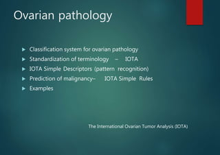
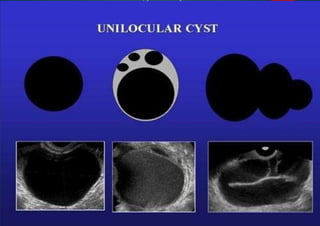
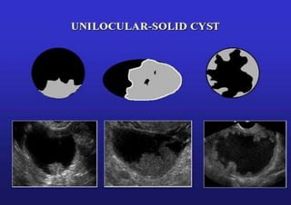
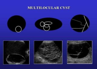
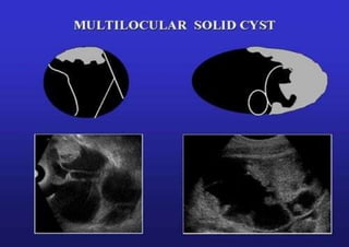
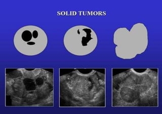
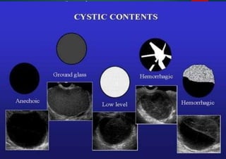
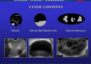
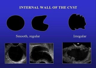
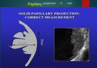
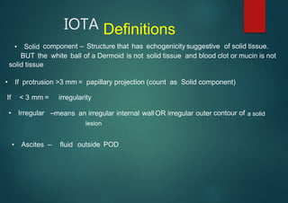
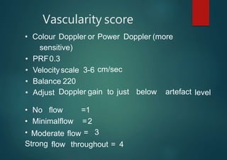
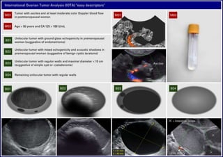
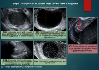
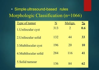
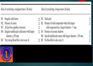
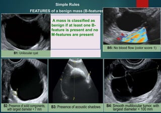
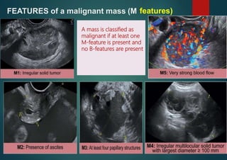
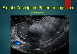
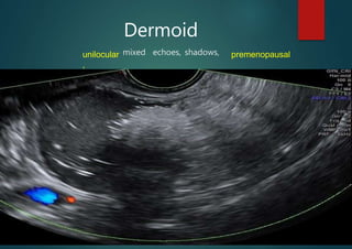
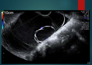
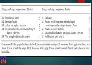
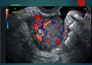
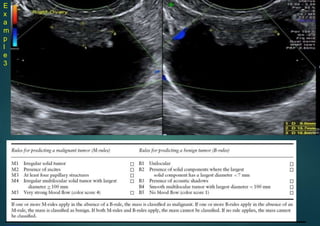
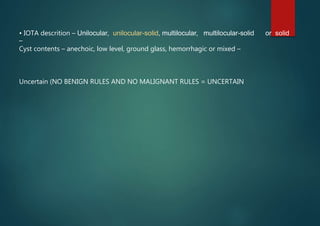
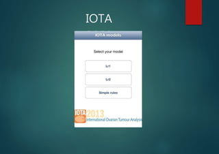
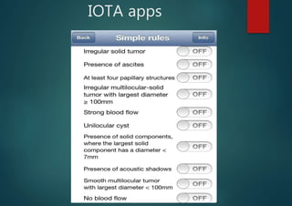
Adnexal mass evaluation (IOTA
Terminology, Simple Descriptors and Simple
Rules)
A.S.M. SUFIAN
HMO , Cumilla Medical College Hospital
(Radiology & Imaging )
Ovarian pathology
 Classification system for ovarian pathology
 Standardization of terminology – IOTA
 IOTA Simple Descriptors (pattern recognition)
 Prediction of malignancy– IOTA Simple Rules
 Examples
The International Ovarian Tumor Analysis (IOTA)
Ovarian cysts and tumors
IOTA
IOTA
Normal
ovaries
Premenopausal VS postmenopausal women
The report reallymatters
• Is it benign or malignant?
• Simple cyst
• Complex cyst
• Can we do better?
By the end of this presentation you will be able to report this!
How to report this abnormality?
Classification system for adnexal
masses
• Benign ovarian
• Benign non-ovarian
• Primary malignant ovarian
• Secondary malignant ovarian
Classification system for adnexal
masses
BENIGN OVARIAN:
ascites and
• Polycystic ovaries
• Functional cysts
• Endometriomas
• Serous cystadenoma
• Mucinous cystadenoma
• Mature teratoma
• Fibroma (rare, can cause Meig’s syndrome:
pleural effusion)
Classification system for adnexal
masses
BENIGN NON-OVARIAN:
• Paratubal cyst
• Hydrosalpinx
• Tubo-ovarian abscess
• Peritoneal pseudocysts
• Appendicular abscess
• Diverticular abscess
• Pelvic kidney
• Ectopic pregnancy
Classification system for adnexal
masses
PRIMARY MALIGNANT OVARIAN
Epithelial carcinoma
– Borderline
– Serous cystadenocarcinoma (commonest ovarian cancer, 50% malignancies)
– Mucinous cystadenocarcinoma (10% ovarian malignancies)
• Borderline variant is pseudomyxoma peritonei -exclude appx primary
– Endometrioid carcinoma (25% ovarian malignancies)(associated with endometrial ca in 20%)
– Clear cell carcinoma
Germ cell tumor
– Malignant teratoma
– Dysgerminoma
• Sex-cord tumor –
Granulosa cell tumor (secretes estrogen)
Classification system for adnexal masses
SECONDARY MALIGNANT OVARIAN
• 10% of ovarian malignancy
Predominantly:
• Breast
• Gastrointestinal carcinoma (Krukenberg tumor)
IOTA definitions
– Unilocular, Unilocular-solid, Multilocular, Multilocular-solid or Solid
– Cyst contents– or hemorrhagic.
anechoic, low level, ground glass, mixed
– Solid material or papillary structures or wall irregularity (presence and size)
– Vascularity
– Shadows
– Ascites
Unilocular
IOTA
Unilocular-solid
IOTA
Multilocular
IOTA
Multilocular-solid
IOTA
Solid
IOTA
Cystic contents
IOTA
Mixed cystic contents
IOTA
Internal wall of the cyst
IOTA
IOTA
-projection >3 mm
Papillary
IOTA Definitions
• Solid component – Structure that has echogenicity suggestive of solid tissue.
BUT the white ball of a Dermoid is not solid tissue and blood clot or mucin is not
solid tissue
• If protrusion >3 mm = papillary projection (count as Solid component)
If < 3 mm = irregularity
• Irregular – contour of
means an irregular internal wallOR irregular outer
• Ascites – fluid outside POD
lesion
a solid
Vascularity score
• Colour Doppler or Power Doppler (more
cm/sec
Doppler gain to just below artefact
sensitive)
• PRF0.3
• Velocityscale 3-6
• Balance 220
• Adjust level
Moderate flow
• No flow =1
• Minimalflow =2
• =
Strong
3
flow throughout = 4
Ovarian pathology
• Classification systemfor ovarian pathology
• Standardization of terminology – IOTA
• IOTA Simple Descriptors (pattern
recognition)
• Prediction of malignancy – IOTA Simple
Rules
• Worked examples
Simple Descriptors
• Certain abnormalities are really obvious:
RECOGNITION -
• Endometrioma
• Benign cystic teratoma / Dermoid Cyst
• Simple cyst or serous cystadenoma
• Functional
• Malignant
Cyst. Eg. Hemorrhagic cyst.
tumour with ascites
PATTERN
BD, benign descriptor; MD, malignant descriptor.
Simple Descriptors of an ovarian mass used to make a diagnosis
When Simple Descriptorsdo not apply:
• If the mass is not instantly recognizable
and Simple Descriptorsdo not applicable:
–Then apply Simple Rules
Ovarian pathology
• Classification systemfor ovarian pathology
• Standardization of terminology – IOTA
• IOTA Simple Descriptors (pattern
recognition)
• Prediction of malignancy – IOTA Simple
Rules
• Worked examples
• Simple ultrasound-based rules
Morphologic Classification (n=1066)
Type of tumor N Malign. %
1.Unilocular cyst 313 2 0.6
2.Unilocular solid 132 44 33
3.Multilocular cyst 196 20 10
4.Multilocular solid 284 116 41
5.Solid tumour
136 84 62
Simple Rules
FEATURES of a benign mass (B-features)
A mass is classified as
benign if at least one B-
feature is present and no
M-features are present
FEATURES of a malignant mass (M features)
A mass is classified as
malignant if at least one
M-feature is present and
no B-features are present
Simple Rules
Rule 1: If one or more M features are present in
absence of B feature(s), the mass is classified as
malignant.
• Rule 2: If one or more B features are present in
absence of M feature(s), the mass is classified as
benign.
• Rule 3: If both M features and B features are present,
or if no B or M features are present, the result is
inconclusive and a second stage test is recommended.
• examples–
premenopausal
Endometrioma
unilocular, ground glass echogenicity,
Simple Descriptors Pattern recognition
premenopausal
unilocular
,
Dermoid
mixed echoes, shadows, premenopausal
How to differentiate
<10 cm
Serous cystadenoma
unilocular, regular walls, <10 cm
Stage 4 ovarian carcinoma with
ascites
Tumor with at least moderate blood flow, ascites, postmenopausal
Exampleswhere Simple descriptor don’t apply
<10cm
Example 1
Simplerules: BENIGN
Mucinous cystadenoma
Example 2
Example 2
Simple rules:MALIGNANT
Metastasis from bowel primary
E
x
a
m
p
l
e
3
• IOTA descrition – Unilocular, unilocular-solid, multilocular, multilocular-solid or solid
–
Cyst contents – anechoic, low level, ground glass, hemorrhagic or mixed –
Uncertain (NO BENIGN RULES AND NO MALIGNANT RULES = UNCERTAIN
Example 3
Simple rules:UNCERTAIN
Borderline ovariantumour
Serous cystadenofibroma
Ovarian Fibroma
most commonly manifest as solid, hypoechoic masses with ultrasound beam attenuation.
As such, they may appear similar to a pedunculated subserosal uterine fibroid
Resources
apps (risk
• IOTA – papers, courses,
calculators), conferences
hKp://www.iotagroup.org
• ISUOG – papers, journal club slides
• GE poster.
• Internet slide and picture.
IOTA
apps
IOTA apps
THANK YOU
&
Jajakallah khair

Ovarian tumor analysis

Editor's Notes

  • #43 Example
  • #58  Video
  • #60 Benign , fibroma
  • #63 video
  • #64 CT scan – Multilobulated solid masses with prominent fibrovascular septa. Post contrast imaging shows enhancement of the septae. Other- calcification may be present in a speckled pattern.
  • #66 video
  • #69 example
  • #70 Papillary serous cystadenocarcinoma