This document discusses the pathogenesis of oral submucous fibrosis (OSF). It begins by describing the characteristics and presentation of OSF. It then discusses several potential factors that may be involved in the development and pathogenesis of OSF, including genetic alterations, infectious agents like viruses and Candida, and carcinogenic agents found in tobacco like areca nut and smoke. The document suggests a multifactoral model may best explain the disease process, involving genetic susceptibility combined with environmental exposures like carcinogens in betel quid and infections. It notes many similarities between OSF and oral cancers in terms of genetic changes and risk factors.
this seminar emphasizes abour osmf,historical background,nearly all classification were covered,its pathogenesis starting from physical changes to cellular changes in oral mucosa and treatment modalities
This document provides information on oral submucous fibrosis (OSMF), including its definition, epidemiology, etiology, clinical features, histopathological classification, and management. OSMF is a precancerous condition characterized by fibrosis of the submucosa and juxta-epithelial inflammatory reaction. It predominantly affects people of Asian descent and is strongly associated with chewing areca nut and consumption of chili peppers. Clinically, it presents as blanching and stiffness of the oral mucosa leading to restricted mouth opening. Histopathological examination can classify the severity of fibrosis. Arecoline in areca nut is the main etiological agent that causes fibrosis through oxidative stress and increased collagen production.
This document discusses oral submucous fibrosis (OSF), a chronic condition characterized by fibrosis of the submucosal tissues and epithelial atrophy of the oral cavity. It causes stiffness of the oral mucosa and trismus. The condition is caused by chewing areca nut and ingesting spicy foods. The pathogenesis involves increased collagen production and decreased breakdown due to areca nut compounds, as well as genetic and immunological factors. Symptoms progress from pigmentation and salivation to dry mouth and trismus. Treatment involves preventing habits, medical management, physiotherapy, and surgery such as excision of fibrous bands and use of flaps like buccal fat pad or nasolabial flaps to
1. Oral submucous fibrosis (OSF) is a chronic disease characterized by inflammation and fibrosis of the oral cavity that causes mouth opening restriction. It is associated with chewing betel nut and is considered a precancerous condition.
2. OSF has multiple treatment approaches including medication, physiotherapy, surgery, and lifestyle changes. Medications include steroids, hyaluronidase, and pentoxifylline to reduce inflammation. Surgeries such as fibrotomy and grafts help improve mouth opening.
3. While various treatments provide temporary relief, recurrence of fibrosis is common. The disease has no definitive cure, though early diagnosis and lifestyle changes may help prevent progression. Close monitoring is also
This document summarizes a presentation on a retrospective study comparing surgical treatment modalities for 100 patients with oral submucous fibrosis. The study compared outcomes of excising fibrous bands and using different grafting techniques, including buccal fat pad graft (group I), tongue flap (group II), nasolabial fold flap (group III), and split skin graft (group IV). The results found that buccal fat pad grafting had the highest mean mouth opening at 1 week post-op, highest post-op pain/function scores at 1 month, and offered ease of surgery and low morbidity. Tongue flap had the lowest post-op mouth opening and caused discomfort due to difficulty with tongue movement.
Mr. Ayaz Baig, a 25-year-old male, presented with a chief complaint of restricted mouth opening and burning sensation in the mouth when consuming spicy foods for the past 1-2 months. On examination, fibrous bands were seen bilaterally on the buccal mucosa, mouth opening was limited to 15mm, and pit and fissure caries were detected. A provisional diagnosis of stage III oral submucous fibrosis bilaterally was made. The treatment plan included stopping tobacco use, nutritional supplements and intralesional injections to reduce inflammation, followed by restorative treatment and regular follow ups.
The document provides an overview of oral submucous fibrosis (OSF), including its definition, epidemiology, etiology, and pathogenesis. Some key points:
- OSF is a chronic disease affecting the oral cavity and oropharynx that results in fibrosis of the submucosal tissues and difficulty opening the mouth.
- It predominantly affects people ages 20-45 in India. Common causes include chewing betel quid and areca nut, which contain alkaloids that stimulate collagen production leading to fibrosis. Tobacco use can also contribute.
- Nutritional deficiencies may play a role by inhibiting normal collagen breakdown. Genetic factors may influence susceptibility by affecting collagen-related genes. Local irritants like ch
This document discusses oral submucous fibrosis (OSF), a chronic disease of the oral cavity that causes fibrosis of the lamina propria and stiffness of the oral mucosa leading to trismus. It is characterized by vesicle formation, juxta-epithelial inflammatory reaction, and fibro-elastic changes. OSF is most common in Southeast Asia and India, has a multifactoral pathogenesis involving chronic irritation from spices and betel nut, and can progress to malignancy in some cases. Staging of OSF severity ranges from mild stomatitis to severe trismus with premalignant or malignant changes. Management involves behavioral therapy to remove irritants, medical therapies like steroids and pentox
This document outlines and compares several existing clinical classification systems for oral submucous fibrosis (OSMF). OSMF is a potentially malignant condition of the oral cavity characterized by fibrosis of the submucosa and restricted mouth opening. The classifications systems are grouped based on whether they were proposed before or after 2000. They stage OSMF based on criteria like mouth opening, area of involvement, presence of precancerous lesions, and clinical symptoms. While no single system is universally accepted, classification helps diagnose and manage OSMF.
This document provides an overview of oral submucous fibrosis (OSF), including its definition, epidemiology, etiology, pathogenesis, clinical features, classification, and medical management. OSF is characterized by fibrous bands in the oral mucosa leading to mouth opening restriction. It is commonly found in India and associated with areca nut chewing. Clinical features include blanching of the oral mucosa, trismus, and palpable fibrous bands. Staging systems exist based on clinical features and mouth opening. Management focuses on eliminating risk factors and includes antioxidants, biogenic stimulants, immune modulators, and blood flow promotors to reduce fibrosis and support healing.
Oral submucous fibrosis is defined as “An insidious chronic disease affecting any part of the oral cavity and sometimes the pharynx. A wide range of treatment including drug therapy, surgical therapy, and physiotherapy have been attempted till date, with varying degrees of benefit, but none have been able to cure this disease.
The treatment depends on the level of clinical involvement. At a very early stage, cessation of the habit is adequate. Medical/surgical treatment is necessary for moderate to severe cases. Surgical treatment is the method of choice in patients with marked limitation of mouth opening or in patients not responding to the conservative management.
This document provides an overview of oral submucous fibrosis (OSF), including its definition, epidemiology, classification, etiology, pathogenesis, clinical features, and histopathology. OSF is a chronic disease characterized by inflammation and fibrosis of the submucosal tissues caused by chewing areca nut. It predominantly affects people from South Asia and is associated with significantly increased risk of oral cancer. The areca nut alkaloid arecoline is the main causative agent, inducing fibrosis through oxidative damage, upregulation of growth factors and cytokines, and inhibition of collagen degradation. Clinically, OSF presents with burning sensation and scarring that results in restricted mouth opening and tongue movement.
This document discusses oral submucous fibrosis (OSF), a chronic disease caused by chewing betel quid that results in fibrosis of the oral cavity and inability to open the mouth fully. It describes the epidemiology, pathogenesis, clinical features and classification system for OSF. Surgical management is often needed for advanced OSF cases. One approach described is resection of fibrotic bands and reconstruction of the surgical defect using a buccal fat pad graft, which provides a well-vascularized tissue that reliably epithelializes the area. The document includes a case report demonstrating successful application of this technique.
Oral submucous fibrosis (OSF) is a chronic disease that causes scarring of the oral mucosa and may progress to cancer. It is characterized by inflammation and fibrosis of the submucosa that results in stiffening of the oral tissues and a burning sensation in the mouth. The disease commonly affects people in India and Southeast Asia and is associated with chewing betel quid and areca nut. Clinical findings include blanching and fibrosis of the oral mucosa that causes restricted mouth opening and difficulty with eating, speaking, and swallowing in advanced cases. Biopsy is required for definitive diagnosis. OSF has been classified clinically and functionally based on the extent of fibrosis and degree of mouth opening limitation.
This document provides an overview of oral submucous fibrosis (OSF), a chronic disease affecting the oral cavity and sometimes pharynx that causes stiffening of the mucosa and trismus. It discusses the definition, epidemiology, etiology, pathogenesis, clinical features, malignant potential, investigations, and treatment of OSF. The main causes are believed to be the chewing of areca nut and consumption of chillies. Arecoline from areca nut stimulates fibrosis through various mechanisms like increased collagen production and decreased breakdown. OSF has a high malignant transformation rate of 7-13% and is considered a precancerous condition. Treatment involves eliminating habits, corticosteroids, hyaluronidase, plac
PATHOGENESIS OF ORAL SUB MUCOUS FIBROSIS(OSMF)RoshanYadav36
This document discusses the pathogenesis of oral submucous fibrosis (OSF). It begins by defining OSF and noting its prevalence in Southeast Asia. The pathogenesis is described as multifactorial, involving local irritants like areca nut chewing and genetic predispositions. Arecoline from areca nuts increases collagen production by stimulating fibroblasts and inhibiting degradation through tannins. Genetic factors like collagen genes may also play a role. Chronic inflammation from betel quid chewing triggers cytokine release and increased growth factors that further stimulate collagen production and inhibit breakdown, ultimately leading to fibrosis.
This document provides an overview of oral submucous fibrosis (OSMF), a potentially malignant condition of the oral mucosa. It discusses the epidemiology, etiology, pathogenesis, clinical features, grading systems, investigations, histopathology, and management of OSMF. The condition is characterized by fibrosis of the lamina propria and epithelial atrophy leading to stiffness of the oral mucosa and trismus. Chronic irritation from spices and nut chewing are significant risk factors. Management involves behavioral changes, medical therapies such as steroids, surgical treatments like grafts, and physiotherapy.
1) Oral submucosal fibrosis is a chronic, progressive scarring condition of the oral cavity caused by chewing betel quid and smoking tobacco.
2) It mainly affects the buccal mucosa, lips, soft palate and retromolar areas, causing blanching and fibrosis that restricts mouth opening.
3) The condition is linked to areca nut components in betel quid and alkaloids like arecoline that stimulate collagen production by fibroblasts. Early detection allows for reversal by stopping the habit. Advanced cases require corticosteroid injections and supplements to improve symptoms.
ORAL SUBMUCOUS FIBROSIS BY DR. C. P. ARYA (B.Sc. B.D.S.; M.D.S.; P.M.S.; R.N....DR. C. P. ARYA
OSMF is a chronic disease characterized by inflammation and fibrosis of the oral cavity that causes stiffness of the oral mucosa and difficulty opening the mouth. It predominantly affects people from India and Southeast Asia and is caused by chewing areca nut-containing products like betel quid and gutkha. Treatment focuses on eliminating risk factors and managing symptoms through corticosteroid injections, hyaluronidase, and physical therapies.
Oral submucous fibrosis (OSMF or OSF) is a chronic, complex, premalignant (1% transformation risk) condition of the oral cavity, characterized by juxta-epithelial inflammatory reaction and progressive fibrosis of the submucosal tissues (the lamina propria and deeper connective tissues). As the disease progresses, the jaws become rigid to the point that the person is unable to open the mouth.
The condition is remotely linked to oral cancers and is associated with areca nut or betel quid chewing, a habit similar to tobacco chewing, is practiced predominantly in Southeast Asia and India, dating back thousands of years.
Advanced Treatment of Oral Submucous Fibrosis by Dr. Amit T. SuryawanshiAll Good Things
This presentation is created by Face Art International Cosmetic Super speciality for “Patient education purpose”. This presentation is presented by Dr. Amit T. Suryawanshi (Oral & Maxillofacial plastic Surgeon, Dental Surgeon & Hair Transplant Specialist from University of Greifswald, Germany) at National & International level in the field of above mentioned Specialities. Hope this would somehow be helpful to you while making your presentations. All the best & your replies will be welcomed! Follow us for Future Journey towards betterment of Humanity.
1) Oral submucous fibrosis is a chronic, premalignant condition that results in fibrosis of the oral cavity and sometimes pharynx.
2) The condition is caused by the chewing of areca nut and results in deposition and stabilization of collagen in the lamina propria through various mechanisms including clonal selection of fibroblasts, increased growth factors like TGF-β, and decreased breakdown of collagen.
3) Genetic and nutritional factors may also play a role, but areca nut is the main etiological agent through its alkaloids, tannins and reactive oxygen species that cause inflammation and fibrosis.
Oral Submucous Fibrosis and its Management.Maxfac Center
Oral Submucous Fibrosis and its various treatment modalities inclusive of both non-surgical and surgical management.
Mentor: Dr Saikat Saha MDS, OMFS, SIliguri, West Bengal, India
Address: MAXFAC Center for Oral and Maxillofacial and Head & Neck Surgery, Siliguri
Email : maxfacmail@gmail.com
This document presents a classification system for oral submucous fibrosis (OSMF) proposed by Deepak Passi et al. in 2017. The classification system grades OSMF on a scale from 1 to 4 based on clinical involvement, mouth opening ability, histopathological findings, and recommended treatment approaches. Grade 1 involves less than one-third of the oral cavity and has inflammatory histological features. Grade 4 involves over two-thirds of the oral cavity, possible malignant transformation, and requires surgical treatment including biopsies. The classification aims to assist clinicians in categorizing and managing OSMF based on disease severity.
Oral submucous fibrosis is a chronic, scarring disease of the oral mucosa characterized by rigidity and restricted mouth opening due to fibroelastic changes in the submucosa. It has an 8% risk of malignant transformation. The precise etiology is unknown but is likely multifactorial, involving factors like areca nut consumption, nutritional deficiencies, autoimmunity, and genetics. Clinical features include burning mouth, restricted opening, and palpable fibrous bands in the mouth. Staging depends on the degree of mouth opening restriction. Management focuses on stopping the habit, injecting steroids, surgically splitting bands, and monitoring for cancer development.
This document reports a case study of a 63-year-old male patient with an unusual presentation of peripheral ossifying fibroma (POF). POFs typically affect younger females in the maxillary anterior gingiva. In this case, the POF occurred in an older male patient in the mandibular anterior gingiva. Histological examination of the excised lesion found features consistent with POF, including calcified deposits resembling cementum or bone within a cellular fibrous connective tissue stroma. While the etiology of POF remains uncertain, this case supports the view that at least some POFs may be true neoplastic lesions rather than purely reactive proliferations.
Basal cell ameloblastoma a rare case report and review of literatureQuách Bảo Toàn
The document reports on a rare case of basal cell ameloblastoma occurring in a 12-year-old patient. Basal cell ameloblastoma is believed to be the rarest histologic subtype of ameloblastoma and typically occurs in adults aged 30-40 years. Only 6 previous cases had been reported in the literature. Microscopic examination of the lesion showed uniform basaloid cells without stellate reticulum, consistent with basal cell ameloblastoma. Considering the rarity of this lesion in such a young patient, this case highlights the importance of histopathological examination for diagnosing odontogenic tumors.
Abstract—This study was aimed to present a case report of a case of peripheral ossifying fibroma which is a rare case. This case was a 30 years non smoker male with the chief complaint of growth of gum tissue, moderately large in the mandibular posterior region. On intraoral examination, a peduncalated growth of 17 x 12 x 6 mm on marginal and attached gingiva with respect to tooth number 47 considerably hard in consistency and movable was seen. The lesion was erythmatous having a smooth non ulcerated surface. It was asymptomatic with no sign of pain. Intra oral periapical radiograph was taken which revealed slight erosion of crest of bone which was later confirmed during surgical excision. The possible reason of crestal bone erosion may be constant pressure of the growth. Differential diagnosis of irritation fibroma, pyogenic granuloma and peripheral giant cell granuloma was considered. However, clinical appearance and consistency was of a hard fibrous growth, which therefore led to a provisional diagnosis of peripheral ossifying fibroma or peripheral odontogenic fibroma.
The document summarizes oral submucous fibrosis (OSMF), a chronic disease characterized by fibrosis of the oral mucosa and sometimes the pharynx. It causes stiffness of the oral cavity and difficulty opening the mouth. The document discusses the definition, history, epidemiology, etiology, clinical findings, classification and malignant transformation risk of OSMF. The main causative factors are believed to be chewing betel nut and areca nut, which contain irritating compounds like arecoline. Chronic irritation and ingestion of spices are also thought to contribute to the pathogenesis.
Odontogenic keratocyst involving maxillary antrum / dental coursesIndian dental academy
The Indian Dental Academy is the Leader in continuing dental education , training dentists in all aspects of dentistry and
offering a wide range of dental certified courses in different formats.
1) Rathke's pouch forms the pituitary gland, which must migrate to the hypothalamus during development. In some individuals, pituitary tissue remains in the roof of the pharynx.
2) Down syndrome results from trisomy 21 and is characterized by midface hypoplasia, reducing nasopharynx and oropharynx volume.
3) Crouzon syndrome is an autosomal dominant craniosynostosis with midface hypoplasia and relative mandibular prognathism, potentially causing airway obstruction.
This document discusses acute gingival infections, focusing on acute necrotizing ulcerative gingivitis (ANUG). It describes the types and historical understanding of ANUG. The disease is caused by a polymicrobial infection involving spirochetes and other bacteria. Risk factors include malnutrition, stress, smoking, and immune system impairment. Clinically, ANUG presents as a progressive painful infection of the gums with ulceration and sloughing of dead tissue.
Acute necrotizing ulcerative gingivitis (ANUG), also known as Vincent's gingivitis or Vincent's infection, is a polymicrobial infection of the gums that results in ulceration, necrosis, and pain. It is caused by an overgrowth of oral bacteria like spirochetes and fusiform bacteria when the immune system is weakened by factors like stress, malnutrition, or HIV infection. ANUG begins as small ulcers on the gums that can spread and deepen over time if left untreated, potentially resulting in bone and tissue loss. Symptoms include bleeding, foul taste, and pain when eating. Treatment focuses on improving oral hygiene and nutrition to restore immune function and resolve the
A 60-year-old male presented with a large intraoral mass on the floor of the tongue. Excision biopsy revealed the mass to be a lipoma, a benign tumor composed of adipose tissue that most commonly develops in subcutaneous tissues but can also occur in deeper tissues like the oral cavity. Lipomas typically occur in patients over 40 years of age and can cause interference with functions if allowed to grow large. Complete surgical excision is the recommended treatment and provides good long-term outcomes without recurrence, as was the case for this patient.
Localized aggressive periodontitis (LAP) and generalized aggressive periodontitis (GAP) are forms of periodontitis that primarily affect younger individuals. LAP typically affects the first molars and incisors, causing rapid attachment loss and bone destruction. GAP affects at least three teeth besides molars and incisors, with periods of destruction followed by remission. Both involve familial factors and bacterial pathogens like P. gingivalis and A. actinomycetemcomitans. Treatment involves non-surgical and surgical therapies along with systemic antibiotics. Frequent maintenance is important for managing the diseases.
The Indian Dental Academy is the Leader in continuing dental education , training dentists in all aspects of dentistry and
offering a wide range of dental certified courses in different formats.for more details please visit
www.indiandentalacademy.com
1) Premalignant lesions of the oral mucosa occur in 1.5-4.5% of the global population and account for 17-35% of new oral cancer cases, with an annual malignant transformation rate of 0.7-2.9%.
2) Common premalignant lesions include leukoplakia, erythroplakia, submucous fibrosis, and lichen planus. Leukoplakia may transform to cancer in 0.13-34% of cases depending on population.
3) Risk factors for oral premalignant lesions include smoking, smokeless tobacco, alcohol, HPV infection, and nutritional deficiencies. Early diagnosis and treatment can prevent malignant transformation
This document provides information on oral submucous fibrosis (OSMF), including its definition, history, pathogenesis, clinical features, and staging classifications. OSMF is a chronic disease affecting the oral cavity and sometimes pharynx, characterized by juxtaepithelial inflammatory reaction and fibrosis of the lamina propria. Chewing betel quid and areca nut releases alkaloids and tannins that cause irritation and inflammation, activating fibroblasts and increasing production and cross-linking of collagen over time, resulting in stiffness and inability to open the mouth. OSMF is considered a precancerous condition due to its association with oral cancer. Staging systems describe progression from initial symptoms to trismus and possible malignancy
Differential diagnosis of white lesions in the form of a combination of various journals.
For easy diagnosis of various white lesions which we usually encounter in our clinics. A diagnosis tree along with various white lesions along with the lesions to which they resemble.
By:
Dr. Sunbul Tabrez
This document provides information on aggressive periodontitis, including its historical background, classification, clinical presentation, epidemiology, etiology, and pathogenesis. Some key points include:
- Aggressive periodontitis is a rare, severe form of periodontitis characterized by early onset and familial aggregation. It includes localized aggressive periodontitis (LAP) and generalized aggressive periodontitis (GAP).
- LAP typically affects first molars and incisors in adolescents/young adults and is associated with A. actinomycetemcomitans infection, while GAP has a more generalized pattern of attachment/bone loss.
- Screening involves measuring attachment loss via probing or radiographic assessment of alveolar bone levels
This document provides an overview of fibrous osseous lesions of the jaws and their classification and management. It begins with an introduction to fibrous osseous lesions and their classification. It then discusses specific lesions in more detail, including fibrous dysplasia, cemento-osseous dysplasia, ossifying fibroma, and others. For each lesion, it covers clinical features, radiographic patterns, differential diagnosis, and treatment approaches. The goal is to review current knowledge around the diagnosis and management of these complex fibro-osseous lesions of the jaws.
This document describes a case report of a peripheral ossifying fibroma (POF) in a 22-year-old female patient. POFs are non-neoplastic reactive lesions of the gingiva caused by local irritants like plaque or food impaction. Histopathological examination showed fibrous connective tissue with bony trabeculae and dystrophic calcification, consistent with POF. Treatment involves complete surgical excision to remove the lesion and any remaining irritants in order to reduce the risk of recurrence.
Peripheral Ossifying Fibroma: A Case Reportiosrjce
This case report describes a peripheral ossifying fibroma (POF) in a 50-year-old male patient. The patient presented with a 3x3 cm swelling in the mandibular left posterior region. Radiographs showed well-marginated radiopaque lesions with radiolucent foci suggestive of calcifications. Histopathological examination revealed variable thickness stratified squamous epithelium with fibrovascular connective tissue containing dense collagen fibers, compressed blood vessels, chronic inflammatory cells, and osseous-like structures, confirming the diagnosis of POF. POF is a benign reactive gingival lesion that is commonly mistaken for other lesions clinically. Complete surgical excision is the recommended treatment due to the lesion's recurrence
The document discusses changes that occur in the periodontium with aging. It states that the prevalence of periodontal diseases increases with age due to changes in tissues over time. Specifically, it mentions that the gingival epithelium thins with age and has reduced keratinization. The gingival connective tissue has fewer cellular elements and increased collagen with aging. The periodontal ligament also has fewer cells and thicker collagen fibers with an irregular structure in older individuals. Cementum thickens with age as well.
Fibro osseous lesions of jaws/oral surgery courses by indian dental academyIndian dental academy
Indian Dental Academy: will be one of the most relevant and exciting training center with best faculty and flexible training programs for dental professionals who wish to advance in their dental practice,Offers certified courses in Dental implants,Orthodontics,Endodontics,Cosmetic Dentistry, Prosthetic Dentistry, Periodontics and General Dentistry.
STUDIES IN SUPPORT OF SPECIAL POPULATIONS: GERIATRICS E7shruti jagirdar
Unit 4: MRA 103T Regulatory affairs
This guideline is directed principally toward new Molecular Entities that are
likely to have significant use in the elderly, either because the disease intended
to be treated is characteristically a disease of aging ( e.g., Alzheimer's disease) or
because the population to be treated is known to include substantial numbers of
geriatric patients (e.g., hypertension).
5-hydroxytryptamine or 5-HT or Serotonin is a neurotransmitter that serves a range of roles in the human body. It is sometimes referred to as the happy chemical since it promotes overall well-being and happiness.
It is mostly found in the brain, intestines, and blood platelets.
5-HT is utilised to transport messages between nerve cells, is known to be involved in smooth muscle contraction, and adds to overall well-being and pleasure, among other benefits. 5-HT regulates the body's sleep-wake cycles and internal clock by acting as a precursor to melatonin.
It is hypothesised to regulate hunger, emotions, motor, cognitive, and autonomic processes.
How to Control Your Asthma Tips by gokuldas hospital.Gokuldas Hospital
Respiratory issues like asthma are the most sensitive issue that is affecting millions worldwide. It hampers the daily activities leaving the body tired and breathless.
The key to a good grip on asthma is proper knowledge and management strategies. Understanding the patient-specific symptoms and carving out an effective treatment likewise is the best way to keep asthma under control.
Nano-gold for Cancer Therapy chemistry investigatory projectSIVAVINAYAKPK
chemistry investigatory project
The development of nanogold-based cancer therapy could revolutionize oncology by providing a more targeted, less invasive treatment option. This project contributes to the growing body of research aimed at harnessing nanotechnology for medical applications, paving the way for future clinical trials and potential commercial applications.
Cancer remains one of the leading causes of death worldwide, prompting the need for innovative treatment methods. Nanotechnology offers promising new approaches, including the use of gold nanoparticles (nanogold) for targeted cancer therapy. Nanogold particles possess unique physical and chemical properties that make them suitable for drug delivery, imaging, and photothermal therapy.
These lecture slides, by Dr Sidra Arshad, offer a simplified look into the mechanisms involved in the regulation of respiration:
Learning objectives:
1. Describe the organisation of respiratory center
2. Describe the nervous control of inspiration and respiratory rhythm
3. Describe the functions of the dorsal and respiratory groups of neurons
4. Describe the influences of the Pneumotaxic and Apneustic centers
5. Explain the role of Hering-Breur inflation reflex in regulation of inspiration
6. Explain the role of central chemoreceptors in regulation of respiration
7. Explain the role of peripheral chemoreceptors in regulation of respiration
8. Explain the regulation of respiration during exercise
9. Integrate the respiratory regulatory mechanisms
10. Describe the Cheyne-Stokes breathing
Study Resources:
1. Chapter 42, Guyton and Hall Textbook of Medical Physiology, 14th edition
2. Chapter 36, Ganong’s Review of Medical Physiology, 26th edition
3. Chapter 13, Human Physiology by Lauralee Sherwood, 9th edition
Osvaldo Bernardo Muchanga-GASTROINTESTINAL INFECTIONS AND GASTRITIS-2024.pdfOsvaldo Bernardo Muchanga
GASTROINTESTINAL INFECTIONS AND GASTRITIS
Osvaldo Bernardo Muchanga
Gastrointestinal Infections
GASTROINTESTINAL INFECTIONS result from the ingestion of pathogens that cause infections at the level of this tract, generally being transmitted by food, water and hands contaminated by microorganisms such as E. coli, Salmonella, Shigella, Vibrio cholerae, Campylobacter, Staphylococcus, Rotavirus among others that are generally contained in feces, thus configuring a FECAL-ORAL type of transmission.
Among the factors that lead to the occurrence of gastrointestinal infections are the hygienic and sanitary deficiencies that characterize our markets and other places where raw or cooked food is sold, poor environmental sanitation in communities, deficiencies in water treatment (or in the process of its plumbing), risky hygienic-sanitary habits (not washing hands after major and/or minor needs), among others.
These are generally consequences (signs and symptoms) resulting from gastrointestinal infections: diarrhea, vomiting, fever and malaise, among others.
The treatment consists of replacing lost liquids and electrolytes (drinking drinking water and other recommended liquids, including consumption of juicy fruits such as papayas, apples, pears, among others that contain water in their composition).
To prevent this, it is necessary to promote health education, improve the hygienic-sanitary conditions of markets and communities in general as a way of promoting, preserving and prolonging PUBLIC HEALTH.
Gastritis and Gastric Health
Gastric Health is one of the most relevant concerns in human health, with gastrointestinal infections being among the main illnesses that affect humans.
Among gastric problems, we have GASTRITIS AND GASTRIC ULCERS as the main public health problems. Gastritis and gastric ulcers normally result from inflammation and corrosion of the walls of the stomach (gastric mucosa) and are generally associated (caused) by the bacterium Helicobacter pylor, which, according to the literature, this bacterium settles on these walls (of the stomach) and starts to release urease that ends up altering the normal pH of the stomach (acid), which leads to inflammation and corrosion of the mucous membranes and consequent gastritis or ulcers, respectively.
In addition to bacterial infections, gastritis and gastric ulcers are associated with several factors, with emphasis on prolonged fasting, chemical substances including drugs, alcohol, foods with strong seasonings including chilli, which ends up causing inflammation of the stomach walls and/or corrosion. of the same, resulting in the appearance of wounds and consequent gastritis or ulcers, respectively.
Among patients with gastritis and/or ulcers, one of the dilemmas is associated with the foods to consume in order to minimize the sensation of pain and discomfort.
PGx Analysis in VarSeq: A User’s PerspectiveGolden Helix
Since our release of the PGx capabilities in VarSeq, we’ve had a few months to gather some insights from various use cases. Some users approach PGx workflows by means of array genotyping or what seems to be a growing trend of adding the star allele calling to the existing NGS pipeline for whole genome data. Luckily, both approaches are supported with the VarSeq software platform. The genotyping method being used will also dictate what the scope of the tertiary analysis will be. For example, are your PGx reports a standalone pipeline or would your lab’s goal be to handle a dual-purpose workflow and report on PGx + Diagnostic findings.
The purpose of this webcast is to:
Discuss and demonstrate the approaches with array and NGS genotyping methods for star allele calling to prep for downstream analysis.
Following genotyping, explore alternative tertiary workflow concepts in VarSeq to handle PGx reporting.
Moreover, we will include insights users will need to consider when validating their PGx workflow for all possible star alleles and options you have for automating your PGx analysis for large number of samples. Please join us for a session dedicated to the application of star allele genotyping and subsequent PGx workflows in our VarSeq software.
The skin is the largest organ and its health plays a vital role among the other sense organs. The skin concerns like acne breakout, psoriasis, or anything similar along the lines, finding a qualified and experienced dermatologist becomes paramount.
Travel Clinic Cardiff: Health Advice for International TravelersNX Healthcare
Travel Clinic Cardiff offers comprehensive travel health services, including vaccinations, travel advice, and preventive care for international travelers. Our expert team ensures you are well-prepared and protected for your journey, providing personalized consultations tailored to your destination. Conveniently located in Cardiff, we help you travel with confidence and peace of mind. Visit us: www.nxhealthcare.co.uk
Pictorial and detailed description of patellar instability with sign and symptoms and how to diagnose , what investigations you should go with and how to approach with treatment options . I have presented this slide in my 2nd year junior residency in orthopedics at LLRM medical college Meerut and got good reviews for it
After getting it read you will definitely understand the topic.
Know the difference between Endodontics and Orthodontics.Gokuldas Hospital
Your smile is beautiful.
Let’s be honest. Maintaining that beautiful smile is not an easy task. It is more than brushing and flossing. Sometimes, you might encounter dental issues that need special dental care. These issues can range anywhere from misalignment of the jaw to pain in the root of teeth.
Know the difference between Endodontics and Orthodontics.
Osmf
1. 2011
Pathogenesis of Oral Submucous Fibrosis
Relationship to Risk Factors Associated With Oral Cancer
Radhakrishna Pillai, PhD,*,t Prabha Balaram, PhD,t
and Kannan Sankara Reddiar, MSct$
Oral submucous fibrosis (OSF) is a chronic disease of the may lead to relative loss of auditory acuity because of
oral mucosa characterized by inflammation and a pro- stenosis of the opening of the Eustachian tube. In ad-
gressive fibrosis of the lamina propria and deeper con- vanced cases, there may be severe trismus, and the to-
nective tissues. It is a condition predominantly seen tally inelastic mucosa is forced against the teeth, leading
among people of Indian origin, and an epidemiologic sur- to chronic ulceration and subsequent infection.
vey done a decade ago showed no less than 250,000 cases
In many cases, the fibrous tissue can be seen to
in India, a figure that must have increased sharply. OSF
is a condition with a high risk of malignant transforma-
extend from the anterior pillars into the soft palate as a
tion; to date, no conclusive etiologic agent has been iden- delicate reticulum of interlacing white strands that later
tified, although plenty of data have been generated on become confluent. The cheeks have a mottled marble-
various aspects of the disease. These include genetic, car- like mosaic appearance, with normal reddish mucosa
cinogenic, immunologic, viral, nutritional, and autoim- intermingling with depigmented diseased mucosa. The
mune possibilities, all of which also have been impli- floor of the mouth becomes pale and thickened, the
cated in the development of oral cancer. This article re- tongue becomes reduced in size and motility, and bands
views existing evidence on the pathogenesis of OSF and of encircling collagen distort the lips. If the fibrosis ex-
its relation to oral cancer and suggests a possible multi- tends down to the esophagus, the patient has progres-
factorial model to explain the disease process. Cancer sive dysphagia.
1992; 69:2011-2020.
Histologically, OSF is characterized by juxtaepithe-
lial fibrosis, along with atrophy or hyperplasia of the
Oral submucous fibrosis (OSF)is a chronic oral mucosal overlying epithelium, keratinizing metaplasia, and ac-
condition that occurs predominantly among Indians cumulation of hyalinized collagen beneath the base-
and people of Indian origin living outside India, occa- ment membrane with a progressive loss of vascularity.
sionally in other Asians, and sporadically in Europeans. Often, a variable infiltration of the lamina propria with
With prevalence rates in India from 0.2% to 1.2%, an chronic inflammatory cells also is e ~ i d e n t (Figs. 1, 2,
~-~
epidemiologic survey done a decade ago indicated no and 3).
less than 250,000 cases in the country.',' The possible precancerous nature of OSF first was
OSF is characterized by inflammation and a pro- described by Payma~ter,~ observed the onset of
who
gressive fibrosis of the lamina propria. The major pre- slowly growing squamous cell carcinomas in one-third
senting complaint is a progressive inability to open the of such patients. These observations were confirmed
mouth because of the accumulation of inelastic fibrous subsequently by Pindborg.' The cause of the disease,
tissue in the juxtaepithelial region of the oral mucosa, however, is still obscure. Hypersensitivity to chili and
along with concomitant muscle degeneration. Patients betel nuts and nutritional deficiencies all have been
describe a gradual onset of burning pain when eating suggested at various times to account for the pathogene-
spicy food that previously had caused no d i ~ t r e s sThe
.~ sis of the disease. However, it appears from current evi-
fibrosis also leads to difficulty in mastication, speech, dence that a combination of various factors could best
and swallowing and pain in the throat and ears. It also explain the pathogenesis of this unique condition.
These factors are discussed below, along with their re-
lationships to oral cancer.
From the *Department of FamiIy and Community Medicine,
University of Arizona, Tucson, Arizona; and the tRegiona1 Cancer
Centre, Trivandrurn, India. Genetic Alterations
4 Recipient of the Senior Research Fellowship of the Council for
Scientific and Industrial Research, India.
Address for reprints: Radhakrishna Pillai, PhD, Regional Cancer
Cancer researchers have amassed a great deal of data
Centre, Trivandmrn 69501 1, Kerala State, India. indicating that changes occur in the genetic control ap-
Accepted for publication July 15, 1991. paratus as cells progress to a malignant state. Almost all
2. 2012 CANCER April 25, 2992, Volume 69, No. 8
Figure 1. Histologic changes in OSF showing atrophic epithelium Figure 3. Histologic changes in OSF showing atrophic epithelium
with loss Of rete Pegs and avascular hYalinized with loss of rete pegs and relatively avascular hyalinized collagen
extending downwards from the basement membrane (low-power extending downwards from the basement membrane (high-power
view; H & E, original magnification X75) view of the subepithelial region; H & E, original magnification
X300).
carcinogenic agents, whether physical (radiation),
chemical, or infectious (viruses), act as mutagens. They euploidy, detected with relative ease when flow
change the structure of the genetic material, producing cytometry is used.16
point mutations, deletions, insertions, or rearrange- Another finding closely allied to genetic abnormali-
m e n t ~ . ~ -The tendency for development of specific
" ties is the role of oncogenic viruses. These viruses carry
types of tumors has been noticed to be inherited in genes that can act in a dominant fashion to cause cells to
many situations, such as retinoblastoma, Cowden's undergo malignant transformation. lo Oncogenes or tu-
syndrome, Gardner's syndrome, and neurofibromato- mor-causing genes often are mutated genes derived
sis.ll-14 Specific chromosomal aberrations have been from apparently normal eukaryotic genes called proto-
found consistently in a number of tumors, such as Bur- oncogene^.'^ A large number of proto-oncogenes have
kitt's lymphoma, acute and chronic leukemias, and been identified in the human genome, whereas mu-
Wilm's t ~ m o r . These abnormalities are mostly trans-
'~ tated amplified and rearranged oncogenes have been
locations and deletions with breakpoints at specific lo- found in a wide variety of malignant It is
cations. Another chromosomal abnormality that ap- interesting that many of the chromosome deletions and
pears ubiquitous to most tumors is the presence of an- translocations characteristic of many cancers have
breakpoints near or at the proto-oncogene location^.^'
Although no one has precisely or even closely iden-
tified a genetic basis for OSF, it cannot be ruled out.
Although hypersensitivity to chili and betel quid often
is explained as a common factor in the development of
OSF, it is difficult to understand why the disease is not
seen in Mexico and South America, where the diet in-
cluding chili intake equals or even exceeds that in India
or the Far East. A proper genetic perspective thus is vital
to explain the condition.
Studies of invasive oral carcinomas have produced
substantial findings, which, if can be shown to also oc-
cur in OSF, could explain its pathogenesis. We had ex-
plained earlier the genetic abnormalities in oral cancers,
including altered oncogene expre~sion.'~ was evi-
This
dent by a fivefold to tenfold increase in amplification of
c-myc, N-myc, and Ki Y U S genes in 20% to 40% of biopsy
Figure 2. Histologic changes in OSF showing atrophic epithelium
with loss of rete pegs and relatively avascular hyalinized collagen specimens studied, with 56% showing amplification of
extending downwards from the basement membrane (high-Dower
x - 1
at least one of the oncogene^.'^ Studies on squamous
view of the epithelium; H & E, original magnification X300). cell carcinoma cell lines show considerable chromo-
3. Pathogenesis of OSFIPillai et al. 201 3
some triploidy and tetraploidy. One such study" found laboratory35 and others36 have shown high titers of
aberrations of chromosome 1 in nine head and neck anti-HSV1 antibody in patients with oral cancer and
carcinoma cell lines examined that mainly involved de- precancer, including OSF, compared with patients with
letions at lq21, lq32, lq13, or lp22. Two known proto- other cancers and normal controls, but efforts to isolate
oncogenes map to these areas. Transfection experi- or characterize virus-specific proteins from lesions were
ments also have been performed with the use of DNA u n s u c c e ~ s f u lHowever, as discussed in the next sec-
.~~
from squamous carcinomas of the head and neck. Al- tion, synergy between HSV, Candida, nutritional status,
though such experiments examining different types of and topical carcinogens may provide interesting clues
solid tumors have found that 15% to 30% of these tu- to the development of oral lesions. Human papilloma-
mors contained transforming gene^,'',^^,^^ studies with virus (HPV) infection also has been associated with cer-
DNA obtained from head and neck cancers indicate vical dysplasia and invasive ~arcinoma.~' relevance
Of
that nearly 100% may have been transforming to oral cancer is the demonstration of HPV structural
gene^.'^,'^ Transforming genes from head and neck antigens and Epstein-Barr virus in "hairy" leukoplakia,
cancers have been passed by transfection through four a condition seen in patients with acquired immune de-
generations of mouse fibroblast^,^^ which then go on to ficiency syndrome.39 Silvermann ef al. have shown a
produce fibrosarcomas when injected into nude mice.24 greater incidence of oral squamous carcinoma in pa-
Changes in DNA content and chromosome num- tients with acquired immune deficiency syndrome or
bers in head and neck carcinomas have been analyzed those at risk of getting it.40 Morphologic virus-like
in several studies.26-28 was seen that tumor cells
It changes have been seen in verrucous carcinoma^,^' and
mostly had an abnormally high DNA content, ranging Loning ef al. recently reported HPV antigens in oral
from 1.1 to 3.3 times the normal diploid In a No
papillomas and leukoplakia~.~' such study has been
unique study of another precancerous lesion of the oral done in OSF, but it could show valuable information
cavity, leukoplakia, Grassel-Pietrusky et ~ 1 . ' found be-
~ such as the possible role of viruses in oral precancers. Is
nign keratoses and mild to moderate leukoplakias to a defect in the immune system necessary for the viral
have normal diploid quantities of DNA, whereas le- infection? Or, vice versa, does viral infection cause im-
sions with dysplasia had aneuploid levels of DNA. mune defects? These questions are discussed further in
Studies such as the ones outlined thus are paving the section on immunology.
the way to a better understanding of the development
of precancers and cancers of the oral cavity. OSF is a Carcinogens
potent oral precancer and may have a strong genetic
link. These aspects may provide answers to the intrigu- One of the best-defined etiologic agents in the patho-
ing questions regarding the disease. genesis of most oral lesions, including OSF, is tobacco,
although it usually is associated with the areca nut
Infectious Agents Associated With Oral Cancer making up the betel quid. In one study, La1 reported
and Their Possible Role in OSF that all patients with OSF, without exception, had a
history of tobacco use.43In a cadaver study of oral epi-
Interest has been focused recently on the increase in thelium, Valentine ef ~ 1 used morphometry to relate
. ~ ~
chronic infections of the oral cavity and their role in the lingual epithelial thickness to levels of alcohol and to-
pathogenesis of premalignant lesions and cancer. Prom- bacco use. They found a reduction in the maturation
inent among the infectious agents postulated to have layer resulting mainly from cell shrinkage and con-
such a role are Candida and viruses. Clinically and ex- cluded that the changes were nonspecific reactions to
perimentally, candidiasis has been associated with epi- local toxic effects of tobacco and alcohol. Polycyclic aro-
thelial h y p e r p l a ~ i a . ~ It -also has been shown to be
~ ~l matic hydrocarbons are the main precarcinogens in to-
present in speckled leukoplakias and has been asso- bacco smoke. They are activated to ultimate carcino-
ciated with OSF.32 The development of precancers into gens in cells by microsomal complex enzymes com-
invasive malignant neoplasms also has been reported to monly referred to as aryl hydrocarbon hydroxylases.
be higher when the latter is associated with candid^.^^ It Making an important observation in this regard, Trell et
is not clear whether Candida has a definitive role in the found that patients with oral cancer had higher
pathogenesis of OSF or its subsequent malignant trans- aryl hydrocarbon hydroxylase inducibility compared
formation, although it has a definite role in nitrosamine with controls. Another type of carcinogen found in to-
production, as discussed later. bacco is N-nitrosonornicotine, which is the predomi-
Herpes simplex viruses (HSV) have long been asso- nant carcinogen found in chewing tobacco. "-nitro-
ciated with cancers: HSVl with lip and oral cancer and sonornicotine is produced by bacterial and enzymatic
HSV2 with uterine cervix cancer.34 Studies from our nitrosation of nicotine and can be found by reaction of
4. 2014 CANCER April 15, 1992, Volume 69, No. 8
salivary nitrates with nornicotine. N-nitrosonornico- atrophy of the mucosa of the upper gastrointestinal
tine levels increased 44% when tobacco was mixed with tract in middle-aged women who were chronically ane-
saliva, and it is important to realize that the N-nitro- mic. The condition was further elaborated by Walden-
sonornicotine extracted from chewing tobacco with sa- strom and Kjellberg,60who showed the significance of
liva is approximately 1000 times that found in cigarette diminished iron stores and the absence of stainable
smoke.46These findings have direct relevance to condi- bone marrow iron. They introduced the term sidero-
tions such as OSF because these patients have histories penic dysphagia to describe this disease complex
of prolonged continuous tobacco chewing. known as the Paterson-Kelly syndrome or Plummer-
There remains, however, some disagreement as to Vinson syndrome. Additional studies,61,62 apart from
whether the tobacco component is in fact the main fac- confirming the importance of the syndrome in the de-
tor of concern in the development of OSF or if the entire velopment of carcinoma of the meso and hypopharynx,
betel quid is dangerous. In reviewing literature, Kha- showed that this also applied to the buccal mucosa,
dim47reported that the addition of tobacco to the betel tongue, and all levels of the esophagus. A number of
quid increased the risk from 4 (without tobacco) to as studies also have reported the development of single or
much as 29 with its addition. Atkinson et ~ l . , ~ however,
' multiple oral cancers in such patient^.^^-^^ The similari-
suggested that the possible etiologic agent may be lime. ties between OSF and sideropenic dysphagia were so
This idea is supported by the observation of an elevated evident that Ramanathan66suggested that OSF may be
oral cancer incidence related to endemic chewing habits considered the Asian analog of the Plummer-Vinson
in Malaysia and Papua New G ~ i n e a , ~ where it is
',~~ syndrome.
customary to use betel nut with lime but without to- Rennie et u1.67,68
have shown that, in human iron-
bacco. The incidence is contrastingly low in Afghanis- deficiency anemia and experimental iron deficiency in
tan and Nigeria, where tobacco is chewed without hamsters, quantitative histologic changes in the oral epi-
lime.50Taking into account these possibilities, we had thelium are demonstrable. The epithelium is atrophic,
suggested earlier51that the lime in the betel quid causes with a reduced maturation compartment but an in-
constant aberration of the oral mucosa, allowing direct creased keratinized compartment. Cell kinetic studies
access to the carcinogens. have shown increased cell production, indicating that,
Also of relevance to OSF is the role of the betel nut. despite the atrophy, the epithelial turnover is rapid.
It has been shown that betel nut extracts5' and, in partic- From this it can be presumed that there may be an in-
ular, the alkaloid component a r e ~ o l i n e ~ ~stimulate
can creased susceptibility to chemical carcinogens due to an
fibroblast proliferation and collagen synthesis in vitro. increased population of susceptible dividing cells and
The flavonoids and tannins from betel nut can stabilize also to a more permeable epithelium. An animal
collagen fibrils and render them resistant to degrada- showed that the development of carcinomas in rats
tion by collagenase. treated with a carcinogen was more rapid in iron- defi-
Most of the carcinogens involved in the malignant cient animals than normal animals.
transformation of the oral mucosa may be acting to- There is no doubt that iron is essential for overall
gether or synergistic to each other. Recent studies have integrity and health of epithelia of the digestive tract,
shown local synergistic effects between HSV infection and its importance may lie in its contribution to normal
and t o b a c ~ o .Yeasts have the capacity to produce
~~,~~ enzymes. Serious impairment of cell-mediated immune
carcinogens, mainly nitrosamines, from precursors and functions also has been found in iron-deficient pa-
could act in association with other carcinogen^.^^ This t i e n t ~Changes in the oral mucosa can occur before
.~~
has direct relevance to oral precancers when one con- significant alterations in erythrocyte morphologic char-
siders the high prevalence of oral infections in such acteristics or hemoglobin levels are observed.71It thus
people. appears that iron can have an important role in the
development of oral precancers, including OSF, and
Nutritional Factors their conversion to cancer.
Although most interest has been shown in the role
Iron metabolism is important in maintaining the health of iron, other nutritional factors also may be involved in
of the oral mucosa, and many disease states, including the pathogenesis of OSF. Deficiencies in folic acid, pyr-
cancers, are associated with iron depletion.57Iron defi- idoxine, and vitamin B1272,73 may be secondary to that
ciency may be the most common deficiency state in the of iron, and hence difficult to estimate. Also of great
world, affecting both affluent and developing coun- interest are the possible protective effects of certain nu-
tries. trients such as vitamin A and beta-carotene. Studies
In 1919, Paterson5' and Kelly59independently de- from India show that 76.2% of patients with oral and
scribed the symptom complex of chronic dysphagia and oropharyngeal cancers have subnormal levels of serum
5. Pathogenesis of OSF/PiJlai et al. 2015
vitamin A.74 similar study from Pakistan showed pa-
A totoxic rather than suppressor T-cells would contribute
tients with oral cancer to have significantly lower levels to an antitumor response.94However, criticism of this
of vitamin A and beta-carotene compared with normal concept stems from the overall skepticism of the im-
controls.75It is of interest that intervention trials with munosurveillance theory as a result of the apparent lack
beta-carotene and vitamin A in patients with oral pre- of antigenicity of malignant or potentially malignant
cancers have resulted in substantial regression of the cells. This would preclude any role for the immune sys-
lesion^.^^,^^ tem (apart from the NK-cell system). Nevertheless,
available evidence does indicate that immune effector
Immunologic Factors to be Considered in OSF cells recognize cancer cells as foreign and the malignant
transformation of oral precancers such as OSF may in-
A most obvious and significant element related to the volve the elicitation of a defective or inappropriate re-
ultimate development of cancer is the status of the im- sponse. An example would be the generation of a sup-
mune system. It has been shown that when immuno- pressor T-cell response rather than one by cytotoxic T-
suppression has been present for significant periods of cells.95 A decrease in the CD4/CD8 ratio among
time, the likelihood of a malignant tumor appearing is tissue-infiltrating cells in oral precancers, as was ob-
enhanced.78This may happen in naturally occurring im- served by Migliorati et is suggestive of this and
munosuppression or when immunosuppression is in- probably represents an imbalance in immunoregula-
duced artificially. The significant role of immunity is tion. Proliferation of suppressor cells rather than helper
becoming appreciated, especially with regard to the cytotoxic cells can lead to immunosuppression and con-
management and prognostication of many tumors. We sequent tissue damage. Evidence from murine and hu-
have conclusively shown immune defects in patients man ~ t u d i e sshows~suppressor cells to be distinct
~~, ~
with squamous cell c a r c i n ~ m a s ~ ~and~ indicated
,~ -~' from cytotoxic cells; the former are Ia positive, whereas
how this could be directly applicable to progno~is.~' We the latter are Ia negative. Most of the T-cell infiltrates
also were able to show consistent immunologic abnor- seen in oral precancerous lesions were CD8-positive
malities in patients with OSF, which could have impli- and Ia-positive cells and, therefore, presumably were
cations for the malignant transformation of the le- suppressor cells.93
sion.51,79,91.92p rofound alterations of peripheral blood Another interesting link between immune re-
T-lymphocyte subsets were found in OSF, with an im- sponses of patients with OSF and a possible viral origin
balance in the ratios of cells bearing the gamma and mu is also possible. Studies of virally induced diseases also
receptors functionally recognized as cells mediating have shown similar immune derangements as those
suppressor and helper functions, re~pectively.~~ These mentioned earlier, reflected by an abnormality of the
findings subsequently were confirmed further with the CD4/CD8 r a t i ~ . ~We~observed a similar situation in
', ~
use of monoclonal antibodies to T-cell surface determi- cancer of the uterine cervix, a tumor with a possible
n a n t ~Helper T-cells play a vital role in the functional
.~~ viral origin." As discussed earlier, a viral origin also
differentiation of B-cells and the production and elabo- has been suggested for oral precancers, including
ration of interleukin-2. Decreased interleukin-2 produc- 0~~.35-38,42 antigens can elicit changes in mononu-
Viral
tion could result in diminished cellular and humoral clear cell phenotypes with the induction of an inappro-
immune responses. priate specific suppressor T-cell response.99The result-
Investigations of the inflammatory infiltrate into ing immunosuppression would allow spread of the
oral precancers have yielded valuable information. viral antigens and associated transformation of the epi-
Migliorati et ~ 1 showed the infiltrate to be mainly T-
. ~ ~ thelium. This has been reported to occur in HPV infec-
cells, although some B-cells and natural killer (NK) cells t i ~ n , and, with the recent detection of HPV proteins in
~'
were present. Cells expressing Ia-like antigens (immune oral tissues,42this is an important consideration.
response associated antigens) were observed in connec- Of the many immune abnormalities seen in OSF
tive tissue, suggestive of the presence of activated T- and other oral precancers, a most significant one could
cells. A notable feature was the absence of monocytes be related to observations on the role of NK-cells." The
and macrophages. NK-cell can lyse tumor-related and virally infected cells
Specific stimulation and variation of T-lymphocyte without prior sensitization and, hence, have a key role
subtypes in oral precancers could be antigen mediated in the control of tumor cells."' The actual killing of
(viral, tumor, or other antigens) and suggest the occur- target cells by NK-cells is dependent on two steps: tar-
rence of complex cell interactions. Stimulation and acti- get cell binding and subsequent lysis."' Normal pat-
vation of helper T-lymphocytes in dysplastic conditions terns of target binding cells were seen in OSF, but with
can promote antibody synthesis against any tumor-as- reduced active killer cells. This is suggestive of a defect
sociated antigens. Furthermore, the proliferation of cy- in the additional processes that lead to target cell lysis.
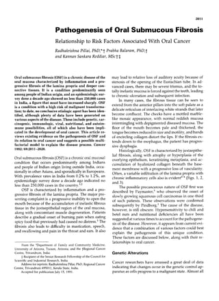
6. 2016 CANCER April 25, 2992, Volume 69,No. 8
Treatment of the effector NK-cells with alpha-inter- Autoimmunity and OSF
feron resulted in highly elevated killer cell activity but
no changes in the target binding cell^.^' This could One of the earliest names by which OSF was identified
imply that inactive target binding cells are seen and that was "idiopathic scleroderma of the mouth," and, in
they could be activated or programmed to kill. We in- view of the female preponderance of patients, its pre-
terpreted this to mean that prekiller cells exist that have sentation in middle life, and histologic similarities, the
killer cell receptors and can bind targets but remain in- analogy seems reasonable. The well-documented find-
active until they interact with appropriate stimuli. ings of clinical, immunologic, and histologic abnormali-
These findings open up a whole new rationale for im- ties in OSF and similar reports in other connective tis-
munologic intervention in the therapeutics of oral pre- sue disorders, such as rheumatoid arthritis, progressive
cancers. Biologic response modifiers such as interferon systemic sclerosis, systemic lupus erythematous, and
possibly could modify the pathogenic course of lesions polymyositis, suggest a fundamental autoimmune basis
such as OSF. Another possible candidate would be for the disease. In many autoimmune diseases, genetic
beta-carotene, with low toxicity and powerful immun- factors are thought to be responsible for abnormalities
omodulatory effects, including many on NK-cells. '02-'04 in immunity. Immune response genes may be linked to
Indeed, the use of this vitamin precursor has resulted in the HLA-DR locus of the major histocompatibility com-
clinical remission of some oral precancerous lesions.77 plex in humans, and associations between this locus
Changes in humoral immunity also have been ob- and autoimmune diseases have been ought."^ The DR
served in our studies on OSF and in heavy chewers of antigens are associated with a susceptibility to diseases
betel quid with tobacco. Increased levels of circulating with an autoimmune aspect in their pathogenesis; this
immune complexes (CIC) were noticed in patients with may be because the immune response genes are situ-
OSF and in those who were heavy chewers of betel ated at or near the D locus on chromosome 6. Because
quid compared with nonchewing normal control^.^' A many of the connective disorders, including rheuma-
repeat study of this in our laboratories further con- toid arthritis and systemic lupus erythematous, have
firmed our finding^."^ Similar findings were seen in been reported to be associated with unique HLA-DR
patients with oral cancer and those with oral kerato-
sis,106,107 Historically, CIC and their constituents have I 1
been prominent as factors alleged to be capable of in-
hibiting cell-mediated immune responses in several
types of cancers. 18'o We also have demonstrated pre-
0-'
nutritional
deficiencies
...................
...................
viously the down regulation of NK-cell activity by autol-
ogous serum containing high levels of CIC.83It is possi-
ble for a similar phenomenon to occur in OSF. Cancer
of the oral cavity in India and in many other regions is ...........
.......... 3
associated closely with chewing betel quid and tobacco. J,J,
The carcinogenic potential of the mixture was discussed
earlier. Chewing of the quid causes constant aberration 8 Betal Quid ....... ............3
of the oral mucosa (particularly by the action of slaked
lime, a major component of the quid). This, combined
J.
. r
4 Changes 3-1
with the poor oral hygiene in these people, gives ample
chance for oral infections, enabling the carcinogenic
Abnormalities
+I
components direct access to the cells. The possibility
exists that the carcinogenic action alters the cellular
components in some way, leading to the production of
low-avidity antibodies forming CIC. Preliminary inves-
tigations indicate high levels of immunoglobulin A-
containing CIC.'06~''oa have observed a similar find-
We
...................
...................
ing in other squamous cell cancers,86and at least one
group has described an immunoglobulin A-like serum Oral Submucus Fibrosis
blocking factor in the sera of patients with nasopharyn-
Figure 4. A multifactorial model for the pathogenesis of OSF. Bold
geal carcinoma."' In addition, Basler et al.'l2 have re- arrows show effects mediated by various factors through the
ported persistent elevation of immunoglobulin A-con- immune system, whereas broken arrows show possible direct
taining CIC in head and neck cancers. effects of the factors on oral mucosa.
7. Pathogenesis of OSF/Pillai et al. 2017
antigen^,"^-"^ a similar association has been sought mation on this disease, which rapidly is becoming an
for OSF. excellent model for studying genetic-environmental-
In an elegant study of 50 unrelated patients of In- immunologic-nutritional interactions in disease patho-
dian origin with OSF, Canniff et ~ 1 . showed increased
"~ genesis.
frequencies of A10, B7, and DR3. It is of interest that in
Indian populations HLA A10, B8, and DR3 occur in
References
positive linkage disequilibrium."' The finding reported
by Canniff et ~ l . , " ~ A10 and DR3 were increased
that
1. Pindborg JJ, Is submucous fibrosis a precancerous condition in
significantly in a group of patients with OSF, suggests
the oral cavity? Int Dent I 1972; 22:474-480.
the possibility that a haplotype encoding these antigens 2. Pindborg JJ. Lesions of the oral mucosa to be considered prema-
is associated with the susceptibility to OSF. It has been lignant and their epidemiology. In: Mackenzie IC, Dabelsteen E,
shown recently that particular haplotypes, rather than Squier CA, eds. Oral Premalignancy. Iowa City: University of
individual antigens, are associated with susceptibility to Iowa Press, 1980; 2-12.
3. Pindborg JJ, Chawla TN, Srivastava AN, Gupta D, Mehrotra
rheumatoid arthritis,"' insulin-dependent diabetes
ML. Clinical aspects of oral submucous fibrosis. Acta Odontol
mellitus,"' and gold-induced nephropathy.'20 The de- Scatld 1964; 221679-691.
fects in cellular immunity seen in OSF51,79,91*92 are fur- 4. Adwani DG. Histopathological studies before and after Kena-
ther suggestive of an autoimmune phenomenon. More- cort in oral submucous fibrosis. MDS Thesis, University of Bom-
over, the presence of autoantibodies against gastric bay, India, 1982.
cells, smooth muscle, and nuclei also has been docu- 5. Paymaster JC. Cancer of the buccal mucosa: Clinical study of
650 cases in Indian patients. Cancer 1956; 9:431-435.
mented.12' Studies on serum from our laboratories also 6 . Kramer IRH. Basic histopathological features of oral premalig-
have shown elevated levels of immunoglobulins G and nant lesions. In: Mackenzie IC, Dabelsteen E, Squier CA, eds.
~,llOa
A similar finding is seen in scleroderma, a disease Oral Premalignancy. Iowa City: University of Iowa Press, 1980;
resembling OSF histologically in that epithelial atrophy 15-34.
and dermal fibrosis are associated with a chronic inflam- 7. Ames BN, Dunston WE, Yamasaki E, Lee FD. Carcinogens are
mutagens: A simple test system combining liver homogenates
matory infiltrate and an increased frequency of HLA- for activation and bacteria for detection. Proc Nati Arnd Sci U S A
DR3 and the haplotypic pair B8/DR3.'228'23 1973; 70:2281-2286.
8. Mc Cann J, Ames BN. Detection of carcinogens as mutagens in
Conclusions the salmonella/microsome test: Assay of 300 chemicals: Discus-
sion. Proc Nut1 Acad Sci U S A 1976; 73:950-959.
9. Hayward WS, Nee1 BG, Astrin SN. Activation of cellular onc
From the data currently available on OSF, it appears
gene by promoter insertion of ALV induced leukosis. Nature
quite clear that the disease is multifactorial, as is the 1981; 290:475-476.
case with oral cancer and most of its precursor lesions. It 10. Bishop J N . Cellular oncogenes and retroviruses. A n n Reu Bio-
also appears that people in whom OSF develops have a cheni 1983; 52:301-325.
genetic predisposition, which could render the oral mu- 11. Cavenee WK, Hansen NF, Nordenskjold Met al. Genetic origins
of mutations predisposing to retinoblastoma. Science 1985;
cosa more susceptible to chronic inflammatory changes
228~501-502.
on exposure to carcinogens. The latter definitely would 12. Swart JGN, Lekkas C, Allard RHB. Oral manifestations in Cow-
include betel quid components including tobacco. As dens syndrome. Oral Surg Oral Med Oral Pathol 1985; 59:264-
discussed earlier, betel nut extracts can stimulate colla- 268.
gen synthesis and fibroblast proliferation.52p53 addi-
In 13. Fader M, Kline SN, Spatz SS, Zubrow HJ. Gardener's syndrome
(intestinal polyposis, osteomas, sebaceous cysts) and a new den-
tion, these extracts also can stabilize the collagen fibrils
tal discovery. Oral Surg Oral Med Oral Pathol 1962; 15:153-157.
and render them resistant to enzymatic degradation. 14. Hope DG, Mulvihill JJ. Malignancy in neurofibromatosis. Adu
The role of genetic abnormalities apart from major his- Neurol 1981; 29:33-43.
tocompatibility complex (MHC) variability still needs to 15. Yunis JJ. The chromosomal basis of human neoplasia. Science
be defined for OSF. Also related to this is the role of 1983; 221~227-236.
16. Greenbaum E, Koss LG, Elequin F, Silver CE. The diagnostic
viruses and their oncogenic potential. It appears that
value of flow cytometric DNA measurements in follicular tu-
immune dysfunction is a common factor and could be mors of the thyroid gland. Cancer 1985; 56:2011-2015.
related to any of the factors discussed so far. Based on 17. Pillai R, Reddiar K, Balaram P. Oncogene expression and oral
these factors, we have suggested a possible model for cancer, I Surg Oncol 1991; 47:102-108.
the pathogenesis of OSF, as illustrated in Figure 4. The 18. Pulciani S, Santos E, Lauver AV, Long LK, Barbacid M. Trans-
forming genes in human tumors. ] Cell Biochern 1982; 20:51-55.
model is indeed speculative and needs to be completed.
19. Willecke K, Schafer R. Human oncogenes. Hun! Genet 1984;
Research into genetic, viral, and immunologic aspects is 66:332-139.
ongoing in our laboratories. These, along with similar 20. Yokota J, Tsunetsugu-Yokata Y, Battifora H, LeFevre C, Cline
approaches elsewhere, should provide valuable infor- MJ. Alterations of tnyc, myb and rasHaprotooncogenes in cancers
8. 2018 CANCER April 25, 2992, Volume 69, No. 8
are frequent and show clinical correlation. Science 1986; A possible viral pathogenesis. Oral Surg Oral Med Oral Pathol
2311261-263, 1985; 59:52-55.
21. Yunis JJ, Soreng AL. Constitutive fragile sites and cancer. Science 42. Loning TH, Reichart P, Staquet MJ, Becker J, Thivolet J. Occur-
1984; 226:1199-1204. rence of papilloma virus structural antigens in oral papillomas
22. Cooper GM, Lane MA. Cellular transforming genes and onco- and leukoplakias. Oral Pathol 1984; 13:463-465.
genesis. Biochem Biophys Acta 1984; 738:9-25. 43. La1 D. Diffuse oral submucous fibrosis. I All India Dent Assoc
23. Krontiris TG, Cooper GM. Transforming activity of human tu- 1953; 26:1-3.
mor DNA. Proc Natl Acad Sci U S A 1981; 78: 1181-1184. 44. Valentine JA, Scott J, West CR, St. Hill CA. A histological analy-
24. Friedman WH, Rosenblum B, Loewenstein P, Thronton H, Kat- sis of the early effects of alcohol and tobacco usage on human
santonis G, Green M. Oncogenes: Preliminary studies in head lingual epithelium. ] Oral Pathol 1985; 14:654-656.
and neck cancer. Latyngoscope 1983; 93:1141-1146. 45. Trell E, Bjorlin G, Andreasson L, Korsgaard R, Mattiasson I.
25. Friedman WH, Rosenblum B, Loewenstein P, Thronton H, Kat- Carcinoma of the oral cavity in relation to aryl hydrocarbon
santonis G, Green M. Oncogenes in laryngeal cancer: Serial pas- hydroxylase inducibility, smoking and dental status. lnt ] Oral
sage of transformed cellular DNA. Otolaryngol Head Neck Surg Surg 1981; 10:93-97.
1985; 93~346-350. 46. Hecht SS, Ornaf RM, Hoffmann D. Chemical studies on tobacco
26. Roa RA, Carey TE, Passamani PP et al. DNA content of human smoke: Nnitrosonornicotine in tobacco: Analysis of possible con-
squamous cell carcinoma lines: Analysis by flow cytometry and tributing factors and biologic implications. ] Natl Cancer Znst
chromosome enumeration. Arch Otolaryngol 1985; 111:565- 1974; 54: 1273-1276.
570. 47. Khadim MI. The effect of pan and its ingredients on oral mu-
27. Grassel-Pietrusky R, Deinlein E, Hornstein OP. DNA ploidy cosa. IPMA 1977; 27:340-344.
rates in oral leukoplakias determined by flow cytometry. 1 Oral 48. Atkinson L, Chester IC, Smyth FG, Ten Seldom REJ. Oral cancer
Patho/ 1982; 11~434-436. in New Guinea: A study in demography and aetiology. Cancer
28. Hauser-Urfer IH, Stauffer J. Comparative chromosome analysis 1964; 4011289-1292.
of nine squamous cell carcinoma lines from tumors of head and 49. Ramanathan K, Ganesan T], Raghavan KV. Carcinoma of the
neck. Cytogenet Cell Genet 1985; 39:35-41. tongue in Malaysians. Mod Med Asia 1978; 14:56-59.
29. Cawson RA, Lehner T. Chronic hyperplastic candidiasis: Candi- 50. Hirayama T. An epidemiological study of oral and pharyngeal
dal leukoplakia. BY Dermatol 1968; 80:9-12. cancer in Central and Southeast Asia. Bull WHO 1966; 34:41-
30. Cawson RA. Induction of epithelial hyperplasia by Candida albi- 44.
cans. Br ] Dermatol 1973; 89:497-500. 51. Balaram P, Pillai R, Abraham T. Immunology of premalignant
31. Russel C, Jones JH. The histology of prolonged candidal infec- and malignant conditions of the oral cavity: 11. Circulating im-
tion of the rat's tongue. I Oral Pathol 1975; 4:330-332. mune complexes. 1Oral Pathol 1987; 16:389-391.
32. Pindborg JJ, Renstrup G, Poulsen HE, Silverman S. Studies in 52. Harvey W, Saitt A, Meghi S, Carniff JP. Stimulation of human
oral leukoplakia: V. Clinical and histological signs of malig- buccal fibroblasts in vitro by betel nut alkaloids. Arch Oral Biol
nancy. Acta Odontol Scand 1963; 21:407-410. 1986; 31145-49.
33. Cawson RA, Binnie WH. Candida, leukoplakia and carcinoma: 53. Kuttan R, Donnely PV, DiFerante N. Collagen treated with (+)
A possible relationship. In: Mackenzie IC, Dabeslsteen E, Squier catechin becomes resistant to the action of mammalian collage-
CA, eds. Oral Premalignancy. Iowa City: University of Iowa nase. Experentia 1981; 37:221-223.
Press, 1980; 59-70. 54. Hirsch JM, Johansson SL, Vahlne A. Effect of snuff and herpes
34. Hollinshed AC, Tarro G. Soluble membrane antigens of lip and simplex virus 1 on rat oral mucosa: Possible associations with
cervical carcinoma: Reactivity with antibody for herpes virus the development of squamous cell carcinoma. Oral Pafhol
non virion antigens. Science 1973; 182713-714. 1984; 13:52-56.
35. Kumari TV, Harikumar T, Prabha B, Vivekanandan S, Vasude- 55. Park NH, Sapp JP, Herbosa EG. Oral cancer induced in ham-
van DM. Detection of antibodies against herpes simplex virus in sters with herpes simplex infection and simulated snuff dipping.
patients with oral cancer. Zndian Cancer 1984; 19:137-140. Oral Surg Oral Med Oral Pathol 1986; 62:164-167.
36. Silverman NA, Alexander CJ, Hollinshed AC, Cheretein PB. 56. Krogh P. The role of yeasts in oral cancer by means of endoge-
Correlation of tumor burden with in vitro lymphocyte reactivity nous nitrosation. Actu Odontol Scand 1990; 48:85-88.
and antibodies to herpes simplex virus in patients with oral 57. Rennie JS, McDonald DG, Dagg JH. Iron and the oral epithe-
cancer. Cancer 1970; 37:135-140. lium. R Soc Med 1984; 77:602-604.
37. Shillitoe EJ, Hwang CBC, Silverman S, Greenspan JS. Examina- 58. Paterson DR. A clinical type of dysphagia. Laryngol 1919;
tion of oral cancer tissue for the presence of the proteins 1 0 4 , 34~289-290.
1CP5, 1 0 6 , 1CP8 and gB of herpes simplex virus type 1. I Natl 59. Kelly AB. Spasm at the entrance of the esophagus. I Larytzgol
Cancer Inst 1986; 76:371-376. 1919; 341285-287.
38. Brescia RJ, Jenson AB, Lancaster WD, Kurman RJ. The role of 60. Waldenstrom J, Kjellberg SR. The roentgenological diagnosis of
human papilloma viruses in the pathogenesis and histologic clas- sideropenic dysphagia. Acta Radiol 1939; 20:618-621.
sification of precancerous lesions of the cervix. H u m Pathol 61. Ahlbom HE. Simple achlorhydric anemia, Plummer Vinson
1986; 17:552-556. syndrome and carcinoma of the mouth, pharynx and esophagus
39. Greenspan JS, Greenspan D, Lennette ET et al. Replication of in women. Br Med 1936; 2:331-334.
Epstein Barr virus within epithelial cells of oral "hairy" leuko- 62. Ahlbom HE. Pradisponierende faktorenfur platten epithelkar-
plakia: An AIDS associated lesion. N Engl ] Med 1985; zinom in mund hals and speiserohre. Acta Radiol 1937; 18:163-
313:1564-1565. 167.
40. Silverman S, Migliorati CA, Lozada-Nur F, Greenspan D, Con- 63. Wynder EL, Fryer JH. Etiologic considerations of Plummer Vin-
ant MA. Oral findings in people with or at high risk for AIDS: A son syndrome. Ann Zntern Med 1958; 259:1106-1109.
study of 375 homosexualmales.]Arn DentAssoc 1986; 112:187- 64. Watts JM. The importance of the Plummer Vinson syndrome in
190. the etiology of carcinoma of the upper gastrointestinal tract.
41. Eisenberg E, Rosenberg B, Krutchkoff D. Verrucous carcinoma: Postgrad Med I 1961; 37:523-526.
9. Pathogenesis of OSFIPillai et al. 2019
65. Shamma MH, Benedict EB. Esophageal webs: A report of 58 cal markers in malignant cervical neoplasia. ] Cancer Res Clin
cases and an attempt at classification. N Engl Med 1958; Oncol 1989; 115:583-591.
259:378-380. 87. Pillai R, Balaram P, Bindu 5,Hareendran NK, Padnabhan TK,
66. Ramanathan K. Oral submucous fibrosis: An alternative hy- Nair MK. Interleukin 2 production in lymphocyte cultures: A
pothesis as to its causes. Med ] Malaysia 1956; 36:243-245. rapid test for cancer associated immunodeficiency in malignant
67. Rennie JS, McDonald DG, Dagg JH. Quantitative analysis of the cervical neoplasia. Cancer Lett 1989; 47:205-210.
human buccal epithelium in iron deficiency anemia. ] Oral 88. Pillai R, Balaram P, Bindu S, Hareendran NK, Padmanabhan
Pathol 1982; 11:39-41. TK, Nair MK. Radiation associated eosinophilia and monocyto-
68. Rennie JS, McDonald DG. Quantitative histological analysis of sis in carcinoma of the uterine cervix: A simple reliable prognos-
the epithelium of the ventral surface of the hamster tongue in tic indicator. Neoplasma 1990; 37:91-96.
iron deficiency. Arch Oral Biol 1982; 27:393-396. 89. Pillai R, Balaram P, Hareendran NK, Bindu 5,Padnabhan TK,
69. Prime SS, McDonald DG, Rennie JS. The effect of iron defi- Nair MK. Clinical and prognostic significance of Concanavalin
ciency on experimental oral carcinogenesis in the rat. B r ] Cancer A induced suppressor cell activity in malignant cervical neopla-
1983; 471413-416. sia. Br / Obstet Gynecol 1990; 97:357-361.
70. Joynson DH, Walker DM, Jacobs A, Dolby AE. Defects of cell 90. Pillai R, Balaram P, Chidambaram S, Padnabhan TK, Nair MK.
mediated immunity in patients with iron deficiency anemia. Development of an immunological staging system to prognosti-
Lancet 1972; 2:1058-1059. cate cervical neoplasia. Gynecol Oncol 1990; 37:200-205.
71. Rennie JS, McDonald DG, Dagg JH. Iron and the oral epithe- 91. Pillai R, Balaram P, Kannan S et al. Clinical implications for oral
lium. ] R Soc Med 1984; 77:602-606. precancers of interferon activation of latent natural killer cells
72. Jacobs A, Cavil1 I. The oral lesions of iron deficiency anemia: and alteration in kinetics of target cell lysis. Oral Surg Oral Med
Pyridoxine and riboflavin status. Br ] Haematol 1968; 14:291- Oral Pathol 1990; 70:458-461.
294. 92. Balaram P, Pillai R, Abraham T. Immunology of premalignant
73. Harrison RJ. Vitamin 812 levels in erythrocytes in hypochromic and malignant conditions of the oral cavity. In: Proceedings of
anemia. ] Clin Pathol 1971; 24:698-670. the Second UAE Cancer Conference. Abu Dhabi, U.A.E.: Emir-
74. Wahi I", Kehar U, Lahiri 8. Factors influencing oral and oropha- ates Medical Association, 1987; 32.
ryngeal cancer in India. BY ] Cancer 1965; 19:646-649. 93. Migliorati CA, Migdnorati EKG, Silvermann S, Greenspan D,
75. Ibrahim NA, Jafarey NA, Zuberi SJ, Plasma vitamin A and caro- Greenspan JS. Phenotypic identification of mononuclear cells in
tene levels in squamous cell carcinoma of the oral cavity and oral premalignant lesions and cancer by monoclonal antibodies.
oropharynx. Clin Oncol 1977; 3:303-306. ] Oral Paihol 1986; 15:352-358.
76. Stich HF, Homby AP, Mathew B, Sankaranarayanan R, Nair 94. Kimura AK, Wigzell H. Development and function of cytotoxic
MK. Response of oral leukoplakias to the administration of vita- T lymphocytes (CTL): In vivo maturation of CTL in the absence
min A. Cancer Lett 1988; 40:93-101. of detectable proliferation results as a normal consequence of
77. Stich HF, Rosin MP, Hornby AP, Mathew B, Sankaranarayanan alloimmunization. ] Immunol 1983; 130:2056-2059.
R, Nair MK. Remission of oral leukoplakias and micronuclei in 95. Robbins DS, Fudenberg HH. Human lymphocyte subpopula-
tobacco/betel quid chewers treated with beta carotene and beta tions in metastatic neoplasia: Six years later. N Engl ] Med 1983;
carotene plus vitamin A. Int ] Cancer 1988; 42~195-199. 308:1595-1596.
78. Urban JL, Schreiber H. Host-tumor interactions in immunosur- 96. Ledbetter JA, Evans RL, Lipinski M, Cunningham-Riddles C,
veillance against cancer. Prog Exp Tumor Res 1988; 32:17-68. Good RA, Herzenberg LA. Evolutionary conservation of surface
79. Pillai R, Balaram P, Abraham T, Nair MK. Lymphocyte popula- molecules that distinguish T lymphocyte helper-inducer and
tions in premalignant and malignant lesions of the oral cavity. suppressor-cytotoxic subpopulations in mouse and man. I Exp
Neoplasma 1987; 34:469-479. Med 1981; 153:310-315.
80. Pillai R, Balaram P, Padmanabhan TK, Nair MK. Monoclonal 97. Reinherz EL, Hussey RE, Schlossman SF. Absence of expression
antibody defined phenotypes of peripheral blood lymphocytes of Ia antigen on human cytotoxic T cells. Immunogenetics 1980;
in cancer of the uterine cervix. Am ] Reprod Zmmunol Microbiol 11:421-423.
1987; 14:141-143. 98. Carney WP, Iaacobiello V, Hersch MS. Functional properties of
81. Balaram P, Pillai R, Padmanabhan TK, Thomas A, Hareendran T lymphocytes and their subsets in cytomegalovirus mononucle-
NK, Nair MK. Immune function in malignant cervical neoplasia. osis. ] Immunol 1983; 130:390-394.
Gynecol Oncol 1988; 31:409-423. 99. Biddison WE, Sharrow SO, Shearer GM. T cell subpopulations
82. Pillai R, Balaram P, Abraham T, Padmanabhan TK, Nair MK. required for human cytotoxic lymphocyte responses to influ-
Natural cytotoxicity and serum blocking in malignant cervical enza virus. ] Immunol 1981; 127:487-491.
neoplasia. A m ] Reprod Immunol Microbiol 1988; 16:159-162. 100. Hanna N . The role of natural killer cells in the control of tumor
83. Pillai R, Balaram P, Padmanabhan TK, Nair MK. Immunological growth and metastases. Biochem Biophys Acta 1984; 70:213-226.
profiles of T lymphocytes in malignant cervical neoplasia. ] Exp 101. Ulberg M, Jondal M. Recycling and target binding capacity of
Clin Cancer Res 1988; 7~251-257. human natural killer cells. ] Exp Med 1981; 153~615-621.
84. Pillai R, Balaram P, Padmanabhan TK, Abraham P, Nair MK. 102. Rhodes J, Stokes P, Abrams P. Human tumor induced inhibition
Interleukin 2 and alpha interferon induced in vitro modulation of interferon action in vitro: Reversal of inhibition by beta caro-
of spontaneous cell mediated cytotoxicity in patients with tene. Cancer Zrnmunol Inmunother 1984; 16:189-192.
cancer of the uterine cervix undergoing radiotherapy. Acta O n c d 103. Prabhala RH, Maxey V, Hicks MJ, Watson RR. Enhancement of
1989; 28~39-44. the expression of activation markers on human peripheral blood
85. Pillai R, Balaram P, Padnabhan TK, Abraham T, Hareendran mononuclear cells by in vitro culture with retinoids and carot-
NK, Nair MK. lmmunocompetence in lung cancer: Relation to enoids. / Leukocyte Biol 1989; 45:249-254.
extent of tumor burden and histological type. Cancer 1989; 104. Abril ER, Rybski JA, Scuderi P, Watson RR. Beta carotene stimu-
64:1851-1858. lates human leukocytes to secrete a novel cytokine. ] Leukocyte
86. Pillai R, Balaram P, Hareendran NK, Bindu S, Padmanabhan Biol 1989; 45~255-259.
TK, Nair MK. Immune reactive proteins as prognostic and clini- 105. Remani P, Ankathil R, Vijayan KK, Beevi VMH, Rajendran R,
10. 2020 CANCER April 15, 1992, Volume 69, No. 8
Vijayakumar T. Circulating immune complexes as an immuno- 114. Gibofsky A, Winchester RJ, Patanoyo M. Disease associations of
logical marker in premalignant and malignant lesions of the oral the Ia like human alloantigens. ] Exp Med 1978; 148:1728-1732.
cavity. Cancer Lett 1988; 40:185-191. 115. Stastny P. Association of the B cell alloantigen DRw4 with rheu-
106. Abraham T, Balaram P. Circulating immune complexes in pa- matoid arthritis. N Engl] Med 1978; 298:869-871.
tients with squamous cell carcinoma of the oral cavity. ltidiati ] 116. Reinertesin JL, Klippel JH, Johnson AH. B lymphocyte alloanti-
Cancer 1987; 24:133-140. gens associated with systemic lupus erythematosus. N Etigl ]
107. Scully C. Immunological abnormalities in oral carcinomas and Med 1978; 299:515-518.
oral keratosis. 1 Cliti Lab Inrrriuriol 1982; 8:113-116. 117. Canniff JP, Batchelor JR, Dodi IA, Harvey W. HLA typing in oral
108. Baldwin RW, Robins RA. Circulating immune complexes in submucous fibrosis. Tissue Antigens 1982; 26:138-142.
cancer. In: Sell S , ed. Cancer Markers. Newark, NJ: Humana, 118. Mittal KK, Naik S , Sansonetti N, Cowherd R, Kumar R, Wong
1980; 507-531. DM. The HLA antigens in Indian Hindus. Tissue Antigetis 1982;
109. Dahlgren C, Elwing H. Inhibition of polymorphonuclear leuco- 20x223-226.
cyte locomotion by antigen-antibody complexes. Inmunology 119. McCluskey 1, Kay I’H, Dawkins RL, Komori K, Christiansen FT,
1980; 49:329-339. McCann UJ. Association of specific MHC supratypes with rheu-
matoid arthritis and insulin dependent diabetes mellitus. Disease
110. Hellstrom KE, Hellstrom I, Nepon JT. Specific blocking factors:
Markers 1983; 1:197-212.
Are they important? Biochetn Biophys Acta 1978; 473:121-148.
120. Batchelor JR, Dodi IA, Woo P, Panayi G, Ansell B, Williams PL.
110a.Balaram P. Unpublished data, 1991. Family study of gold induced nephritis in patients with rheuma-
111. Sundar SK, Abhilashi DV, Kamaraju L et a / . Sera from patients toid arthritis. In: Albert E, Baur M, eds. Histocompatibility Test-
with undifferentiated nasopharyngeal carcinoma contain a fac- ing. Berlin: Springer Verlag, 1984; 375-378.
tor that abrogates specific Epstein Barr virus antigen induced 121. Canniff JP, Harvey W. The aetiology of oral submucous fibrosis.
lymphocyte response. Znt ] Cancer 1982; 29:407-412. Itit Oral Surg 1981; 10:163-167.
112. B a s h MW, Maxim PS, Veltri RW. Circulating IgA immune 122. Kallenberg CGM, Van der Voort-Beelen JM, D’Maro J, The TH.
complexes in head and neck cancer, nasopharyngeal carcinoma, Scleroderma: Increased frequency of B8/DR3 in scleroderma
lung cancer and colon cancer. Fed Proc 1984; 43:1929. and association of the haplotype with impaired cellular immune
113. Griffing WL, Moore SB, Luthra HS. Associations of antibodies response. CIin Exp Infrnunol 1981; 43:478-485.
to native DNA with HLA DRw3: A major histocompatibility 123. Whiteside TL, Medsger JR, Rodnan GP. HLA DR antigens in
complex linked human immune response gene. ] Exp Med 1980; progressive systemic sclerosis (scleroderma). RheurnatoI 1983;
152:319-325. 10:128-131,