1) The study found that IKKα, like IKKβ and NEMO/IKKγ, is required for the activation of NF-κB target genes in response to TNFα and IL-1 stimulation in mouse embryonic fibroblasts.
2) DNA microarray analysis identified many known and novel NF-κB dependent target genes that were regulated by all three subunits of the IKK complex.
3) Some NF-κB target genes were dependent on the IKKs even in the absence of extracellular stimuli, suggesting the IKK complex also regulates basal levels of NF-κB activity.
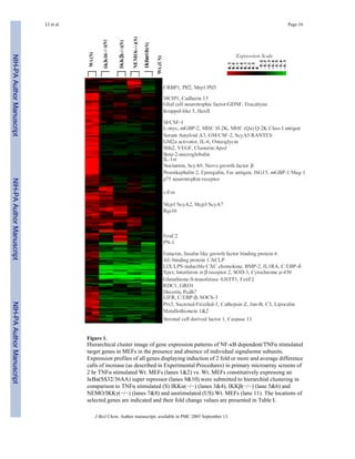
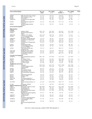
This document summarizes a study investigating the oncoprotein EVI1 and its regulation of the carbonic anhydrase 3 (CA3) gene. Microarray analysis identified CA3 as a major target of EVI1. The study aims to validate changes in CA3 expression and determine the mechanism and significance of EVI1-mediated transcriptional regulation of CA3. Results show that EVI1 suppresses CA3 expression at both the mRNA and protein levels by decreasing CA3 promoter activity. This suppression of the antioxidant CA3 gene leads to increased caspase activity and apoptosis in cancer cells expressing EVI1.
Rin-like (Rinl) is a novel member of the RIN family of proteins that serve as guanine nucleotide exchange factors (GEFs) for Rab GTPases. Rinl preferentially binds and catalyzes GDP/GTP exchange on Rab5a and Rab22, implicating it in endocytic processes regulated by these Rab proteins. Rinl localizes to neuromuscular synapses and interacts with the receptor tyrosine kinase MuSK, a key regulator of neuromuscular synapse development. Overexpression of Rinl affects both fluid-phase and EGFR receptor-mediated endocytosis. Rinl is closely associated with the actin cytoskeleton and thus may recruit Rab5
This study examined the role of IKKα kinase activity in regulating NF-κB target gene expression using gene expression profiling. Rescuing wild-type IKKα in IKKα knockout mouse embryonic fibroblasts globally restored expression of canonical NF-κB targets. Surprisingly, rescuing a kinase-dead IKKα mutant also rescued expression of approximately 28% of these genes, both independently of and dependently on TNFα stimulation. Thus, IKKα regulates NF-κB target gene expression through both kinase-dependent and independent mechanisms in both stimulus-dependent and independent manners. Five distinct classes of IKKα/NF-κB-dependent genes were identified.
- Antibodies are Y-shaped proteins produced by B cells that recognize and bind to pathogens. They have great diversity and specificity that allows them to target a wide range of antigens.
- Antibodies are composed of two heavy chains and two light chains that form an antigen binding site. The variable regions of the heavy and light chains are responsible for binding to different antigens.
- Monoclonal antibodies are produced from a single clone of B cells and have identical antigen specificity, allowing them to be used as standardized reagents for research and medical purposes. They are produced by fusing B cells with myeloma cells to form immortal hybridomas.
Immunoglobulins, also known as antibodies, are Y-shaped glycoproteins produced by B cells that function to recognize and bind to foreign objects like antigens or pathogens. There are five classes of immunoglobulins - IgG, IgM, IgA, IgD, and IgE - which differ in size, sugar content, and biological function. Each immunoglobulin molecule contains two light chains and two heavy chains that form variable and constant regions. The variable regions are responsible for binding to antigens while the constant regions mediate different immune functions.
The document summarizes a study investigating the role of extracellular ATP and the P2X7 receptor (P2X7R) in activating inflammasome signaling in neutrophils. The study finds that:
1) ATP induces IL-1β secretion from mouse and human neutrophils in a dose-dependent manner through activation of the P2X7R and downstream NLRP3 inflammasome.
2) Mouse neutrophils express functional cell surface P2X7Rs, as ATP stimulation induces sustained calcium influx and IL-1β release, which is blocked by P2X7R antagonists.
3) ATP-induced IL-1β secretion and caspase-1 activation requires the NLRP3 inflammasome machinery of NLRP3
The document summarizes the complement system, which consists of over 20 proteins that work together to eliminate pathogens. It operates through three pathways - the classical, lectin, and alternative pathways. The classical pathway requires antibodies, while the alternative pathway can be activated independently of antibodies by pathogens. All three pathways involve a cascade of proteins that activate each other, ultimately forming the membrane attack complex that lyses the target cell membrane. The complement system also has roles beyond direct lysis, such as opsonization, inflammation, and activation of the immune response.
Signal transduction pathways allow plants to respond to environmental stimuli. Receptors on the plasma membrane or inside the cell detect stimuli and activate secondary messengers like calcium or G-proteins. These messengers activate effector molecules like protein kinases that modulate gene expression and trigger responses like tolerance to abiotic stresses. Manipulating these pathways could help protect plants from stresses under climate change.
Signal transduction in plant defense responsesVINOD BARPA
Signal transduction a Process by which a cell converts one kind of signal into another. Plant disease resistance and susceptibility are gov¬erned by the combined genotypes of host and pathogen and depend on a complex exchange of signals and responses occurring under given environmental con¬ditions. During the long process of host-pathogen co-evolution, plants have developed various elaborate mechanisms to ward off pathogen attack. Whereas some of these defense mechanisms are preformed and provide physical and chemical barriers to hinder pathogen infection, others are induced only after pa¬thogen attack. Similar to animal immune responses, induced plant defense responses involve a network of signal transduction and the rapid activation of gene expression following pathogen infection. They do not have immune system and locomotary organs to escape environmental challenges and biotic stresses. In plant, nature has provided them some preformed and inducible defense resistance. Host recognition of invading pathogen is often determined by the so called “gene for gene” interaction between avirulence (avr) gene of pathogen and corresponding resistance (R) gene of host (Flor, 1971) which encode receptor for the recognition of specific elicitor or ligand encoded directly or indirectly by pathogen avr gene. Recent studies have revealed intriguing parallels between animal and plant defense responses as demonstrated by the structural and functional conservation of some of their signal transduction processes. Furthermore, signaling components such as G proteins, NADPH oxidase, H202, salicylic acid (SA, and aspirin), mitogen-activated protein kinases (MAPK), and transcription factors have been shown to be associated with or participate in both animal and plant defense responses, suggesting the presence of con¬served signaling pathways for host defenses in diverse higher eukaryotes.
The document examines how long-term low-dose exposure of human urothelial cells to sodium arsenite activates the lipocalin-2 (LCN2) gene via promoter hypomethylation. It finds that the LCN2 promoter in arsenic-exposed cells is hypomethylated compared to unexposed cells, leading to increased LCN2 expression. This overexpression of LCN2 enhances cellular transformation, anti-apoptotic activity, and inflammatory responses in the arsenic-exposed cells through mechanisms involving NF-κB and increased iron and reactive oxygen species levels.
Cell signaling is part of any communication process that governs basic activities of cells and coordinates all cell actions. The ability of cells to perceive and correctly respond to their microenvironment is the basis of development, tissue repair, and immunity, as well as normal tissue homeostasis
Strucure, functions and genetics of immunoglobulinsJESSE OWAKI
The power-point contains summerised concept on Structures, Functions and Genetics of Immunoglobulins.
It is to help my fellow undergraduate students to have a basic understanding on the topic.
Kindly contact me for more materials. Thank you.
Immunoglobulins are glycoprotein molecules that are produced by plasma cells in response to an immunogen and which function as antibodies. The immunoglobulins derive their name from the finding that they migrate with globular proteins when antibody-containing serum is placed in an electrical field.
This document reports on a study examining how carbon monoxide (CO) induces heme oxygenase-1 (HO-1) expression and inhibits endothelial cell apoptosis triggered by endoplasmic reticulum (ER) stress. The key findings are:
1) CO activates the transcription factor Nrf2 through phosphorylation of the protein kinase R-like ER kinase (PERK), leading to increased HO-1 expression.
2) CO-induced PERK activation results in phosphorylation of eukaryotic translation initiation factor 2α and expression of activating transcription factor 4.
3) CO prevents ER stress-induced expression of X-box binding protein 1 and cleavage of activating transcription factor 6.
4) CO inhibits
- Immunoglobulins are bifunctional proteins that must interact with a small number of specialized molecules like Fc receptors and complement proteins, while also recognizing an infinite array of antigenic determinants.
- They achieve this through a domain structure that provides both structural conservation and infinite variability. Immunoglobulin domains form a beta-barrel structure and are found in many proteins beyond antibodies.
- Within the variable regions, hypervariable loops provide the diversity in antigen recognition, supported by a conserved framework structure. The combination of loops and framework allows recognition of countless antigens.
The 5' terminal uracil of let-7a is critical for the recruitment of mRNA to A...David W. Salzman
This document investigates the interaction between let-7a microRNA, Argonaute2 protein, and mRNA targets. It finds that recombinant Argonaute2 is sufficient to direct let-7a-guided cleavage of a fully complementary mRNA target in vitro. Additionally, it determines that the 5' terminal uracil of let-7a is critical for recruitment of the mRNA target to the let-7a-Argonaute2 complex. Mutation of this 5' uracil inhibits formation of the ternary let-7a-Argonaute2-mRNA complex, but does not affect formation of the binary let-7a-Argonaute2 complex. This suggests the 5' urac
Catterall molecular mechanism of calcium channel regulation in theramezali
This document summarizes a study that reconstituted the molecular mechanism of calcium channel regulation during the fight-or-flight response. The researchers formed an autoinhibitory signaling complex composed of CaV1.2 channels, the distal C-terminal domain, auxiliary subunits, and AKAP15. They observed a 3.6-fold range of CaV1.2 channel activity from minimum to maximum levels. Basal activity required phosphorylation of two residues, whereas further stimulation only required one residue. The study defined a framework for CaV1.2 channel regulation and identified key phosphorylation sites.
The document discusses various mechanisms of gene expression regulation in prokaryotes and eukaryotes. In prokaryotes, gene expression often occurs in operons, clusters of genes that are regulated as a single unit. A key example is the lactose operon in E. coli, which codes for proteins involved in lactose metabolism. In eukaryotes, gene expression can be regulated at multiple stages including transcription, RNA processing, and translation. Regulatory mechanisms include transcription factors, histone modification, and alternative splicing of mRNA.
This document discusses the structure and function of antibodies (immunoglobulins). It describes their basic components, including heavy and light chains linked by disulfide bonds. The five major classes of antibodies in humans are IgG, IgM, IgA, IgD, and IgE, which have different structures and functions. IgG is the most abundant antibody and can bind antigens, fix complement, and interact with immune cells via Fc receptors to mediate important immune responses like opsonization and antibody-dependent cellular cytotoxicity.
The document discusses the complement system, which consists of over 30 proteins that work together to help antibodies clear pathogens from the body. It summarizes the three complement activation pathways: classical, lectin, and alternative. The classical pathway is activated by antigen-antibody complexes, the lectin pathway by mannose-binding proteins, and the alternative pathway through spontaneous activation of C3. All three pathways result in the formation of C3 and C5 convertases that cleave complement proteins, and ultimately form the membrane attack complex to lyse cells. The complement system also promotes inflammation, chemotaxis, opsonization, and activation of B cells.
Antibody structure , functions and classificationPuneetKohli19
1. Antibodies are Y-shaped proteins called immunoglobulins that are produced by plasma cells in response to antigens like bacteria or viruses.
2. Immunoglobulins are composed of light and heavy chains that make up the variable and constant regions, and there are 5 major classes - IgG, IgA, IgM, IgD, and IgE that have different structures and functions.
3. IgG is the most abundant antibody and activates the complement system, IgA protects mucosal surfaces, IgM is the first antibody produced in responses and activates complement, IgD function is not fully known but involved in B cell activation, and IgE triggers allergic reactions by binding mast cells and bas
This document provides an overview of immunoglobulin structure and function:
- Immunoglobulins are Y-shaped molecules composed of two heavy chains and two light chains that recognize antigens. They exist as both membrane-bound and secreted forms.
- The B cell receptor (BCR) is a membrane-bound immunoglobulin that plays a critical role in B cell development and activation. Engagement of the BCR triggers downstream signaling pathways.
- Immunoglobulins exhibit tremendous diversity through V(D)J recombination during B cell development, somatic hypermutation after antigen exposure, and class switching which allows different effector functions.
Antibodies, also known as immunoglobulins, are glycoproteins produced by B cells that bind to specific antigens. There are five classes of immunoglobulins - IgG, IgM, IgD, IgA, and IgE - which differ in their heavy chains. Immunoglobulins have two primary functions: antigen binding and effector functions like complement fixation and binding to immune cells. Their structure consists of two light chains and two heavy chains that form a Y-shape, with constant and variable regions that determine the class and allow binding to different antigens.
1. Cellular signal transduction involves signaling molecules, receptors, and intracellular signal transduction pathways that allow cells to respond to changes in their external environment.
2. Signaling molecules like hormones, neurotransmitters, cytokines, and gas molecules bind to membrane or intracellular receptors to activate downstream signaling pathways.
3. The main intracellular signaling pathways include the cAMP/PKA pathway, Ca2+/PKC pathway, cGMP/PKG pathway, and tyrosine kinase pathways which result in phosphorylation of target proteins and regulation of gene expression.
This document summarizes the efficient synthesis of novel 3-aminocyclobut-2-en-1-ones, which are potent antagonists of VLA-4. The synthesis involves the condensation of cyclobuta-1,3-diones with a phenylalanine-derived primary amine. This produces vinylogous amide derivatives in good yields. The cyclobuta-1,3-diones are prepared through cycloaddition of ketene intermediates, generated from acid chlorides, with various alkynes. The resulting 3-aminocyclobut-2-en-1-ones can then be functionalized at C-2 through reaction with electrophilic reagents
This document provides an overview of antibodies (immunoglobulins). It discusses the basic structure of antibodies, including their Y-shape and polypeptide chains. There are five classes of antibodies (IgG, IgA, IgM, IgD, IgE) which have different properties and functions. The document also describes how antibodies carry out their effector functions, such as activating the complement system, opsonization to promote phagocytosis, and neutralization of toxins and pathogens.
1. Kinases and phosphatases work together to regulate cell signaling through phosphorylation and dephosphorylation of proteins. Kinases add phosphate groups while phosphatases remove them, allowing rapid and transient signaling responses.
2. There are three main classes of protein kinases - serine/threonine kinases, tyrosine kinases, and mixed kinases. Key kinase families discussed include PKA, PKC, calcium/calmodulin dependent kinases, receptor tyrosine kinases, and MAP kinases.
3. Phosphatases counteract kinase activity to terminate signaling responses in a timely manner. Important phosphatase families are PP1, PP2A, and calcineurin
Roop Amrit Fairness Gel is an ayurvedic skin fairness product that can be purchased from the website www.roopamritfairnesscream.com. Customers can call 08287057757 for free home delivery of Roop Amrit, which is described as a 100% ayurvedic fairness gel.
1. Sustained activation of the NF-κB pathway through expression of a constitutively active IKKβ mutant (IKKβca) leads to transient growth arrest in murine fibroblasts.
2. This growth arrest is associated with repression of key cell cycle effectors regulated by the transcription factors E2F and FoxM1.
3. The repression of E2F and FoxM1 target genes and growth arrest caused by IKKβca expression is linked to NF-κB activation and involves enhanced expression of the cell cycle inhibitor p21 as well as chromatin remodeling mediated by histone deacetylases (HDACs).
The document provides an introduction to various Islamic financing concepts, including Musharakah, Mudarabah, Murabahah, Ijarah, and Salam/Istisna.
Musharakah refers to a partnership where two or more parties contribute capital to a business and share profits and losses. Mudarabah is a partnership where one party provides capital and the other manages the business, with profits shared according to a predetermined ratio. Murabahah involves the sale of an asset at a pre-agreed markup over cost. Ijarah is a leasing contract where the lessor transfers use of an asset to the lessee for a rental fee but retains ownership.
504 interpretação de textos apostila amostraIury Alberth
- O texto discute a decisão judicial que determinou que as empresas Light e Cerj também paguem bônus aos consumidores que reduziram o consumo de energia entre 100 e 200 kWh, fazendo justiça.
- A Câmara de Gestão da Crise de Energia vinha evitando encarar a questão dos bônus, preocupada com os recursos que teria que desembolsar, apesar do presidente já ter dito que os bônus seriam pagos.
- O texto critica a burocracia brasileira por seu desrespeito ao cidadão
11.vol. 0002www.iiste.org call for paper no. 2_c reinig & ca tilt _pp176-197Alexander Decker
This document analyzes media releases from 2006 from Australia's four major national banks (ANZ, CBA, NAB, and WBC) to investigate how they communicate corporate social responsibility (CSR) to different stakeholders. A content analysis was conducted to determine the extent and nature of CSR-related media releases, as well as their intended audiences. The findings indicate over one-third of the banks' media releases discussed CSR, predominantly around community involvement. Most CSR-related media releases targeted customers and communities as their intended audiences.
WebXpress Business Intelligence using Transport Management Solutions WebXpress.IN
The document provides a sales dashboard that analyzes customer group and business type performance, regional sales, route profits, routes with specific profit margins, and cooling analysis of dockets. It also includes contact information.
This document proposes creating a virtual fitness lab in Second Life to support physical education instruction and encourage physical activity. The lab would have 3 modules: 1) instruction on fitness testing, 2) practice simulations of tests, and 3) a community space where users can create avatars, complete pre/post-tests, and track fitness progress. The goal is to help students learn exercise concepts hands-on through virtual simulation without expensive equipment or endangering subjects, while also motivating real-world fitness. Challenges include planning, funding, maintenance, and community collaboration.
"With each new year of continuous operations in the asymmetric environments of OIF/OEF comes new lessons learned, new advancements made in the military healthcare community, and new challenges for the future. Medical advancements in treatment, training, and technology are helping to improve the level of care our warriors can receive from Level 1 to CONUS.
IDGA’s Battlefield Healthcare summits have become the highly recognized and respected forum where members of the military healthcare community can come together for advanced and focused discussions on up to date clinical treatment techniques, lessons learned from the battlefield, and panel discussions that also allow participants to earn continuing education (CME and CE) credits.
"
The document outlines the process for transitioning to a new data management system, including identifying goals, defining needs, exploring options, selecting a system, implementing it, and ongoing usage and support. The key steps are:
1) Identifying goals and defining needs through stakeholder interviews;
2) Considering ways to improve current processes;
3) Researching options by demoing 3-5 systems and checking references;
4) Implementing the selected system, customizing it, and migrating data;
5) Training users, defining usage guidelines, and providing ongoing support; and
6) Measuring success and checking in regularly to ensure goals are being met. The entire process typically takes 1
Share the spirit of the Holy Month with five different Ramadan nights’ settings. NewLevel Egypt will handle your corporate events for Iftar or Sohor! The variety of venues includes historical sites like the Citadel or Pyramids Area as well as parks for enchanted open air Ramadan events, and luxury hospitality when you opt for a 5* hotel. Contact NewLevel Egypt, and leave the rest to us.
The document is a curriculum vitae for Mohamed Labib Salem, PhD that outlines his professional experience and qualifications. It details that he is currently a visiting professor at the Medical University of South Carolina and a professor of immunology at Tanta University in Egypt. It lists his roles directing multiple research centers and prior positions, and outlines his research focusing on cancer immunotherapy. The CV provides information on his education, publications, awards, teaching experience, and overall research performance.
This document discusses the hypothesis that the small ubiquitin-like modifier (SUMO) modification of IκBα, an inhibitor of the NF-κB transcription factor, results in its localization in the nucleus where it functions as a synergy control factor. The hypothesis is that SUMO-IκBα localizes to the nucleus and creates a dynamic nuclear pool that inhibits NF-κB-dependent transcription. A series of experiments are proposed to test this hypothesis, including examining the localization and dynamics of SUMO-IκBα in primary non-transformed cells using cellular fractionation, pulse-chase assays, and FRET.
1) The study found that pancreatic cancer cell lines with constitutive activity of the RelA transcription factor overexpressed the anti-apoptotic protein Bcl-xl. 2) The bcl-xl promoter contains two NF-kB binding sites (kB/A and kB/B) that interact with different NF-kB complexes - kB/A binds RelA/p50 heterodimers while kB/B binds p50/p50 homodimers. 3) Inhibition of RelA activity through expression of a dominant negative IkBa mutant reduced expression of Bcl-xl, indicating Bcl-xl is a target gene regulated by RelA/p50 complexes.
REPRODUCTIONREVIEWThe role of nuclear factor kappa B in hu.docxaudeleypearl
REPRODUCTIONREVIEW
The role of nuclear factor kappa B in human labour
Tamsin M Lindström and Phillip R Bennett
Parturition Research Group, Institute of Reproductive and Developmental Biology, 3rd Floor IRDB,
Hammersmith Campus, Imperial College, Du Cane Road, London W12 0NN, United Kingdom
Correspondence should be addressed to T M Lindström; Email: [email protected]
Abstract
Preterm birth remains the leading cause of perinatal mortality and morbidity, largely as a result of a poor understanding of
the precise mechanisms controlling labour onset in humans. Inflammation has long been recognised as a key feature of both
preterm and term labour, with an influx of inflammatory cells into the uterus and elevated levels of pro-inflammatory cyto-
kines observed during parturition. Nuclear factor kappa B (NF-kB) is a transcription factor family classically associated with
inflammation. Accumulating evidence points to a role for NF-kB in the physiology and pathophysiology of labour. NF-kB
activity increases with labour onset and is central to multiple pro-labour pathways. Premature or aberrant activation of NF-kB
may thus contribute to preterm labour. The current understanding of NF-kB in the context of human labour is discussed here.
Reproduction (2005) 130 569–581
Introduction
Human labour is the culmination of fetal maturation, fetal
membrane rupture, placental separation, cervical ripening
and dilatation, and coordinated uterine contractions. The
precise mechanisms controlling the onset of labour in
humans remains one of the fundamental unsolved ques-
tions in reproductive biology. Understanding this process
is of great importance given that premature delivery com-
plicates up to 10% of pregnancies and is associated with
up to 70% of neonatal morbidity and mortality. Inflam-
mation is now widely accepted to be a key feature of
human labour. Microarray and suppression subtractive
hybridization studies have demonstrated the up-regulation
of a large panel of pro-inflammatory genes in both fetal
membranes (Marvin et al. 2002, Bethin et al. 2003) and
myometrium (Chan et al. 2002, Charpigny et al. 2003)
with labour onset. Extensive work over the last two dec-
ades has implicated cytokines and prostaglandins in the
parturition process. Parturition is characterised by an
influx of inflammatory cells into the uterus (Young et al.
2002) and increased expression of pro-inflammatory cyto-
kines in both preterm and term labour: elevated levels of
interleukin-1b (IL-1b), IL-6 and tumour necrosis factor-a
(TNF-a) are detected in amniotic fluid (Romero et al.
1992, Opsjln et al. 1993, Romero et al. 1993, Maymon
et al. 1999), while IL-1b and IL-6 expression is also
increased in amnion (Dudley et al. 1996, Keelan et al.
1999), myometrium and choriodecidua (Keelan et al.
1999, Osman et al. 2003) and cervicovaginal secretions
(Steinborn et al. 1996, Tanaka et al. 1998). These
pro-inflammatory cytokines are thought to contribute to
labour onset by stimulating ...
This document discusses several transcription factors involved in hematopoiesis and lymphocyte development, including HOXB4, PU.1, E2A, EBF, Pax5, Bcl-6, and Blimp-1. It describes how these transcription factors regulate gene expression and cellular differentiation at different stages of blood cell development, often through complex regulatory networks and interactions with other transcription factors. Mutation or misregulation of these transcription factors can lead to blood cell disorders or cancers.
1) TNF enhances IL-6 induced STAT3 signaling in HEK-Blue IL-6 cells through the TAK1 pathway. TNF increases the sensitivity of cells to IL-6 without inducing IL-6 secretion.
2) The TAK1 inhibitor 5Z-7-oxozeaenol blocks the TNF-enhanced IL-6 response but not the basal IL-6 response, suggesting TNF acts through the TAK1/NFkB pathway.
3) JAK and STAT3 inhibitors block both basal and TNF-enhanced IL-6 signaling, indicating IL-6 signals through JAK/STAT3.
1) Activation of the NF-κB signaling pathway in macrophages induces production of pro-inflammatory mediators during infection. This study examines how a novel microtubule-depolymerizing compound called NT-07-16 affects NF-κB signaling.
2) The study finds that NT-07-16 reduces the translocation of NF-κB from the cytoplasm to the nucleus in activated macrophages by decreasing the interaction between NF-κB and microtubules.
3) This suggests that NT-07-16's anti-inflammatory effects are partly due to microtubule depolymerization limiting the amount of NF-κB that reaches the nucleus and reduces transcription of pro-inflammatory cytokines.
This study investigated the mechanism of biphasic NF-κB activation in response to proinflammatory cytokines. The results show that:
1) MEKK3 is essential for the rapid activation of NF-κB, whereas MEKK2 controls the delayed activation.
2) MEKK3 is involved in forming the IκBα:NF-κB/IKK complex that regulates the transient phase, while MEKK2 participates in assembling the IκBβ:NF-κB/IKK complex for the persistent phase.
3) Different MAP3K kinases and IκB isoforms are involved in specific complex formation with IKK and NF-κB to regulate the biphasic NF-κB activation induced by cytokines
The NF-κB/Rel family consists of signal-responsive transcription factors that are activated by signals like cytokines and bacterial LPS. They exist as inactive complexes in the cytoplasm bound to inhibitory IκB proteins. Upon stimulation, IκB is degraded and NF-κB dimers translocate to the nucleus to activate target genes involved in immune, inflammatory, and stress responses. The family includes proteins containing Rel homology domains that form homo- and heterodimers and are regulated by two main signaling pathways, one dependent on IKKβ and the other on IKKα and NIK.
This study investigated the relationship between Twist-2 and liver endotoxin tolerance. Rats were pretreated with a low dose of lipopolysaccharide (LPS) to induce endotoxin tolerance (ETT group) or saline (NETT group). Twist-2 expression was found to be higher in the ETT group liver tissues and Kupffer cells compared to the NETT group. TNF-α levels were lower in the ETT group, indicating Twist-2 may act as a negative regulator of cytokine signaling by repressing the NF-κB dependent cytokine pathway. Knockdown of Twist-2 using shRNA increased TNF-α levels and NF-κB activation in Kupffer cells, supporting Twist-2's
This study aimed to investigate mechanisms regulating expression of the tumor suppressor DAPK1 in chronic lymphocytic leukemia (CLL) cells. The researchers found that ZIPK, an interacting partner of the transcription factor ATF6, plays a critical role in IFN-γ-induced autophagy by promoting DAPK1 expression. Knockdown of ZIPK in cells prevented autophagy stimulation by IFN-γ. Additionally, primary CLL cells showed low levels of transcripts for proteins involved in autophagy induction like C/EBP-β, ATF6, and DAPK1, suggesting dysregulation of the pathway. Understanding how DAPK1 expression is controlled could help develop therapies
A study investigated how a pyrrole-based microtubule-depolymerizing compound called NT-07-16 reduces pro-inflammatory signaling in macrophages. The compound was found to decrease the translocation of the NF-κB transcription factor from the cytoplasm into the nucleus of macrophages activated by lipopolysaccharide. This resulted in less NF-κB available in the nucleus to interact with pro-inflammatory gene promoters. The compound appeared to reduce the interaction of the NF-κB p65 subunit with microtubules, likely by depolymerizing microtubules. This provides evidence that the anti-inflammatory effects of NT-07-16 are partly due to decreased transport of NF-κB into the nucleus through disruption of the microtubule network in
This document summarizes research on NF-kB signaling in cancer, specifically gastric cancer. It describes the background, structure and activation processes of NF-kB. Experiments show that NF-kB1 and RELA are upregulated in gastric cancer cell lines and tumors, and their knockdown inhibits tumor growth in vitro and in vivo. Furthermore, miR-508-3p is identified as a tumor suppressor that directly targets and downregulates NF-kB1 in gastric cancer. Re-expression of NF-kB1 partly reverses the tumor suppressive effects of miR-508-3p overexpression. In conclusion, downregulation of miR-508-3p contributes to canonical NF-kB activation in gastric tumorigenesis.
Epidermal growth factor and its receptor tyrosine kinaseGedion Yilma
The document discusses epidermal growth factor (EGF) signaling and the EGF receptor. It notes that EGF is involved in normal cell processes like development, differentiation, and wound healing. The EGF receptor belongs to the ErbB family of receptor tyrosine kinases and plays a key role in signaling pathways regulating cell proliferation, survival, and apoptosis. Overexpression or abnormal activation of the EGF receptor and other ErbB family members is implicated in many epithelial cancers.
Cytokines regulate immune and inflammatory responses through the JAK-STAT pathway. CIS and SOCS proteins negatively regulate this pathway. There are 8 CIS/SOCS family proteins that contain an SH2 domain and SOCS box. CIS and SOCS2 inhibit cytokine signaling by competing for STAT binding sites. SOCS1 and SOCS3 inhibit JAK kinases through their KIR domain and target interacting proteins for degradation via their SOCS box. Studies using gene knockout mice have revealed the physiological roles of each CIS/SOCS family member in immune regulation and inflammatory diseases.
Klf2 is an essential factor that sustains ground state pluripotency cell st...Jia-Chi Yeo, PhD
This study investigates the molecular mechanisms underlying ground state pluripotency in mouse embryonic stem cells (mESCs) cultured under 2i conditions (dual inhibition of Mek and Gsk3 signaling pathways). The authors find that Klf2 protein levels are post-translationally regulated in mESCs - Mek/Erk signaling leads to phosphorylation and proteasomal degradation of Klf2, while inhibition of Mek with PD0325901 stabilizes Klf2 protein. Klf2-null mESCs can survive under normal culture conditions but not under 2i, demonstrating that Klf2 is essential for ground state pluripotency mediated by 2i. The study defines the Mek/
Direct conversion of neurons to fibroblastssyed shafiq
The document summarizes a research article that was published in Nature in 2010. The study found that fibroblasts could be directly converted into functional neurons by infecting the cells with viruses containing five transcription factor genes (Ascl1, Brn2, Myt1l, Zic1, and Olig2). The resulting induced neuronal (iN) cells displayed neuronal morphology, membrane properties, and ability to form functional synapses similar to primary neurons. Further experiments showed that Ascl1 alone or Ascl1 combined with Brn2 and Myt1l were able to generate iN cells, demonstrating the key factors required for direct neuronal conversion.
1) Loss of the kinase NEK1 in mice leads to abnormal retention of the cohesin component SMC3 on chromosome arms during meiotic prophase I.
2) Mass spectrometry analysis found that WAPL, a protein involved in cohesin removal, has abnormal elevated phosphorylation at a specific residue in NEK1-deficient mice.
3) This suggests NEK1 may regulate WAPL and cohesin removal during meiosis, though not directly as its loss leads to increased rather than decreased WAPL phosphorylation. NEK1 likely acts through another protein to phosphorylate or dephosphorylate WAPL.
1) The document describes the inflammatory response, including leukocyte recruitment to sites of infection through selectins and integrins, as well as the roles of macrophages, T cells, B cells, and cytokines in maintaining inflammation.
2) Ion channels and lipid mediators such as eicosanoids are also involved in modulating the inflammatory response through cell signaling pathways.
3) Chronic inflammation can contribute to conditions like atherosclerosis and cardiovascular disease through effects on monocyte migration and vascular cell proliferation.
1. The document discusses research into how inhibition of TXNRD or SOD1 can overcome resistance to β-lapachone that is mediated by NRF2. Stable cell lines were generated for experiments using lentiviral transduction.
2. Western blot and fluorescence techniques were used to analyze protein expression and localization. Nuclear isolation was also utilized. Results showed that overexpression of KEAP1 or NRF2 led to resistance to β-lapachone. Silencing NRF2 reduced this resistance.
3. Depletion of catalase did not affect β-lapachone sensitivity, but inhibition of TXNRD with auranofin increased DNA damage and cell death in response to β-lap
The DIONESUS algorithm provides a scalable and accurate method to reconstruct dynamic phosphoproteomic networks and reveal new drug targets. The document describes an experiment where the DIONESUS algorithm was applied to phosphorylation kinetics data from panels of growth factors, small molecule inhibitors, and antioxidants tested on A431 cells. Inhibition of SRC family kinases or PLCγ pathways attenuated EGF-stimulated cell death, while inhibition of PI3K, autocrine signaling pathways, or antioxidants decreased cell viability. The phosphorylation profiles informed dynamic network modeling to infer signaling pathways and potential drug targets.
PERTURBAÇÕES INTUITIVAS DE 1901 CONFISCO (Baseados em conclusões do - b b...
nihms3030
1. IKKα, IKKβ and NEMO/IKKγ are each required for the NF-κB
mediated inflammatory response program
Xiang LI1,+, Paul E. MASSA2,4,+, Adedayo HANIDU1, Gregory W. PEET1, Patrick ARO4, Ann
Savitt3, Sheenah MISCHE1, Jun LI1,*, and Kenneth B. MARCU2,3,4,*
1 Department of Biology, Boehringer Ingelheim Pharmaceuticals, 900 Ridgebury Rd., P.O. Box 368,
Ridgefield, CT 06877-0368;
2 Genetics Graduate Program,
3 Microbiology,
4 Biochemistry and Cell Biology Depts., SUNY @ Stony Brook, Stony Brook, NY 11794-5215;
Summary
The IKKβ and NEMO/IKKγ subunits of the NF-κB activating signalsome complex are known to be
essential for activating NF-κB by inflammatory and other stress-like stimuli. However, the IKKα
subunit is believed to be dispensable for the latter responses and instead functions as an in vivo
mediator of other novel NF-κB dependent and independent functions. In contrast to this generally
accepted view of IKKα’s physiological functions, we demonstrate in mouse embryonic fibroblasts
(MEFs) that, akin to IKKβ and NEMO/IKKγ, IKKα is also a global regulator of TNFα and IL-1
responsive IKK signalsome-dependent target genes including many known NF-κB targets such as
Serum amyloid A3, C3, IL-6, IL-11, IL-1RA, VEGF, Ptx3, β2 microglobulin, IL-1α, Mcp-1 & 3,
RANTES, Fas antigen, Jun-B, c-Fos, M/CSF and GM/CSF. Only a small number of NF-κB
dependent target genes were preferentially dependent on IKKα or IKKβ. Constitutive expression of
a trans-dominant IκBα super repressor (IκBαSR) in wild type MEFs confirmed that these signalsome
dependent target genes were also dependent on NF-κB. A subset of NF-κB target genes were IKK
dependent in the absence of exogenous stimuli suggesting that the signalsome was also required to
regulate basal levels of activated NF-κB in established MEFs. Overall, a sizeable number of novel
NF-κB/IKK dependent genes were identified including Secreted Frizzled, Cadherin 13,
Protocadherin 7, C/EBPβ&δ, Osteoprotegerin, FOXC2&F2, BMP-2, p75 neurotrophin receptor,
Caspase 11, Guanylate binding proteins 1 and 2, ApoJ/Clusterin, Interferon (α & β) receptor 2,
Decorin, Osteoglycin, Epiregulin, Proliferins2&3, Stromal Cell derived factor and Cathepsins B, F
and Z. SOCS-3, a negative effector of STAT3 signaling was found to be an NF-κB/IKK induced
gene, suggesting that IKK mediated NF-κB activation can coordinately illicit negative effects on
STAT signaling.
Introduction
The NF-κB transcription factors are pivotal regulators of gene expression programs
culminating in stress-like responses and the genesis of innate and acquired immunity {reviewed
in (1–4)}. A host of extracellular stimuli including inflammatory cytokines, viral and bacterial
infections, oxidative and DNA damaging agents, UV light and osmotic shock can all result in
NF-κB activation (1,3–5). NF-κB transcription factors bind to DNA as hetero- or homodimers
*Corresponding authors Tel#631-632-8553 (KBM); Tel#203-798-5714 (JL), Fax#631-632-9730 (KBM); Fax#203-791-6906 (JL), E
mail: kmarcu@ms.cc.sunysb.edu and jli@rdg.boehringer-ingelheim.com.
+These authors contributed equally to this work
NIH Public Access
Author Manuscript
J Biol Chem. Author manuscript; available in PMC 2005 September 13.
Published in final edited form as:
J Biol Chem. 2002 November 22; 277(47): 45129–45140.
NIH-PAAuthorManuscriptNIH-PAAuthorManuscriptNIH-PAAuthorManuscript
2. that are selectively derived from five possible subunits (RelA/p65, c-Rel, RelB, p50 and p52)
with each binding to half of a conserved 10 base pair consensus sequence (GGGRNWTYCC)
(1,5). While the RelA/p65 and p50 subunits are ubiquitously expressed, the p52, c-Rel and
RelB subunits are more functionally important in specific differentiated cell types (1,6).
Cytoplasmic p50/p65 heterodimers, c-Rel homodimers and RelB are bound to IκBs (inhibitors
of NF-κB) thereby sequestering them in the cytoplasm of most cells that are not experiencing
a stress-like response (1,5). Activators of NF-κB mediate the site-specific phosphorylation of
two amino terminal serines on each IκB which makes the nearby lysines targets for
ubiquitination thereby resulting in IκB proteasomal destruction. NF-κB is then free to
translocate to the nucleus and bind DNA leading to the activation of a host of inflammatory
response target genes (1,5). Recent evidence has shown that NF-κB subunits dynamically
shuttle between the cytoplasm and the nucleus but a dominant acting nuclear export signal in
IκBα ensures their transport back to the cytoplasm. It was recently shown that nuclear retention
of RelA/p65 is regulated by reversible acetylation with its acetylated form being severely
compromised for interaction with IκBα (7).
In contrast to RelA/p65, c-Rel and RelB, the NF-κB p50 and p52 subunits are derived from
p105 and p100 precursor proteins by removal of carboxy-terminal IκB-like domains, which
possess the inhibitory properties of IκBs (8–10), with the processing of these precursor proteins
being initiated by signal induced phosphorylation. Even though NF-κB is largely considered
to be a transcriptional activator, under certain circumstances it can also be involved in directly
repressing gene expression {reviewed in (1,5)}. In the latter scenario direct repression can
result if activation domain deficient homodimers of the NF-κB p50 and p52 subunits bind to
NF-κB target sequences instead of activating p50/p65 heterodimers (11–13). The IκB
homologue Bcl3, an abundant nuclear IκB-like protein that is not degraded by NF-κB activating
pathways, has been reported to have diverse effects on the binding of p50 or p52 homodimers
to DNA depending on its state of phosphorylation, concentration and association with nuclear
cofactors (14–17). Bcl-3 readily forms ternary complexes with DNA bound p50 and p52
homodimers and in that context function like a transcriptional activator, with its activation
potential enhanced by interaction with the Tip60 histone acetylase (15,17–20). Complexes of
Bcl3/p50 homodimers were recently shown to contribute to the transcriptional activation of
the survival promoting Bcl2 NF-κB target gene (21). Bcl3-p50 complexes form with the same
kinetics as p50/p65 heterodimers but are independent of p50/p65 release from IκBα,
implicating a p105 proteolysis pathway in their production (10).
IκB phosphorylation is mediated by a high molecular weight signalsome complex consisting
of two IκB kinases (IKKα and IKKβ, also known as IKK1/CHUK and IKK2 respectively) and
a non-catalytic regulatory subunit (NEMO/NF-κB essential modulator, also known as IKKγ/
IKKAP-1/FIP-3) {reviewed in (3,4,22)}. Two molecules of NEMO are believed to orchestrate
the assembly of the IKKs into the high molecular weight signalsome complex partially by
binding to specific carboxy-terminally conserved residues of both IKKα and IKKβ termed the
NEMO binding domain (NBD) (23–26). NEMO may also facilitate the recruitment of IκBα to
the IKK complex (27). The two catalytic IKK subunits differentially respond via NEMO to an
array of signal induced upstream kinase activities culminating in the coordinated
phosphorylation of a pair of serines in their MAPK-like T activation loops by an unknown
mechanism.
The roles of the IKKs in NF-κB activation have been investigated in mice lacking IKKβ,
IKKα or NEMO (28–33). Akin to mice genetically deficient for the NF-κB p65 subunit (34),
murine embryos genetically null for either IKKβ or NEMO succumbed to severe liver apoptosis
in utero due to a virtually complete block in NF-κB activation (28–30,35–37). These IKKβ
and NEMO KO animals were severely if not completely deficient for both cytokine mediated
IκB degradation and nuclear NF-κB DNA binding activity (28–30,35–37). In contrast to the
LI et al. Page 2
J Biol Chem. Author manuscript; available in PMC 2005 September 13.
NIH-PAAuthorManuscriptNIH-PAAuthorManuscriptNIH-PAAuthorManuscript
3. IKKβ and NEMO KO mice, IKKα null animals died prenatally due to severe skin, limb and
skeletal abnormalities caused by a block in the terminal differentiation of epidermal
keratinocytes (31–33). Furthermore, IKKα null embryos appeared to be phenotypically normal
for both cytokine induced IκBα degradation, NF-κB nuclear translocation and NF-κB DNA
binding activity (32,33). Subsequent work revealed that IKKα (independent of both its kinase
activity and NF-κB) controls the production of a soluble factor that induces keratinocyte
differentiation (38). Activation of IKKβ depends upon signal induced phosphorylation of
serines 177 and 181 in its T activation loop (39,40). However, unlike IKKβ, a catalytic inactive
mutant of IKKα with alanines replacing serines 176 and 180 in its analogous T activation loop
(IKKαAA) had no demonstrable affect on the activity of IKK holocomplexes to induce IκBα
degradation or NF-κB DNA binding activity in response to inflammatory cytokine stimuli in
either transfected cells (40) or in fibroblasts derived from IKKαAA/AA knockin mice (41). It
is important to note in this context that several reports of IKKα not affecting NF-κB activation
in response to inflammatory stimuli in cells derived from IKKα (−/−) or IKKαAA/AA knockin
mice did not address whether nuclear NF-κB was fully transcriptionally competent in these
mutant cells (32,33,41). However, there appear to be some inconsistencies with this generally
accepted view, as two groups reported some deficiencies in NF-κB transcriptional competence
in IKKα (−/−) embryonic fibroblasts even though IκBα degradation and NF-κB DNA binding
activity appeared to be normal in these cells (31,42). Nevertheless, this body of work has led
to the well accepted tenet that IKKβ alone is essential for NF-κB activation by inflammatory
response mediators (22,25,43,44). More recently and in keeping with its separate and distinct
functions from IKKβ, IKKα has been shown to possess at least two additional novel in vivo
functions: (a) it is essential for B lymphocyte maturation (45) and Peyers patch formation via
an LTβR and NIK dependent signaling pathway (46) wherein it is required to target the cytokine
induced processing of the NF-κB2 (p100) precursor to produce functional NF-κB p52 subunit
(47), and (b) it is required for the proliferation of mammary epithelial cells in response to
RANK ligand but not TNFα signaling to activate cyclin D1 (41). Independent of these studies,
IKKβ was reported to phosphorylate an IκB-like destruction motif in the p50’ p105 precursor,
which produces a recognition site for βTrCP-containing SCF ubiquitin ligases with subsequent
polyubiquitination of p105 causing its complete proteasomal destruction and the induced
release of DNA binding p50 homodimers (10,48), providing additional support for the notion
that IKKβ and IKKα have distinct roles in NF-κB activation.
In this report, we have addressed the roles of each subunit of the NF-κB activating signalsome
in the global control of NF-κB target genes. By employing DNA microarrays we show that
IKKα is just as critical as IKKβ and NEMO/IKKγ for the global activation of NF-κB dependent,
TNFα and IL-1 responsive genes. These experiments also identified a number of novel NF-
κB dependent genes including a variety of regulatory factors, growth promoting activities and
inflammatory response mediators. A significant fraction of the IKK/NF-κB target genes were
dependent on the IKKs for their relative levels of expression in the absence of extracellular
NF-κB activating stimuli, suggesting that the signalsome is also involved in positively
regulating the basal levels of NF-κB responsive genes in MEFs.
Experimental Procedures
Tissue Culture
Wild type MEFs and mutant IKKα (−/−), IKKβ (−/−) and NEMO/IKKγ (−/−) MEFs (kindly
provided by Dr. Michael Karin, UC San Diego) were routinely cultured in growth media (GM)
consisting of DMEM, 2 mM glutamine, 10% fetal bovine serum, 100 U/ml penicillin and 100
μg/ml streptomycin. In most experiments the endogenous IKK complex was activated by either
human TNFα (10 ng/ml) (Invitrogen) or IL-1β (50 ng/ml) (Pharmingen) signaling for 2 hr with
exceptions notes in the text. In some experiments de novo cellular protein synthesis was
LI et al. Page 3
J Biol Chem. Author manuscript; available in PMC 2005 September 13.
NIH-PAAuthorManuscriptNIH-PAAuthorManuscriptNIH-PAAuthorManuscript
4. inhibited by addition of 100 μM anisomycin (SIGMA) to block translational initiation prior to
the addition of TNFα followed by co-incubation with TNFα and anisomycin for up to 2 hr. A
trans-dominant IκBα (SS32/36AA) super repressor (IκBαSR), with serines 32 and 36 mutated
to alanines was introduced into wild type MEFs by retroviral infection as previously described
(49). Following infection with a recombinant murine retrovirus harboring an IκBαSR-IRES-
Neomycin expression cassette, neomycin resistant MEF populations were obtained after 6–8
days of selection in 800 μg/ml Geneticin (Invitrogen) (49).
Probe preparation, chip hybridization and data analyses
Total cellular RNAs were extracted from cell lysates with an RNeasy kit (Qiagene). Purified
RNAs were converted to double-stranded cDNA with a Double Stranded cDNA synthesis kit
(Invitrogen) and an oligo-dT primer containing a T7 RNA polymerase promoter (GENSET).
Biotin-labeled cRNAs were generated from the cDNA samples by an in vitro transcription with
T7 RNA polymerase (Enzo kit, Enzo Diagnostics). The labeled cRNAs were fragmented to an
average size of 35 to 200 bases by incubation at 94°C for 35 min. Hybridization (16 hr), washing
and staining protocols have been described {Affymetrix Gene ChipR Expression Analysis
Technical Manual; (50)}.
We employed Affymetrix MG-U74Av2 chips which includes ~6,000 functionally
characterized sequences of the murine UniGene database in addition to ~6,000 EST clusters.
Chips were stained with streptavidin-phycoerythrin (Molecular Probes) and scanned with a
Hewlett-Packard GeneArray Scanner. DNA microarray chip data analysis was performed using
MAS4.0 software (Affymetrix). The quantitation of each gene expressed was calculated from
the hybridization intensities of 16 pairs of perfectly matched (PM) and mismatched (MM)
control probes (51) (Affymetrix Inc.). The average of the differences (PM minus MM) for each
gene-specific probe family was calculated. The software computes a variety of different
parameters to determine if an RNA molecule is present or absent (Absolute Call) and whether
each transcript’s expression level has changed between the baseline and experimental samples
(Difference Call). For a comparative chip file (such as TNFα stimulated Wt. MEF vs. Wt. MEF/
IκBαSR), the experimental file {Wt.(S)}(S = stimulated) was compared to the baseline file
{IκBαSR(S)}. To minimize false positives, the following criteria were selected for significant
changes for each primary screen: (1) the change in the average difference across all probe sets
was at least 2 fold; (2) for induced genes, a difference call of “increase” or “marginal increase”
should be present, and an absolute call of “presence” should be associated with the experimental
file; (3) for repressed genes, a difference call of “decrease” or “marginal decrease” should be
present, and an absolute call of “presence” should be associated with the baseline file.
Hierarchical clustering was performed with the Cluster program (available at http://
rana.lbl.gov/) as described previously (52) and all genes showing at least 2-fold
induction in the primary screen Wt. MEF vs. Wt. MEF/IκBαSR were included. The
average difference values (representing the quantity of mRNA, see “above”) of the
selected genes were subtracted by the average difference value of the Wt. unstimulated
sample, normalized by genes to the magnitude (sum of the squares of the values) of
a row vector to 1.0. The normalized data were clustered by average linkage clustering
analysis of Y axis (genes) using an uncentered correlation similarity metric, as
described in the program Cluster. Average difference values of 50 or less were set to
50 before Wt. unstimulated centering and normalization. The clustered data were
visualized by the program TreeView (available at http://rana.lbl.gov/).
RT-PCRs and TaqMan Real-Time Quantitative PCR
RT-PCRs were performed as previously described (49,53). To establish their relative qualities,
serial dilutions of cDNAs were amplified with β-actin and Gapdh specific primers for internal
LI et al. Page 4
J Biol Chem. Author manuscript; available in PMC 2005 September 13.
NIH-PAAuthorManuscriptNIH-PAAuthorManuscriptNIH-PAAuthorManuscript
5. standardization. Similarly, linear response ranges were determined for each gene to semi-
quantify their expression levels. In all cases, the sizes of PCR products corresponded to those
expected for each gene. PCR primer pairs were 22–24 mers and their nucleotide sequences
will be provided upon request.
TaqMan Real-time quantitative PCR was based on the fluorogenic 5’ nuclease assay
(54). The same total RNA samples that were used to prepare probes for microarray
hybridization were treated with DNase I followed by the RNeasy Mini protocol for
RNA cleanup (Qiagen). The TaqMan probe consists of an oligonucleotide with a 5’-
reporter dye (FAM) and a 3’-quencher dye (TAMRA). To measure the gene copy
numbers of the target transcript, cloned plasmid DNA or mouse genomic DNA was
serially diluted and used to produce a standard curve as described elsewhere (55).
Data from TaqMan PCR analyses were normalized based on mRNA copy numbers
of Gapdh using the TaqMan rodent Gapdh control reagents (Applied Biosystems).
Results
We previously reported that IL-1 or LPS signaling via NEMO in a differentiating pre-B cell
line induced a host of novel NF-κB dependent target genes but surprisingly also coordinately
down-modulated a large group of genes which were also dependent on NF-κB for their
repression (49). However, in this latter cell context we were not able to determine the individual
roles of the IKKα or IKKβ signalsome subunits for the stimulation or repression of these novel
NF-κB/NEMO dependent genes (49). Therefore we sought an alternative biological system
whereby we could determine the individual contributions of each IKK subunit for the global
cellular response to NF-κB activating pro-inflammatory stimuli. Thus we began a series of
DNA microarray analyses with mutant MEFs genetically null for either IKKα, IKKβ or NEMO/
IKKγ and examined their genomic responses to TNFα stimulation in comparison to wild type
MEFs and MEFs constitutively expressing a trans-dominant IκBα super repressor mutant.
To be certain that only signalsome target genes that were dependent on NF-κB would be
evaluated, we introduced a trans-dominant IκBα super repressor (IκBαSR) by retroviral
transduction into Wt. MEFs and performed two independent primary DNA microarray screens
with and without 2 hr of TNFα stimulation. These primary screens reproducibly identified ~400
NF-κB dependent target genes that were stimulated 2 fold or more and which also exhibited
average difference calls of increase in addition to appropriate present or absent absolute calls
(see Methods). Up to 150 of the stimulated genes were effected 5 fold or more. Parallel
secondary microarray screens comparing Wt. MEFs to mutant MEFs that were null for either
IKKα, IKKβ or NEMO/IKKγ were subsequently performed to determine their requirements
for each signalsome subunit. As with the IκBαSR screens each of the latter IKK subunit screens
were performed two independent times with excellent reproducibility. The screens also
identified a sizeable number of novel NF-κB and IKK dependent repressed genes, which will
be described in detail in a subsequent report. The expression status of each IKK subunit in the
mutant MEF lines was verified by RT-PCR with primer pairs within the targeted exons and
also by western blotting. As previously reported (29,32,37) and as expected, the IKKα (−/−),
IKKβ (−/−) and NEMO (−/−) MEF lines were only null for the expression of the targeted IKK
subunit gene (data not shown).
Most NF-κB dependent genes require both IKKα and IKKβ for their expression
One hundred induced genes were chosen from the two primary Wt. MEF vs. Wt.MEF/
IκBαSR screens and assembled into functional categories (see Table I). A significant number
of the induced genes were known NF-κB positively regulated genes including: Serum Amyloid
A3, IL-6, IL-11, ISG15, IL-1RA, VEGF, Ptx3, β2 microglobulin, IL-1α, Mcp-3, RANTES,
Mcp-1, Fas ligand, Jun-B, c-Fos, M/CSF and GM/CSF {reviewed in (56)}. Most of these
LI et al. Page 5
J Biol Chem. Author manuscript; available in PMC 2005 September 13.
NIH-PAAuthorManuscriptNIH-PAAuthorManuscriptNIH-PAAuthorManuscript
6. known NF-κB dependent genes required all signalsome subunits for their activity. The activity
of a synthetic NF-κB promoter driven luciferase reporter gene in each IKK null line also showed
low to negligible activity in response to TNFα stimulation compared to wild type MEFs (data
not shown). A hierarchical clustering image of all genes induced 2 fold or more in both
independent microarray screens is presented in Figure 1. Genes labeled Wt. (S) in the first two
lanes of Figure 1 were induced in response to TNFα signaling, while their expressions were
inhibited in the two screens of MEFs constitutively expressed IκBαSR as noted in the ninth
and tenth lanes of the figure. The eleventh and last lane of the hierarchical figure is an
unstimulated (US) Wt. MEF control that has been preset as described in Experimental
Procedures to allow the reader to better visualize the effects on specific gene clusters.
Comparisons of the first and the last lanes of Figure 1 reveal that most genes were dependent
on TNFα for the relative levels of expression to varying degrees. However a subset of genes
that were dependent on basal levels of NF-κB for their activity but displayed no significant
TNFα induced stimulation were also present. Surprisingly, a portion of the latter TNFα
independent genes were nevertheless dependent on one or more signalsome subunits for their
expression (also see Table I).
Of great significance, the vast majority of the NF-κB induced genes were co-dependent on
IKKα, IKKβ and NEMO/IKKγ for their expression with exceptional classes of genes that either
required IKKα without a significant need for IKKβ and vice versa (see Fig. 1 and Table I).
Similar results were obtained with an independent source of wild type MEFs (data not shown).
Genes whose expression levels were not significantly altered by the loss of IKKα, IKKβ or
NEMO/IKKγ are listed as no change (NC) calls in Table I. A small group of probable IKKα
independent/IKKβ dependent genes clustered together in Figure 1 (see Rgs16 and Mcp-1/
ScyA2 and Mcp-3/ScyA7). Similarly, a small number of NF-κB targets with the apparent
properties of IKKβ independent/IKKα dependent genes also clustered together (exemplified
by FoxC2 and PN-1 in Figure 1). A small subset of probable NEMO independent genes were
also present (see CRBP1, Plf2, Mrp1/Plf3, RDC1 in Table I) and three of these genes clustered
together in Figure 1 as well (see CRBP1, Plf2 and Mrp1/Plf3). This latter class of NEMO
independent genes were also part of the group of TNFα independent NF-κB targets. As shown
in Table II, three additional microarray screens revealed that 44 out of the 100 selected genes
in Table I were also dependent on each IKK subunit for their response to IL-1. Sixteen of the
eighteen genes in Table I which were not dependent on TNFα were also not stimulated by IL-1.
Mcp-1 and HexII, two IKK dependent genes which were not affected by TNFα, were stimulated
by IL-1. In keeping with the TNFα results (see Table I), the induction of Mcp-1 & 3 by IL-1
were more dependent on IKKβ and NEMO/IKKγ than IKKα (see Table II). In addition, Decorin
was less dependent on NEMO in the response to both TNFα and IL-1. To further assess the
importance of IKKα for the stimulation of NF-κB target genes by TNFα, we also performed
TNFα stimulations for 4, 8 and 12 hours. As shown in Table III, 39 of the 82 genes in Table
I, which showed evidence of TNFα inducibility, remained dependent on IKKα for their
TNFα induction. It is also important to note that Wt. (S) vs. Wt (US) comparisons showed that
the 39 genes in Table III represent all of the genes in Table I which remained significantly
responsive to TNFα for more than 2 hours. So in actuality none of the genes in Table I
selectively lose their IKKα dependence during prolonged exposures to TNFα. Given that the
NF-κB pathway is known to attenuate its own activity by inducing the expression IκBs, a fall
off in the ability of TNFα to persistently maintain the induced expression levels of a number
of IKK/NF-κB target genes is not that surprising (1,5).
We chose several examples of genes that were co-dependent on NF-κB and the signalsome for
their induced expression for re-examination by semi-quantitative RT-PCR or quantitative
TaqMan real time PCR. As shown in Figure 2, TaqMan PCRs were performed for ISG15 and
RANTES with and without 2 hours of TNFα or IL-1 stimulation. RANTES and ISG15 were
strongly stimulated by either TNFα or IL-1 in wild type MEFs. However, their expression was
LI et al. Page 6
J Biol Chem. Author manuscript; available in PMC 2005 September 13.
NIH-PAAuthorManuscriptNIH-PAAuthorManuscriptNIH-PAAuthorManuscript
7. reduced to negligible levels if not strongly inhibited in the IKKα, IKKβ or NEMO/IKKγ null
cells. In keeping with the TaqMan results RT-PCRs conducted with primer pairs specific for
IL-6, C3, SOCS-3, IL-1RA and ISG15 show that they are critically dependent on IKKα and
IKKβ for their expression (see Figure 3). Performing TNFα stimulations along with anisomycin
to block translation initiation revealed that up to 50% of the TNFα dependent IKK and NF-
κB dependent genes were likely to be direct targets of the NF-κB/IKK signaling pathway (data
not shown).
A significant portion of the NF-κB target genes in MEFs were surprisingly dependent on the
IKK subunits in the absence of extracellular NF-κB activating stimuli. Examples of the latter
class of genes included: PLF2&3, L-Myc, Caspase 11, FOXF2, RDC-1, Lipocalin, IL-1RA,
Mcp-1, CRBP1, Entactin and P450 (see Table I and Figure 1). A larger subset of genes exhibited
partial dependence on TNFα signaling for their relative levels of expression but nevertheless
remained extremely dependent on the IKKs in the absence of a stimulus. These latter results
imply that the IKKs are required to maintain basal NF-κB activity to ensure the differential
expression of specific subsets of NF-κB target genes. It is important to note in this context that
constitutively activated IKKs have been observed in specific types of human lymphoid
malignancies (57–59). Thus it will be important to determine in subsequent work if these IKK
dependent/signal independent genes are also present in primary MEFs or whether this is a
physiological property of immortalized, established cells.
Discussion
IKKα plays general role in the global induction of NF-κB dependent inflammatory response
genes
In this study, we specifically sought to address the contributions of each component of the
signalsome in NF-κB regulated gene expression by examining their individual effects on a host
of specific NF-κB chromosomal target genes in mouse embryo fibroblasts in response to
TNFα stimulation. We found that IKKα is equally important as IKKβ and NEMO/IKKγ for
the expression of NF-κB dependent, induced genes in these cells. Indeed many known NF-κB
target genes such as IL-6, RANTES, Fas antigen, C3, Mcp-3, Ptx3, MIP-1γ, c-Fos, Serum
amyloid A3, ISG-15, VEGF, IL-11, IL-1α, GM/CSF2, M/CSF1, Proenkephalin, GRO1, β2
Microglobulin and several other MHC molecules were not stimulated by TNFα nor IL-1 in the
absence of IKKα. This is the first study revealing an essential role for IKKα in the global control
of NF-κB-dependent gene expression in response to two major inflammatory response
cytokines. Indeed, the largest subset of NF-κB dependent genes were those involved in
inflammatory, stress or immune-like responses which exhibited a strong co-dependency on
IKKα and IKKβ with few exceptions (see Table I and Figure 1). A small number of the NF-
κB dependent chromosomal targets also revealed preferential dependencies on IKKα or
IKKβ, indicating that their roles in NF-κB activation appear to be target gene dependent in the
same cellular background and in response to the same extracellular signal. In addition, the
unexpected presence of NF-κB/IKK dependent, signal independent genes suggests that the
signalsome may also play a role in maintaining the activities of genes regulated by basal levels
of activated NF-κB at least in immortalized, established MEFs. Since IKKα has been reported
to be dispensable for the liberation of NF-κB DNA binding activity, it is important to interpret
our findings in the context of a number of other reports revealing important IκB independent
aspects of NF-κB activation, which lend support to our findings that IKKα (in addition to
IKKβ) plays a pleotropic role in NF-κB mediated inflammatory responses.
Even though it is well established that IKKβ is essential for the release of NF-κB from IκB and
the subsequent acquisition of NF-κB DNA binding activity (22,28–30,40,43,60), a number of
studies have revealed additional levels of control in the cytokine and IKK mediated control of
NF-κB activation that are independent of its liberation from IκB. Inhibitors of
LI et al. Page 7
J Biol Chem. Author manuscript; available in PMC 2005 September 13.
NIH-PAAuthorManuscriptNIH-PAAuthorManuscriptNIH-PAAuthorManuscript
8. phosphatidylcholine specific phospholipase C and protein kinase C were initially reported to
block the activation of NF-κB by TNFα and IL-1 signaling without effecting IκBα degradation
or NF-κB DNA binding activity (61). Subsequently, similar results were reported for the
mechanism of phosphatidylinositol-3-OH kinase (PI3K) and PI3K-activated kinase B/Akt
dependent NF-κB activation by IL-1 signaling, which was shown to involve the
phosphorylation of the RelA/p65 activation domain (62). Several other studies suggesting that
NF-κB transcriptional competence was regulated independent of IκB revealed that the catalytic
subunit of protein kinase A (PKAc) phosphorylated RelA/p65 thereby facilitating its binding
to the transcriptional co-activators CREB binding protein (CBP) and its p300 homolog (63–
66). TNFα induced p65/RelA transactivation was blocked by specific inhibitors of the p38
stress and mitogen-activated protein kinases (MAPK) (67) and TNFα mediated p65
phosphorylation was localized to serine 529 within the p65 transcriptional activation domain
(TAD) (68). Multiple serines within c-Rel’s carboxy-proximal TAD were subsequently shown
to be necessary for TNFα-induced c-Rel activation; and PI3K and ζPKC were also identified
as two putative downstream effectors, whose activities were both necessary for c-Rel
transactivation activity (69,70). Continuing along the same theme, PI3K- and Akt- dependent
signaling pathways were reported to stimulate the p65 TAD via IKKβ and were also
functionally and mechanistically correlated with Akt’s anti-apoptotic activity (71). RelA/p65
serine 536 was subsequently shown to be phosphorylated by IKKβ in vitro and in vivo (39,
72). The molecular requirements for Akt mediated activation of the p65 TAD were furthered
dissected to reveal that: (a) p65 TAD serines 529 and 536 were both required by Akt signaling,
which operated at least in part via IKKβ; and (b) Akt and IL-1 signaling also activated p38 in
an undefined IKKα dependent pathway, which appeared in part to facilitate p65 engagement
with the CBP/p300 co-activator (73). Akt activation in vivo requires PIP3 (phosphatidylinositol
3,4,5-triphosphate), a natural product of PI3K activity; and PIP3 is down-regulated by PTEN,
a lipid phosphatase and tumor suppressor (74). PTEN was initially reported to inhibit TNFα
induced NF-κB transactivation and DNA binding activity (75,76). However, a more recent
study showed that PTEN only inhibited p65 transactivation and not NF-κB DNA binding,
which was rescued by over-expression of activated forms of PI3K, Akt or Akt and IKK
providing additional support for the controversial role of the PI3K-Akt pathway in uniquely
controlling NF-κB transactivation potential (77). Interestingly, more recent efforts have shown
that efficient IL-1 and Akt mediated NF-κB transactivation appears to require both IKKα and
IKKβ, which are also co-dependent on each other for p65 TAD phosphorylation (42). Taken
together, the above findings reveal that important gaps remain in our knowledge regarding the
physiological significance and controversial mechanisms of action of the IKK complex for
establishing the transcriptional competence of DNA bound NF-κB. Furthermore, our findings,
that IKKα plays an unexpectedly general role in the global competence of NF-κB, indicate that
IKKα is likely to be a direct or indirect contributor to a number of these phenomenon.
Novel NF-κB dependent genes encoding regulators of gene expression, differentiation and
cellular fate
In addition to revealing an unappreciated critical role for IKKα in the expression of
NF-κB dependent genes associated with inflammatory/stress like responses, we also
identified novel cellular regulator that were subject to NF-κB control. FoxF2 (78) and
FoxC2 (79), members to the Forkhead/Winged Helix family of DNA binding
transcription factors, were both dependent on NF-κB for their levels of expression.
The highly related C/EBPβ and C/EBP/δ leucine zipper class transcription factors,
which contribute to the activation of inflammatory responses (80), were coordinately
dependent on NF-κB and the IKKs. Two members of the cadherin superfamily of
glycoproteins, (non-classical proto-cadherin 7/Pcdh7 and Cadherin 13/T-Cadherin)
(81,82), were TNFα dependent targets of the IKKs in MEFs. Cadherins contain
multiple extracellular cadherin/EC domains that promote homophilic adhesive
LI et al. Page 8
J Biol Chem. Author manuscript; available in PMC 2005 September 13.
NIH-PAAuthorManuscriptNIH-PAAuthorManuscriptNIH-PAAuthorManuscript
9. interactions which are believed to play critical roles in embryonic morphogenesis and
in neuronal circuitry of the brain (83). Osteoprotegerin (84,85) and BMP-2 (86),
mediators of bone resorption and formation respectively were both NF-κB targets
with BMP-2 being dependent on IKKα and IKKβ, while Osteoprotegerin did not
appear to be significantly dependent on IKKβ in response to TNFα signaling.
Nocturin, a probably circadian clock effector in mammalian cells (87), was strongly
dependent on each subunit of the signalsome for its induction by TNFα induction in
MEFs. Secreted frizzled, a Wnt binding secreted protein bearing homology to the
ligand binding domain of the frizzled family of transmembrane receptors (88), was
highly dependent on each signalosome subunit for its expression which was enhanced
by TNFα. Expression of L-myc, a member of the c-Myc family of oncoproteins (89),
was surprisingly dependent on the IKKs and NF-κB in the absence of a stimulus.
Positive and negative effectors of cellular proliferation and mortality were amongst the genes
dependent on IKK/NF-κB signaling
Genes encoding proteins that directly influence cellular growth required the IKKs and NF-κB
for their expression. Epiregulin (an EGF-like autocrine growth factor for kerotinocytes) (90),
Granulin/epithelin precursor/GEP (a potent MEF specific growth factor that functions
independent of insulin-like growth factor receptor) (91), Stromal cell derived growth factor (a
potent lymphocyte chemotactic chemokine activity produced by stromal cells) (92) and
Leukemia inhibitory factor receptors (93) were amongst the TNFa responsive IKK dependent
genes. However, Proliferin 2/PLF2 and Proliferin 3/PLF3/Mrp1 (94,95), prolactin related
hormones with angiogenic properties which also stimulate endothelial cell chemotaxis,
belonged to a subset of genes which were dependent on IKKα and IKKβ in the absence of
TNFα stimulation in MEFs. Several neurotrophic activities including the p75 neurotrophin
receptor, nerve growth factor β and glial cell-derived neurotrophic factor (GDNF) were also
dependent on the IKKs and TNFα signaling for their levels of expression and p75 was also
responsive to IL-1 signaling.
Several negative effectors of cellular growth, cell cycle progression, cellular viability or
inflammatory reactions were also surprisingly dependent on IKKα and IKKβ for their TNF
stimulation including Clusterin/ApoJ, BMP-2 and Schlafen 2 and the p75 neurotrophin
receptor, while Caspase 11 appeared to be dependent on IKKα and NEMO in the absence of
extracellular NF-κB stimuli. BMP-2 has been reported to promote apoptosis in a SMAD
independent, protein kinase C dependent pathway by increasing the Bax/Bcl2 ratio and
increasing the release of cytochrome C from mitochondria. Enforced expression of Schlafen
2 in transgenic mice has been reported to block double positive thymocyte maturation and to
retard fibroblast cell growth in vitro (96). Nerve growth factor has been reported to illicit pro-
apoptotic effects on neuroblastoma cells via the p75 neurotrophin receptor, while it can also
promote a survival response upon signaling via the homologous TrkA neurotrophin receptor
(97). The p75NTR has also been shown to promote apoptosis by binding to beta amyloid
peptide, an effect which is enhanced by IL-1 signaling (98). Because IKK mediated NF-κB
activation by TNFα has been shown to promote neuronal cell survival (99), subsequent
activation of p75 by NF-κB could also be a double edged sword, contributing under some
physiological situations to apoptotic responses. Caspase 11/Ich-3 is a member of the ice/ced
family of death promoting proteins (100). It is dramatically induced by mediators of septic
shock and promotes apoptosis, which can be abrogated by the Bcl-2 survival factor (100).
Therefore, under some physiological circumstances IKK mediated NF-κB activation can have
unexpected inhibitory effects on cellular growth, cell cycle progression and cellular viability.
Clusterin/ApoJ, a molecular chaperone-like glycoprotein, has been well documented to
accumulate at the sites of tissue remodeling and degeneration in various disease states (101,
LI et al. Page 9
J Biol Chem. Author manuscript; available in PMC 2005 September 13.
NIH-PAAuthorManuscriptNIH-PAAuthorManuscriptNIH-PAAuthorManuscript
10. 102). ApoJ was also repressed in proliferating cells and its over-expression was recently shown
to impede cell cycle progression of transformed cells in vitro (103). Clusterin/ApoJ was also
recently reported to act in an anti-inflammatory capacity in vivo by regulating immune complex
metabolism and clearance with ApoJ deficient mice exhibiting enhanced kidney aging due to
immune complex deposition (104). Therefore, induction of Cluterin/ApoJ by NF-κB could
conceivably protect against immune complex mediated inflammatory reactions in vivo.
Important regulators of signal transduction and metabolic pathways requiring NF-κB/IKK
signaling for their expression
Components of NF-κB independent signal transduction and metabolic pathways were also
amongst the novel target genes downstream of IKK mediated NF-κB activation. SOCS-3, a
negative regulator of STAT3 signaling (105), was a TNFα dependent NF-κB/IKK target,
revealing a novel type of regulatory cross talk wherein NF-κB has the potential to
simultaneously inhibit STAT signaling pathways. SOCS-3 was also recently shown to be an
intracellular effector of IL-10 induced anti-inflammatory responses in macrophages, where it
was capable of blocking the LPS induced expression of a number of NF-κB target genes
including IL-6, TNFα and GM-CSF (106). Consequently, the activation of SOCS-3 by NF-
κB/IKK could conceiveable represent a novel mechanism to attentuate NF-κB induced
inflammatory responses. In addition to SOC-3, intracellular effectors of other NF-κB
independent signaling pathways were also found to be NF-κB/IKK dependent. GBP1/Mag-1
and mGBP2, 65-kDa GTPases which were known to be amongst the genes activated in the
cellular response to IFN-γ (107,108), were both found to be strongly dependent on NF-κB and
each IKK for its induction by TNFα and mGBP1/Mag-1 was also dependent on IKKα for its
stimulation by IL-1. Interestingly, Interferon (alpha and beta) receptor 2 (the murine homolog
of the human interferon alpha receptor) was also found to be dependent on IKKα and IKKβ
for its TNFα stimulation (109). Rgs16, a negative regulator of G- protein-coupled receptor
(GPCR) signaling induced in response to bacterial infection (110,111) that we had previously
shown was dependent on NEMO and NF-κB in a differentiating pre-B cell line (49) was
dependent on NEMO but not significantly on either IKKα or IKKβ in MEFs. RDC1, an orphan
G-protein coupled receptor and a novel HIV/SIV co-receptor (112), was dependent on NF-κB
and IKKα for its TNFα independent expression but did not appear to be independent of NEMO
nor IKKβ. MCIP1, (myocyte-enriched calcineurin interacting protein) which is located in the
Down syndrome critical region and can act as a blocker of calcineurin signaling (113), was
dependent on IKKα and IKKβ for its TNF induced expression. The 22-kDa subunit of
Cytochrome b-558/p22-Phox, an essential component of the phagocytic NADPH-oxidase
responsible for superoxide generation and absent in inherited chronic granulomatours disease
(CGD) (114,115), was dependent on IKKα but not IKKβ for its stimulation by TNFα while it
was dependent on both catalytic IKKs for its activation by IL-1 signaling. Ceruloplasmin/
Ferroxidase, a copper and iron binding oxidoreductase which is upregulated in acute phase
inflammatory responses (116,117), was highly dependent on the IKKs for its expression and
stimulation by TNFα and IL-1. NAGLU (α-Nacetylglucosaminidase), which is required for
heparin sulfate degradation and known to be responsible for the rare autosomal recessive
disorder Sanfilippo syndrome (118), required IKKα and IKKβ for its response to TNFα. In
addition, the GM2 activator, which plays an essential role in the lysosomal degradation of
GM2 gangliosides and is the causal deficiency of neurodegenerative Tay-Sachs and Sandhoff
diseases (119), required each IKK for its response to TNFα and IL-1. Cholesterol 25-
hydroxylase, which synthesizes 25-hyroxycholesterol (a co-repressor that reduces cholesterol
biosynthesis and blocks sterol regulatory element binding protein processing) (120), was
amongst the IKK/NF-κB/TNFα dependent genes. Co-ordinate TNFα induction of Decorin and
Osteoglycin, two members of the lecuine rich repeat family of proteoglycans (121), was also
dependent on each IKK subunit; and Decorin was also responsive to IL-1 signaling. Finally,
LI et al. Page 10
J Biol Chem. Author manuscript; available in PMC 2005 September 13.
NIH-PAAuthorManuscriptNIH-PAAuthorManuscriptNIH-PAAuthorManuscript
11. three Cathepsin cysteine proteinases (Cathepsins B, F and Z) (122–124) were also co-
ordinately dependent on IKKα and IKKβ for their stimulation by TNFα and IL-1.
To summarize we have shown that akin to IKKβ and NEMO/IKKγ, IKKα is also a critically
important regulator of inflammatory response gene targets. Future work will in part be directed
towards identifying the IKKα protein targets that are responsible for its essential role in the
regulation of NF-κB dependent, inflammatory response genes. Given these observations, it
will also be important to identify the IKKα independent, IKKβ and NEMO/IKKγ specific target
genes that are responsible for their unique roles in maintaining cellular survival in vivo. In light
of our findings, IKKα could represent a more efficacious target for pharmaceutical intervention
than either IKKβ or NEMO/IKKγ to block the unwanted, deleterious side effects of chronic
and acute inflammatory reactions without compromising cell viability.
Acknowledgements
The assistance of Ms. Darlene Balzarano with a portion of the RT-PCRs assays is greatly appreciated. We also thank
Dr. Michael Karin for providing us with the IKKα, IKKβ and NEMO/IKKγ null MEFs. This research was supported
in part by an NIH grant awarded to KBM.
References
1. Baldwin A Jr. Annu Rev Immunol 1996;14:649–683. [PubMed: 8717528]
2. May MM, Ghosh S. ImmunolToday 1998;19:80–88.
3. Mercurio F, Manning AM. Oncogene 1999;18:6163–6171. [PubMed: 10557108]
4. Barkett M, Gilmore TD. Oncogene 1999;18:6910–6924. [PubMed: 10602466]
5. Ghosh S, May MJ, Kopp EB. Annu Rev Immunol 1998;16:225–260. [PubMed: 9597130]
6. Liou HC, Sha WC, Scott ML, Baltimore D. Mol Cell Biol 1994;14:5349–5359. [PubMed: 8035813]
7. Chen LF, Fischle W, Verdin E, Greene WC. Science 2001;293:1653–1657. [PubMed: 11533489]
8. Liou HC, Nolan GP, Ghosh S, Fujita T, Baltimore D. EMBO J 1992;11:3003–3009. [PubMed:
1639070]
9. Hatada EN, Naumann M, Scheidereit C. Embo J 1993;12:2781–2788. [PubMed: 8334994]
10. Heissmeyer V, Krappmann D, Wulczyn FG, Scheidereit C. Embo J 1999;18:4766–4778. [PubMed:
10469655]
11. Kang SM, Tran AC, Grilli M, Lenardo MJ. Science 1992;256:1452–1456. [PubMed: 1604322]
12. Plaksin D, Baeuerle PA, Eisenbach L. J Exp Med 1993;177:1651–1662. [PubMed: 8496683]
13. Brown AM, Linhoff MW, Stein B, Wright KL, Baldwin AS Jr, Basta PV, Ting JP. Mol Cell Biol
1994;14:2926–2935. [PubMed: 8164652]
14. Wulczyn FG, Naumann M, Scheidereit C. Nature 1992;358:597–599. [PubMed: 1501714]
15. Bours V, Franzoso G, Azarenko V, Park S, Kanno T, Brown K, Siebenlist U. Cell 1993;72:729–739.
[PubMed: 8453667]
16. Nolan GP, Fujita T, Bhatia K, Huppi C, Liou HC, Scott ML, Baltimore D. Mol Cell Biol
1993;13:3557–3566. [PubMed: 8497270]
17. Dechend R, Hirano F, Lehmann K, Heissmeyer V, Ansieau S, Wulczyn FG, Scheidereit C, Leutz A.
Oncogene 1999;18:3316–3323. [PubMed: 10362352]
18. Fujita T, Nolan GP, Liou HC, Scott ML, Baltimore D. Genes Dev 1993;7:1354–1363. [PubMed:
8330739]
19. Pan J, McEver RP. J Biol Chem 1995;270:23077–23083. [PubMed: 7559449]
20. Hirano F, Tanaka H, Hirano Y, Hiramoto M, Handa H, Makino I, Scheidereit C. Mol Cell Biol
1998;18:1266–1274. [PubMed: 9488441]
21. Kurland JF, Kodym R, Story MD, Spurgers KB, McDonnell TJ, Meyn RE. J Biol Chem
2001;276:45380–45386. [PubMed: 11567031]
22. Karin M. Oncogene 1999;18:6867–6874. [PubMed: 10602462]
LI et al. Page 11
J Biol Chem. Author manuscript; available in PMC 2005 September 13.
NIH-PAAuthorManuscriptNIH-PAAuthorManuscriptNIH-PAAuthorManuscript
12. 23. Krappmann D, Hatada EN, Tegethoff S, Li J, Klippel A, Giese K, Baeuerle PA, Scheidereit C. J Biol
Chem 2000;275:29779–29787. [PubMed: 10893415]
24. Li XH, Fang X, Gaynor RB. J Biol Chem 2001;276:4494–4500. [PubMed: 11080499]
25. Hatada EN, Krappmann D, Scheidereit C. Current Opinion in Immunology 2000;12:52–58. [PubMed:
10679399]
26. May MJ, D’Acquisto F, Madge LA, Glockner J, Pober JS, Ghosh S. Science 2000;289:1550–1554.
[PubMed: 10968790]
27. Yamamoto Y, Kim DW, Kwak YT, Prajapati S, Verma U, Gaynor RB. J Biol Chem 2001;276:36327–
36336. [PubMed: 11470788]
28. Li Q, Van Antwerp D, Mercurio F, Lee KF, Verma IM. Science 1999;284:321–325. [PubMed:
10195897]
29. Li ZW, Chu W, Hu Y, Delhase M, Deerinck T, Ellisman M, Johnson R, Karin M. J Exp Med
1999;189:1839–1845. [PubMed: 10359587]
30. Tanaka M, Fuentes ME, Yamaguchi K, Durnin MH, Dalrymple SA, Hardy KL, Goeddel DV.
Immunity 1999;10:421–429. [PubMed: 10229185]
31. Li Q, Lu Q, Hwang JY, Buscher D, Lee KF, zpisua-Belmonte JC, Verma IM. Genes Dev
1999;13:1322–1328. [PubMed: 10346820]
32. Hu Y, Baud V, Delhase M, Zhang P, Deerinck T, Ellisman M, Johnson R, Karin M. Science
1999;284:316–320. [PubMed: 10195896]
33. Takeda K, Takeuchi O, Tsujimura T, tami S, Adachi O, Kawai T, Sanjo H, Yoshikawa K, Terada N,
Akira S. Science 1999;284:313–316. [PubMed: 10195895]
34. Beg AA, Sha WC, Bronson RT, Ghosh S, Baltimore D. Nature 1995;376:167–170. [PubMed:
7603567]
35. Rudolph D, Wen-Chen Y, Wakeham A, Rudolph B, Nallainathan D, Potter J, Elia AJ, Mak TW.
Genes and Dev 2000;14:854–862. [PubMed: 10766741]
36. Schmidt-Supprian M, Bloch W, Courtois G, Addicks K, Israel A, Rajewsky K, Pasparakis M. Mol
Cell 2000;5:981–992. [PubMed: 10911992]
37. Makris C, Godfrey VL, Krahn-Senftleben G, Takahashi T, Roberts JL, Schwarz T, Feng L, Johnson
RS, Karin M. Mol Cell 2000;5:969–979. [PubMed: 10911991]
38. Hu Y, Baud V, Oga T, Kim KI, Yoshida K, Karin M. Nature 2001;410:710–714. [PubMed: 11287960]
39. Mercurio F, Zhu H, Murray BW, Shevchenko A, Bennett BL, Li J, Young DB, Barbosa M, Mann M,
Manning A, Rao A. Science 1997;278:860–866. [PubMed: 9346484]
40. Delhase M, Hayakawa M, Chen Y, Karin M. Science 1999;284:309–313. [PubMed: 10195894]
41. Cao Y, Bonizzi G, Seagroves TN, Greten FR, Johnson R, Schmidt EV, Karin M. Cell 2001;107:763–
775. [PubMed: 11747812]
42. Sizemore N, Lerner N, Dombrowski N, Sakurai H, Stark GR. J Biol Chem 2002;277:3863–3869.
[PubMed: 11733537]
43. Karin M, Ben-Neriah Y. Annu Rev Immunol 2000;18:621–663. [PubMed: 10837071]
44. Ghosh, S., and Karin, M. (2002) Cell 109 Suppl, S81–96
45. Kaisho T, Takeda K, Tsujimura T, Kawai T, Nomura F, Terada N, Akira S. J Exp Med 2001;193:417–
426. [PubMed: 11181694]
46. Matsushima A, Kaisho T, Rennert PD, Nakano H, Kurosawa K, Uchida D, Takeda K, Akira S,
Matsumoto M. J Exp Med 2001;193:631–636. [PubMed: 11238593]
47. Senftleben U, Cao Y, Xiao G, Greten FR, Krahn G, Bonizzi G, Chen Y, Hu Y, Fong A, Sun SC,
Karin M. Science 2001;293:1495–1499. [PubMed: 11520989]
48. Heissmeyer V, Krappmann D, Hatada EN, Scheidereit C. Mol Cell Biol 2001;21:1024–1035.
[PubMed: 11158290]
49. Li J, Peet GW, Balzarano D, Li X, Massa P, Barton RW, Marcu KB. J Biol Chem 2001;276:18579–
18590. [PubMed: 11279141]
50. Mahadevappa M, Warrington JA. Nat Biotechnol 1999;17:1134–1136. [PubMed: 10545926]
51. Lockhart DJ, Dong H, Byrne MC, Follettie MT, Gallo MV, Chee MS, Mittmann M, Wang C,
Kobayashi M, Horton H, Brown EL. Nat Biotechnol 1996;14:1675–1680. [PubMed: 9634850]
LI et al. Page 12
J Biol Chem. Author manuscript; available in PMC 2005 September 13.
NIH-PAAuthorManuscriptNIH-PAAuthorManuscriptNIH-PAAuthorManuscript
13. 52. Eisen MB, Spellman PT, Brown PO, Botstein D. Proc Natl Acad Sci U S A 1998;95:14863–14868.
[PubMed: 9843981]
53. McKenzie FR, Connelly MA, Balzarano D, Muller JR, Geleziunas R, Marcu KB. Mol Cell Biol
2000;20:2635–2649. [PubMed: 10733566]
54. Livak KJ, Flood SJ, Marmaro J, Giusti W, Deetz K. PCR Methods Appl 1995;4:357–362. [PubMed:
7580930]
55. Li X, Wang X. Brain Res Brain Res Protoc 2000;5:211–217. [PubMed: 10775843]
56. Pahl HL. Oncogene 1999;18:6853–6866. [PubMed: 10602461]
57. Kordes U, Krappmann D, Heissmeyer V, Ludwig WD, Scheidereit C. Leukemia 2000;14:399–402.
[PubMed: 10720133]
58. Davis RE, Brown KD, Siebenlist U, Staudt LM. J Exp Med 2001;194:1861–1874. [PubMed:
11748286]
59. Hinz M, Loser P, Mathas S, Krappmann D, Dorken B, Scheidereit C. Blood 2001;97:2798–2807.
[PubMed: 11313274]
60. Baud V, Liu ZG, Bennett B, Suzuki N, Xia Y, Karin M. Genes Dev 1999;13:1297–1308. [PubMed:
10346818]
61. Bergmann M, Hart L, Lindsay M, Barnes PJ, Newton R. J Biol Chem 1998;273:6607–6610. [PubMed:
9506955]
62. Sizemore N, Leung S, Stark GR. Mol Cell Biol 1999;19:4798–4805. [PubMed: 10373529]
63. Gerristen ME, Williams AJ, Neish AS, Moore S, Shi Y, Collins T. Proc Natl Acad Sci U S A
1997;94:2927–2932. [PubMed: 9096323]
64. Perkins ND, Felzien LK, Betts JC, Leung K, Beach DH, Nabel GJ. Science 1997;275:523–527.
[PubMed: 8999795]
65. Zhong H, Suyang H, Erdjument-Bromage P, Tempst P, Ghosh S. Cell 1997;89:413–424. [PubMed:
9150141]
66. Zhong H, Voll RE, Ghosh S. Mol Cell 1998;1:661–671. [PubMed: 9660950]
67. Vanden Berghe W, Plaisance S, Boone E, De Bosscher K, Schmitz ML, Fiers W, Haegeman G. J
Biol Chem 1998;273:3285–3290. [PubMed: 9452444]
68. Wang D, Baldwin AS Jr. J Biol Chem 1998;273:29411–29416. [PubMed: 9792644]
69. Martin AG, Fresno M. J Biol Chem 2000;275:24383–24391. [PubMed: 10823840]
70. Martin AG, San-Antonio B, Fresno M. J Biol Chem 2001;276:15840–15849. [PubMed: 11278885]
71. Madrid LV, Wang CY, Guttridge DC, Schottelius AJ, Baldwin AS Jr, Mayo MW. Mol Cell Biol
2000;20:1626–1638. [PubMed: 10669740]
72. Sakurai H, Chiba H, Miyoshi H, Sugita T, Toriumi W. J Biol Chem 1999;274:30353–30356.
[PubMed: 10521409]
73. Madrid LV, Mayo MW, Reuther JY, Baldwin AS Jr. J Biol Chem 2001;276:18934–18940. [PubMed:
11259436]
74. Cantley LC, Neel BG. Proc Natl Acad Sci U S A 1991;96:4240–4245. [PubMed: 10200246]
75. Koul D, Yao Y, Abbruzzese JL, Yung WK, Reddy SA. J Biol Chem 2001;276:11402–11408.
[PubMed: 11278366]
76. Gustin JA, Maehama T, Dixon JE, Donner DB. J Biol Chem 2001;276:27740–27744. [PubMed:
11356844]
77. Mayo MW, Madrid LV, Westerheide SD, Jones DR, Yuan XJ, Baldwin AS Jr, Whang YE. J Biol
Chem 2002;277:11116–11125. [PubMed: 11799112]
78. Miura N, Kakinuma H, Sato M, Aiba N, Terada K, Sugiyama T. Genomics 1998;50:346–356.
[PubMed: 9676429]
79. Fang J, Dagenais SL, Erickson RP, Arlt MF, Glynn MW, Gorski JL, Seaver LH, Glover TW. Am J
Hum Genet 2000;67:1382–1388. [PubMed: 11078474]
80. Caivano, M., Gorgoni, B., Cohen, P., and Poli, V. (2001) J Biol Chem
81. Yoshida K, Yoshitomo-Nakagawa K, Seki N, Sasaki M, Sugano S. Genomics 1998;49:458–461.
[PubMed: 9615233]
LI et al. Page 13
J Biol Chem. Author manuscript; available in PMC 2005 September 13.
NIH-PAAuthorManuscriptNIH-PAAuthorManuscriptNIH-PAAuthorManuscript
14. 82. Takeuchi T, Misaki A, Liang SB, Tachibana A, Hayashi N, Sonobe H, Ohtsuki Y. J Neurochem
2000;74:1489–1497. [PubMed: 10737605]
83. Yagi T, Takeichi M. Genes Dev 2000;14:1169–1180. [PubMed: 10817752]
84. Simonet WS, Lacey DL, Dunstan CR, Kelley M, Chang MS, Luthy R, Nguyen HQ, Wooden S,
Bennett L, Boone T, Shimamoto G, DeRose M, Elliott R, Colombero A, Tan HL, Trail G, Sullivan
J, Davy E, Bucay N, Renshaw-Gegg L, Hughes TM, Hill D, Pattison W, Campbell P, Boyle WJ, et
al. Cell 1997;89:309–319. [PubMed: 9108485]
85. Lomaga MA, Yeh WC, Sarosi I, Duncan GS, Furlonger C, Ho A, Morony S, Capparelli C, Van G,
Kaufman S, van der Heiden A, tie A, Wakeham A, Khoo W, Sasaki T, Cao Z, Penninger JM, Paige
CJ, Lacey DL, Dunstan CR, Boyle WJ, Goeddel DV, Mak TW. Genes Dev 1999;13:1015–1024.
[PubMed: 10215628]
86. Hay E, Lemonnier J, Fromigue O, Marie PJ. J Biol Chem 2001;276:29028–29036. [PubMed:
11395480]
87. Wang Y, Osterbur DL, Megaw PL, Tosini G, Fukuhara C, Green CB, Besharse JC. BMC Dev Biol
2001;1:9. [PubMed: 11394964]
88. Rattner A, Hsieh JC, Smallwood PM, Gilbert DJ, Copeland NG, Jenkins NA, Nathans J. Proc Natl
Acad Sci U S A 1997;94:2859–2863. [PubMed: 9096311]
89. Legouy E, DePinho R, Zimmerman K, Collum R, Yancopoulos G, Mitsock L, Kriz R, Alt FW. Embo
J 1987;6:3359–3366. [PubMed: 2828024]
90. Shirakata Y, Komurasaki T, Toyoda H, Hanakawa Y, Yamasaki K, Tokumaru S, Sayama K,
Hashimoto K. J Biol Chem 2000;275:5748–5753. [PubMed: 10681561]
91. Zanocco-Marani T, Bateman A, Romano G, Valentinis B, He ZH, Baserga R. Cancer Res
1999;59:5331–5340. [PubMed: 10537317]
92. Bleul CC, Fuhlbrigge RC, Casasnovas JM, Aiuti A, Springer TA. J Exp Med 1996;184:1101–1109.
[PubMed: 9064327]
93. Tanaka M, Hara T, Copeland NG, Gilbert DJ, Jenkins NA, Miyajima A. Blood 1999;93:804–815.
[PubMed: 9920829]
94. Groskopf JC, Syu LJ, Saltiel AR, Linzer DI. Endocrinology 1997;138:2835–2840. [PubMed:
9202225]
95. Toft DJ, Rosenberg SB, Bergers G, Volpert O, Linzer DI. Proc Natl Acad Sci U S A 2001;98:13055–
13059. [PubMed: 11606769]
96. Schwarz DA, Katayama CD, Hedrick SM. Immunity 1998;9:657–668. [PubMed: 9846487]
97. Bono F, Lamarche I, Bornia J, Savi P, Della Valle G, Herbert JM. FEBS Lett 1999;457:93–97.
[PubMed: 10486571]
98. Perini G, Della-Bianca V, Politi V, Della Valle G, Dal-Pra I, Rossi F, Armato U. J Exp Med
2002;195:907–918. [PubMed: 11927634]
99. Mattson MP, Culmsee C, Yu Z, Camandola S. J Neurochem 2000;74:443–456. [PubMed: 10646495]
100. Wang S, Miura M, Jung Y, Zhu H, Gagliardini V, Shi L, Greenberg AH, Yuan J. J Biol Chem
1996;271:20580–20587. [PubMed: 8702803]
101. Silkensen JR, Schwochau GB, Rosenberg ME. Biochem Cell Biol 1994;72:483–488. [PubMed:
7654321]
102. Rosenberg ME, Silkensen J. Int J Biochem Cell Biol 1995;27:633–645. [PubMed: 7648419]
103. Bettuzzi S, Scorcioni F, Astancolle S, Davalli P, Scaltriti M, Corti A. Oncogene 2002;21:4328–
4334. [PubMed: 12082621]
104. Rosenberg ME, Girton R, Finkel D, Chmielewski D, Barrie IA, Witte DP, Zhu G, Bissler JJ,
Harmony JAK, Aronow BJ. Mol Cell Biol 2002;22:1893–1902. [PubMed: 11865066]
105. Starr R, Willson TA, Viney EM, Murray LJ, Rayner JR, Jenkins BJ, Gonda TJ, Alexander WS,
Metcalf D, Nicola NA, Hilton DJ. Nature 1997;387:917–921. [PubMed: 9202125]
106. Berlato C, Cassatella MA, Kinjyo I, Gatto L, Yoshimura A, Bazzoni F. J Immunol 2002;168:6404–
6411. [PubMed: 12055259]
107. Wynn TA, Nicolet CM, Paulnock DM. J Immunol 1991;147:4384–4392. [PubMed: 1753106]
108. Boehm U, Guethlein L, Klamp T, Ozbek K, Schaub A, Futterer A, Pfeffer K, Howard JC. J Immunol
1998;161:6715–6723. [PubMed: 9862701]
LI et al. Page 14
J Biol Chem. Author manuscript; available in PMC 2005 September 13.
NIH-PAAuthorManuscriptNIH-PAAuthorManuscriptNIH-PAAuthorManuscript
15. 109. Uze G, Lutfalla G, Bandu MT, Proudhon D, Mogensen KE. Proc Natl Acad Sci U S A 1992;89:4774–
4778. [PubMed: 1533935]
110. Beadling C, Druey KM, Richter G, Kehrl JH, Smith KA. J Immunol 1999;162:2677–2682. [PubMed:
10072511]
111. Panetta R, Guo Y, Magder S, Greenwood MT. Biochem Biophys Res Commun 1999;259:550–556.
[PubMed: 10364456]
112. Shimizu N, Soda Y, Kanbe K, Liu HY, Mukai R, Kitamura T, Hoshino H. J Virol 2000;74:619–
626. [PubMed: 10623723]
113. Rothermel B, Vega RB, Yang J, Wu H, Bassel-Duby R, Williams RS. J Biol Chem 2000;275:8719–
8725. [PubMed: 10722714]
114. Parkos CA, Dinauer MC, Walker LE, Allen RA, Jesaitis AJ, Orkin SH. Proc Natl Acad Sci U S A
1988;85:3319–3323. [PubMed: 3368442]
115. Dinauer MC, Pierce EA, Bruns GA, Curnutte JT, Orkin SH. J Clin Invest 1990;86:1729–1737.
[PubMed: 2243141]
116. Aldred AR, Grimes A, Schreiber G, Mercer JF. J Biol Chem 1987;262:2875–2878. [PubMed:
3818625]
117. Klomp LW, Farhangrazi ZS, Dugan LL, Gitlin JD. J Clin Invest 1996;98:207–215. [PubMed:
8690795]
118. Tessitore A, Villani GR, Di Domenico C, Filocamo M, Gatti R, Di Natale P. Hum Genet
2000;107:568–576. [PubMed: 11153910]
119. Liu Y, Hoffmann A, Grinberg A, Westphal H, McDonald MP, Miller KM, Crawley JN, Sandhoff
K, Suzuki K, Proia RL. Proc Natl Acad Sci U S A 1997;94:8138–8143. [PubMed: 9223328]
120. Lund EG, Kerr TA, Sakai J, Li WP, Russell DW. J Biol Chem 1998;273:34316–34327. [PubMed:
9852097]
121. Matsushima N, Ohyanagi T, Tanaka T, Kretsinger RH. Proteins 2000;38:210–225. [PubMed:
10656267]
122. Qian F, Frankfater A, Chan SJ, Steiner DF. DNA Cell Biol 1991;10:159–168. [PubMed: 2012677]
123. Santamaria I, Velasco G, Pendas AM, Fueyo A, Lopez-Otin C. J Biol Chem 1998;273:16816–16823.
[PubMed: 9642240]
124. Santamaria I, Velasco G, Pendas AM, Paz A, Lopez-Otin C. J Biol Chem 1999;274:13800–13809.
[PubMed: 10318784]
LI et al. Page 15
J Biol Chem. Author manuscript; available in PMC 2005 September 13.
NIH-PAAuthorManuscriptNIH-PAAuthorManuscriptNIH-PAAuthorManuscript
16. Figure 1.
Hierarchical cluster image of gene expression patterns of NF-κB dependent/TNFα stimulated
target genes in MEFs in the presence and absence of individual signalsome subunits.
Expression profiles of all genes displaying induction of 2 fold or more and average difference
calls of increase (as described in Experimental Procedures) in primary microarray screens of
2 hr TNFα stimulated Wt. MEFs (lanes 1&2) vs. Wt. MEFs constitutively expressing an
IκBα(SS32/36AA) super repressor (lanes 9&10) were submitted to hierarchial clustering in
comparison to TNFα stimulated (S) IKKα(−/−) (lanes 3&4), IKKβ(−/−) (lane 5&6) and
NEMO/IKKγ(−/−) (lanes 7&8) and unstimulated (US) Wt. MEFs (lane 11). The locations of
selected genes are indicated and their fold change values are presented in Table I.
LI et al. Page 16
J Biol Chem. Author manuscript; available in PMC 2005 September 13.
NIH-PAAuthorManuscriptNIH-PAAuthorManuscriptNIH-PAAuthorManuscript
17. Figure 2.
TaqMan real-time PCR validations of selected induced hits from gene chip screenings. Total
cellular RNAs were isolated from wild type and mutant MEFs with and without stimulation
by TNFα and or IL-1 for 2 hours. RT and PCR was carried out using TaqMan quantitation
(showing mRNA copy numbers detected in 40 ng total RNA). The copy numbers of gene
transcripts were determined according to DNA standard and normalized with Gapdh (see
Experimental Procedures). All TaqMan PCR reactions of each individual sample were
performed in triplicate, then the copy numbers and standard error were determined.
LI et al. Page 17
J Biol Chem. Author manuscript; available in PMC 2005 September 13.
NIH-PAAuthorManuscriptNIH-PAAuthorManuscriptNIH-PAAuthorManuscript
18. Figure 3.
Semi-quantitative RT-PCRs reveal the IKKα and IKKβ requirements of selected MEF genes
within 2 hours of TNFα stimulation. All RT-PCRs were performed in the linear response range
for each transcript (in comparison to a GAPDH reference control) and products were resolved
on 6% PAGE and revealed by ethidium bromide staining. RT-PCRs were performed at least
three independent times with similar results as described in Experimental Procedures.
LI et al. Page 18
J Biol Chem. Author manuscript; available in PMC 2005 September 13.
NIH-PAAuthorManuscriptNIH-PAAuthorManuscriptNIH-PAAuthorManuscript
19. NIH-PAAuthorManuscriptNIH-PAAuthorManuscriptNIH-PAAuthorManuscript
LI et al. Page 19
Table I
Signalsome subunit requirements of selected genes in MEFs that are dependent on NF-κB for their activity. One
hundred representative genes with fold change values greater than or equal to 2, that were dependent on NF-κB
for maintaining their relative expression levels, were selected from a primary Wt (+ TNFα) Vs. IκBαSR
(+TNFα) screen. Genes were grouped in categories based upon their physiological functions or properties. Gene
accession numbers are in the far left column adjacent their names and descriptions. In the first data column, fold
changes of genes identified in two independent primary microarray screens of Wt. MEFs compared to Wt. MEFs
constitutively expressing the IκBα(SS32/36AA) super repressor are provided. In data columns 2–4, the
dependencies of each of these genes on IKKα, NEMO/IKKγ and IKKβ were determined in six independent
microarray screens wherein Wt. MEFs were compared to mutant MEFs null for the individual IKK subunits.
Fold change values from duplicate screenings were listed together. Dependence on one or more signalsome
subunits was stringently evalutated by adhering to two criteria: wild type MEF absolute calls of “Present” (P)
and “Increase” (I) average difference calls. Each screen was performed with cells stimulated for 2 hr with 10 ng/
ml of human TNFα. Genes which were dependent on basal NF-κB for their expression were identified by
performing independent microarray screens in the absence of TNFα stimulation. Redundancy hits (corresponding
to different oligo regions of the same gene) are noted in the gene description column. NC denotes a “No Change”
average difference call, indicating no significant dependence on that IKK subunit. Results of exposure to
TNFα are presented in the indicated column as follows: - (no significant effect), +/− (~2–5 fold stimulation) and
+ (>10 fold stimulated). Two independent screens were conducted in all cases with similar results.
Genes and Descriptions Wt vs. Iκ
BαSR
Wt vs. IKKα
(−/−)
Wt vs.
NEMO(−/−)
Wt vs. IKKβ
(−/−)
TNFα
Inflammation/Stress and Immune-like responses
X03505 Serum amyloid A 3 450.7 / 471.9 321.0 / 269.0 452.0 / 269.0 207.0 / 489.0 +
X81627 Lipocalin 2/24p3 (2 hits) 374.6 / 417.3 315 / 199.8 374.1 / 200.1 254.0 / 408.5 −
X16490 Plasminogen activator inhibitor
type II/PAI-2
189.9 / 138.8 92.7 / 66.2 131.8 / 54.4 42.4 / 41.3 +
X54542 Interleukin 6 65.5 / 50.8 53.7 / 24.6 64.9 / 23.8 19.8 / 18.8 +
U49513 ScyA9/MIP-1γ 48.4 / 35.7 39 / 22 12.5 / 7.4 10.3 / 12.8 +
K02782 Complement component 3 29.4 / 50.1 48.7 / 84.3 59.1 / 118.9 107.0 / 128.9 +/−
D14077 Clusterin/ApoJ 24.3 / 25.9 14.2 / 16.7 15.8 / 19.3 31.0 / 31.2 +
U03421 Interleukin 11 16.2 / 11.8 16.1 / 7.7 18.4 / 8.2 19.8 / 13.0 +
M55544 Guanylate binding protein 1/
mGBP-1/mag-1
14.8 / 15.4 8.7 / 17.0 8.1 / 19.5 16.5 / 17.8 +
X70296 Protease-nexin I/PN-1 14.4 / 25.2 12.7 / 9.3 14.0 / 34.6 NC / NC +
X56602 15 kDa Interferon-stimulated
protein/ISG15
13.9 / 13.1 13.8 / 7.0 16.3 / 7.3 13.7 / 13.8 +
L32838 Interleukin 1 receptor
antagonist/IL-1RA
13.1 / 17.4 236.4 / 116.8 284.6 / 112.5 139.9 / 156.6 −
M95200 Vascular endothelial growth
factor/VEGF
10.7 / 12.3 7.8 / 10.5 16.3 / 24.9 5.7 / 5.7 +
U38261 Superoxide dismutase 3,
extracellular/EC-SOD
8.7 / 6.0 10.3 / 12.0 3.8 / 8.0 3.4 / 3.5 −
U29678 CCR1 8.4 / 6.4 4.1 / 7.6 3.9 / 8.5 6.6 / 8.2 +
U49430 Ceruloplasmin/Ferroxidase 8.1 / 8.6 185.5 / 99.8 53.2 / 36.0 113.6 / 124.6 +/−
U43084 Interferon-ind. Prot. with
tetratricopeptide repeats
8.0 / 3.8 4.9 / 8.6 5.9 / 10.3 7.3 / 9.9 +
X83601 Pentaxin related gene/ptx3 7.8 / 11.9 57.2 / 83.4 33.8 / 56.2 14.9 / 18.9 +
AB031386 Clast1/LR8 7.2 / 10.7 11.9 / 12.2 6.4 / 6.8 14.2 / 10.2 +/−
M14639 Interleukin 1 alpha 6.8 / 7.1 6.0 / 4.3 6.3 / 4.9 7.0 / 7.2 +
AI323667 Immunoresponsive 2/IRG1 6.6 / 4.9 2.6 / 3.4 3.8 / 5.5 4.4 / 9.3 +
AJ007970 GTP binding protein 2/mGBP2 6.4 / 6.2 24.3 / 65.2 16.1 / 36.3 57.3 / 88.4 +
X70058 ScyA7/MARC/Mcp-3 6.2 / 7.2 NC / NC 5.3 / 5.6 8.9 / 9.1 +
U27267 LIX/LPS-
inducible CXC chemokine
5.7 / 8.6 22.7 / 21.9 25.5 / 32.5 137.1 / 198.9 −
AI841894 ERG2 homolog/p27-like 4.6 / 6.0 3.7 / 2.8 3.9 / 5.1 7.9 / 8.1 +
L24118 TNF alpha induced protein 2/
B94
4.4 / 4.3 4.0 / 4.2 34.3 / 20.2 73.4 / 42.2 +
X81584 Insulin-like growth factor
binding protein 6
4.2 / 4.3 89.8 / 104.2 7.0 / 5.6 316 / 192.5 +
AF022371 Interferon activated gene 203 3.8 / 2.8 2.9 / 3.6 2.7 / 2.8 3.5 / 4.0 +
M27034 MHC class I D2d antigen 3.8 / 4.0 10.5 / 10.9 4.7 / 16.1 16.3 / 14.0 +/−
M18837 Beta-2-
microglobulin (Qb-1) gene
3.7 / 3.9 8.2 / 6.3 5.6 / 4.0 17.9 / 12.1 +
AF065947 ScyA5/RANTES (2 hits) 3.6 / 3.7 108.7 / 45.0 130.7 / 44.3 129.7 / 88.9 +
M69069 Histocompatibility 2, D region
locus 1
3.4 / 3.0 19.9 / 15.8 12.8 / 17.6 13.9 / 17.4 +/−
V00746 MHC Class I, H2-K 3.1 / 3.0 10.2 / 9.0 9.1 / 8.9 11.7 / 10.2 +/−
J Biol Chem. Author manuscript; available in PMC 2005 September 13.