1. Photo & Graphics Group
Regulation of Death associated protein kinase 1 (DAPK1) expression
in chronic lymphocytic leukemia
Angela C. DiNardo¹, Padmaja Gade², and Dhan Kalvakolanu²
Nathan Schnaper Internship Program ¹, Department of Microbiology and Immunology² , University of Maryland School of Medicine , Baltimore 21201
Abstract
IFN-γ Induced Expression of DAPK1
Experimental Rationale
Acknowledgements
https://www.landesbioscience.com/journals/autophagy/2012AUTO0283R.pdf
Figure 2. The induction of DAPK1. IFN-γ, an immune responsive cytokine,
stimulates autophagic activity. Upon ER stress within the cell, Activating
transcription factor 6 (ATF6) is dismembered from the ER and translocated to the
Golgi apparatus for proteolytic cleavage. After modification, it enters the nucleus and
interacts with the phosphorylated form of CAAT/Enhancer binding protein β (C/EBP-
β) at the CRE/ATF site to promote expression of Dapk1. DAPK1 is essential to
autophagy.
Background: Previous evidence has shown ZIPK interacting with ATF6
at the Dapk1 promoter upon stimulation by IFN-γ. DAPK1 is essential to
the process of autophagy.
Hypothesis: ZIPK is necessary for driving IFN-γ stimulated
autophagy.
Figure 4. ZIPK knockdown reduces LC3 puncta levels. BEAS 2B cells were
transfected with scrambled shRNA (control) and ZIPK shRNA. Microtubule-
associated LC3 was detected using monoclonal antibodies. DNA is stained with
DAPI. Control cells treated with IFN-γ display an abundance of LC3 puncta,
indicating high levels of autophagic activity compared to ZIPK knockdown cells.
Figure 5. Calculation of mean LC3 puncta. Numbers were obtained from
immunofluorescent stain in figure 4. Compared to the scrambled control, ZIPK
knockdown cells treated with IFN contain significantly lower levels of mean
autophagic puncta.
Preliminary Data
Western Blot Protocol
ZIPK shRNA
Puro Resistance Puro Resistance
Scrambled shRNA
• LC3 protein resides at certain length in
transfer membrane
• 1 rabbit Ab binds to LC3 antigen⁰
• 2 goat α rabbit Ab (contains fluorophore)⁰
binds to primary
• Infrared fluorescence is detected by
LiCor imaging
Protein
Protein antigen
Antibody
3. Probe for protein of interest
Plasmid Types
2. Grow cells, extract protein, run
on acrylamide gel
1. Transfect cells
• Cell type: BEAS2B
• Puromycin was used to select for
transfected cells
• Refer to figure 6 for plasmid types
Figure 6. Diagram of plasmids for
transfection. Each plasmid contained the
gene for shRNA to decrease protein
expression.
Steps:
4. Measure relative protein levels
Figure 7. Diagram of protein-antibody binding.
Results #1
Figure 8. ZIPK reduces LC3-II levels. Conversion from LC3-I to LC3-II
is a process indicative of IFN-stimulated autophagy. LC3 secondary
binds to both LC3-I (17 kDa) and LC3-II (14 kDa). Data indicates lower
LC3-II levels in cells transfected with sh-ZIPK RNA. Actin (control)
indicates relative protein loading amount.
Figure 9. ZIPK reduces autophagic activity. Relative autophagic flux
was calculated for the samples using the previous blot. Data indicates
there is no significant change in flux upon IFN-γ stimulation in ZIPK
shRNA-transfected samples.
Figure 7.
Figure 8.
Clinical Application
♦CLL (chronic lymphocytic leukemia)is characterized by slow progression.
♦Previous evidence suggests a loss of DAPK1 expression in CLL cells due
to forms of epigenetic regulation, such as methylation of the Dapk1
promoter. However, other mechanisms leading to its suppression could
exist.
♦Therefore, we sought to identify any defects in the interactions between
transcription factors that promote Dapk1 expression
♦To accomplish this, we isolated B-cells from patients with CLL to observe
relative total RNA levels that are associated with DAPK1 and autophagy
Overview of Autophagy
Figure 1. The autophagic process. Upon protein signaling, double-membraned
structures arising from the ER and mitochondria form phagophores around misfolded
proteins and damaged organelles. Phagophores mature to microtubule-associated light
chain 3 (LC3)-containing autophagosomes. Autophagosomes then fuse with lysosomes
and degrade internalized material through the activity of lysosomal hydrolases.
CEBPB
B
C
elllineP
1P
2P
4
P
5P
8P
9P
11E
SP
16P
17P
18P
19P
20P
22P
23P
26P
28P
30P
31P
32P
33
0
5
10
15
20 None
IFN-γ
Patient samples
Relativeabundance
ATF6
B
C
elllineP
1P
2P
4
P
5P
8P
9P
11E
SP
16P
17P
18P
19P
20P
22P
23P
26P
28P
30P
31P
32P
33
0
1
2
3
20
25
30
35
40
45
50 None
IFN-γ
Patient samples
Relativeabundance
ZIPK
B
C
elllineP
1P
2P
4
P
5P
8P
9P
11E
SP
16P
17P
18P
19P
20P
22P
23P
26P
28P
29P
30P
31P
32P
33
0
1
2
3
4
5
10
20
30
40 None
IFN-γ
Patient samples
Relativeabundance
Interferons play a critical role in the inhibition of tumor growth by initiating
downstream signaling pathways that promote innate and specific immunity. The
Death-associated protein kinase 1 (DAPK1) is an anti-metastatic protein that
controls cell cycle, apoptosis, and macroautophagy. Previously, our lab has
identified the transcription factor CAAT/Enhancer binding protein (C/EBP-β) as a
key regulator of the IFN-γ induced expression of DAPK1. The ER stress-induced
Activating transcription factor 6 (ATF6) interacts with C/EBP-β to promote the
expression of DAPK1. Several studies have confirmed a loss of DAPK1
expression in tumor cells, including those in chronic lymphocytic leukemia (CLL).
CLL is characterized by slow progression. Although one previous study
suggested that loss of DAPK1 expression in CLL cells is probably due to a
mutation in the upstream enhancer, this appears not to be a universal
mechanism. Therefore, understanding the mechanisms that control DAPK1
expression may aid in the development of targeted therapeutics that prevent CLL
growth. Recently, our lab has identified Zipper-interacting protein kinase
(ZIPK/DAPK3) as a putative interacting partner of ATF6 which promotes Dapk1
expression in response to IFN-γ. We hypothesize that ZIPK plays a critical role in
the activation of DAPK1-dependent autophagy through promoter-binding. To test
this, we depleted ZIPK in the cells to study its impact on DAPK1-dependent
autophagy. IFN-γ failed to induce autophagy in the ZIPK depleted cells when
compared to the controls. In agreement with these observations, primary CLL
obtained exhibited dysfunctions in the IFN-activated collaboration between
C/EBP-β, ATF6 and ZIPK. Consequently, DAPK1 expression was inhibited and
resulted in a loss of growth control in CLL. Together, these studies identified
novel regulatory mechanisms that control DAPK1 and tumor suppression.
DAPK1
B
C
elllineP
1P
2P
4
P
5P
8P
9P
11E
SP
16P
17P
18P
19P
20P
22P
23P
26P
28P
30P
31P
32P
33
0
1
2
3
5
10
15
20
None
IFN-γ
Patient samples
Relativeabundance
Results #2
We have determined:
•ZIPK plays a critical role in autophagy because when it is knocked down, there is no
change in autophagic flux upon IFN-γ treatment.
•The RNA transcripts of proteins essential to autophagy (i.e. C/EBP-B, ATF6, DAPK1)
are expressed at significantly low levels in CLL cells
Based on this information, we would like to:
•Determine the abundance of transcription factors involved in the expression of
DAPK1 in CLL cells
•Use ChIP to observe if transcription factors are truly binding to the Dapk1 promoter
•Study the interactions between C/EBP-β, ATF6, and ZIPK at a protein level in B CLL
cells
Figure 17. The big picture. This diagram illustrates upstream signals driving DAPK1
activation, induction mechanisms leading to increase in Dapk1 expression, as well as the
cellular outcomes of DAPK1.
qRT-PCR protocol
Patient diagnosed
with CLL
Obtain patient sample;
isolate lymphocytes using
Histopaque® columns
Isolate B-cells
from collection
Extract RNA,
convert to cDNA
Perform q-RT
PCR
Figure 11. Diagram of separated layers
in Histopaque® column
Technique requires centrifugation through
a density gradient
Whole blood is layered into sterile
aqueous medium (density 1.077 g/mol at
25 C) containing ficoll and sodium⁰
diatrizoate
PBMC interface is collected and washed
with PBS, then incubated in media
B-cells (non-adherent) contained in
supernatant are isolated
To further purify, a cocktail of magnetic
antibodies are added to tag, pellet, and
remove any remaining non-B cells in
collection
Separation layers
PBMCs
Ficoll interface
WBCs, RBCs
Methodology
Figure 10. Flowchart of experimental
approach. B cells were isolated from whole
blood from patients diagnosed with CLL.
Whole RNA was extracted using the Qiagen
RNeasy Plus Mini Kit. RNA was converted to
cDNA using Life Technologies Invitrogen kit.
cDNA samples were amplified using gene
specific primers. SYBR green (binds to
double stranded DNA) was used to measure
amplification in real time.
None IFN-γ
B-Cell Line
Observe levels of
DAPK1, C/EBP-β,
ATF6, and ZIPK
Patient #30
Patient #31
Patient #32
Patient #33
Figure 12. Experimental samples.
Relative total RNA was obtained from
the B-Cells of Patients #30-#33.
DAPK1, C/EBP-β, ATF6 and ZIPK
levels were determined using
quantitative real time PCR.
Experimental Samples
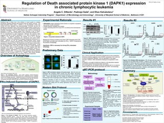
Figures 13-16. Relative RNA abundance. RPL-32 was used as a basis of
comparison for the RNA levels of DAPK1, C/EBP-β, ATF6, and ZIPK. The
delta-delta Ct method was used to calculate relative RNA abundance. Current
samples are boxed in. Other samples were conducted in previous experiments.
Data indicates an overall reduction in the total RNA levels of all four groups
when compared to the B-Cell control. However, some patients do not exhibit
any defects in the RNA transcript of interest.
Special thanks to: Dr. Padmaja Gade, instructor/mentor; Dr. Dhan Kalvakolanu, mentor; Dr. Amy
Kimball, clinical collaborator; Dr. Bret Hassel, NSIP program director
The Nathan Schnaper Summer Internship Program
Marlene and Stewart Greenebaum Cancer Center
University of Maryland Medical Center
National Institutes of Health Grant CA78282
• ZIPK/DAPK3 (Zipper interacting protein kinase) is a regulator of apoptosis
and smooth muscle contraction
• Shuttles between cytoplasm and nucleus
• Is seen interacting with STAT3 and ATF4 to enhance their transcriptional
activity
Figure 3. The protein domains of ZIPK
Summary
Nature Reviews Microbiology (2004) 2:301