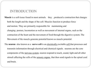
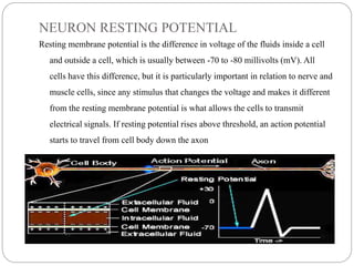
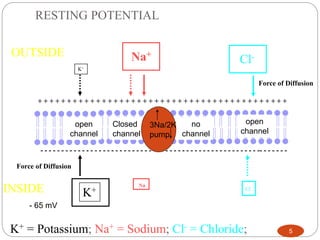
This document discusses neuron and muscle potentials. It begins by introducing muscles and neurons, explaining that muscles contract to produce movement while neurons transmit electrical and chemical signals. It then describes how neuron potentials arise from the electrochemical activity of neurons and the resting potential difference across the neuron membrane. When a neuron is stimulated, this polarity is reversed, generating an action potential. A similar process occurs in muscles - the action potential travels to the neuromuscular junction and causes the release of acetylcholine, generating a muscle action potential and contraction.
2. INTRODUCTION
Muscle is a soft tissue found in most animals. they , produced a contraction that changes
both the length and the shape of the cell. Muscles function to produce force
and motion. They are primarily responsible for maintaining and
changing posture, locomotion as well as movement of internal organs, such as the
contraction of the heart and the movement of food through the digestive system. The
Movement of the muscle generate potential known as muscle potential.
The neuron also known as a nerve cell is an electrically excitable cell that processes and
transmits information through electrical and chemical signals. neurons are the core
components of the nervous system. neuron respond to touch, sound, light and all other
stimuli affecting the cells of the sensory organs that then send signals to the spinal cord
and brain.
3. NEURON POTENTIAL
The neuron potential result from the electrochemical activity of excitable cells
also known as neuron. These cells are surrounded by body fluids having a high
Cl concentration. Neuron acts as a constant current source when stimulated,
and creates an ionic current within the body fluid. This current induces
electrical potentials within the human body. This potential decrease in
amplitude with increasing distance from the excitable cell.
Resting neurons maintain a difference in charge across their cell membrane with
negative charges inside, and a positive charges outside. When neuron is
stimulated this polarity is reversed. The neuron potential is divided into
resting potential and action potential which can be explain as follow
4. NEURON RESTING POTENTIAL
Resting membrane potential is the difference in voltage of the fluids inside a cell
and outside a cell, which is usually between -70 to -80 millivolts (mV). All
cells have this difference, but it is particularly important in relation to nerve and
muscle cells, since any stimulus that changes the voltage and makes it different
from the resting membrane potential is what allows the cells to transmit
electrical signals. If resting potential rises above threshold, an action potential
starts to travel from cell body down the axon
6. INITIALIZATION OF ACTION POTENTIAL
Stimulation of a neuron opens some of the membrane proteins (a.k.a. Na+gates)
allows to pass freely into the cells free flow of Na+ into the cell causes a
reversal of membrane polarity polarity reversal is called the action
potential Stimulation of neuron due to environmental changes opens some of
the membrane protein there by allowing Na+ to pass feely into the cell. The
free flow of into the cell causes a reversal of membrane polarity. This
polarity reversal is called action potential.
This result to the changes in potential from -70mV to about 40mV. spike in voltage
causes the K+ gate open and Na+ gate to close . This make K+ ions rush out of
the neuron. The inside becomes negative again. This is repolarization.
So many K+ ions get out that the charge goes below the resting potential. While the
neuron is in this state it cannot react to additional stimuli. This is termed as
refractory period which last for about 0.5.
7. The sequence of depolarization and repolarization generates a small electrical current
in this localized area. The current affects the nearby protein channels for Na+ and
causes them to open. When the adjacent channels open, Na+ions flood into that area
of the neuron and an action potential occurs. This in turn will affect the areas next
to it and the impulse passes along the entire neuron. The electric current pas.ses
outward over the membrane in all directions.
8.
9. MUSCLE POTENTIAL
The action potential will continue to flow along the axon until it reaches
neuromuscular junction as can be shown below
10. The neuromuscular junction consist of two part. Ie presynaptic terminal on the side of
the axon, and the postsynaptic terminal on the side of the muscle, between them
existed a gap called synaptic cleft. To this end, a chemical known as acetylcholine
will be released( also known as neurotransmitter) this will allow the action potential
to cross the gap and move to the target muscle fibre.
11. MUSCLE FIBRE EXCITATION
Each muscle fibre has one neuromuscular junction, receiving input from just
one efferent neuron . The neuro transmitter allows Na+ ions to enter the
cell, causing a depolarizing excitatory postsynaptic potential (EPSP) that is
above the threshold potential, this triggered an action potential in the muscle
fibre. the EPSP is always well above threshold. This means that under normal
circumstances, an action potential in a somatic efferent neuron always elicits an
action potential in the muscle fibre. Consider the diagram below.
12. The above figure shows a muscle cell EPSP in response to a single action
potential in a somatic efferent neuron . The amount of Ach(neuro transmitter)
released with one neuronal action potential is enough to depolarize the muscle
fibre well above the threshold for eliciting an action potential. The degree that
the EPSP exceeds threshold is known as the safety factor.
The term 'safety factor' refers to the ability of neuromuscular transmission to
remain effective under various physiological conditions and stresses.
13. This is a result of the amount of transmitter released per nerve impulse being
greater than that required to trigger an action potential in the muscle fibre.
The safety factor is a measure of this excess of released transmitter. This
means that any action potential in the neuron will be enough to triggered
action potential in the muscle fibre(also known as sarcolemma).
a single action potential in a motor neuron can activate hundreds of muscle
fibers in synchrony, the resulting currents sum to generate a potential known
as the compound action potential, which is the summed action potentials
of all the muscle fibers in the motor unit. This is also termed as the muscle
potential as can be shown in the figure below.
14. The action potential in the muscle will cause the muscle contraction( also known as
twitch). The twitch is divided into three period.
1. Latent period
brief delay between the stimulus and the muscle contraction The latent period is
less than 2milliseconds in humans
15. 2. Period of contraction
3. Period of relaxation
This can be as shown in the figure below
16. If the muscle is allowed to relax completely before each stimulus than the muscle
will contract with the same force.
If the muscle is stimulated again before it has completely relaxed, then the force
of the next contraction increases.
i.e. stimulating the muscle at a rapid frequency increases the force of contraction.
This is called summation