neuro_module_part_2.pdf interpretation d'un ct scan
1.
NEURORADIOLOGY
DIL part 2
Anapproach to head CT interpretation
K. Agyem MD, G. Hall MD, D. Palathinkal MD, Alexandre
Menard
March/April 2015
2.
OVERVIEW
• Introduction toNeuroimaging - DIL part 1
• Basic Brain Anatomy - DIL part 1
• Standardized Approach to Image Interpretation - DIL part 2
• Common Pathology
• Bleeds (Hemorrhages) - DIL part 3
• Strokes (Infarcts) DIL part 4
• Masses (Tumors) part 5
3.
APPROACH TO CT
•The most important thing is to use the same
approach every time! We will go through an
example approach.
• Use different window settings (ie brain vs bone
windows).
• Use different planes (axials, coronals and
sagittals).
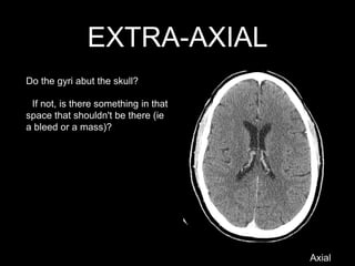
EXTRA-AXIAL
Axial
Do the gyriabut the skull?
If not, is there something in that
space that shouldn't be there (ie
a bleed or a mass)?
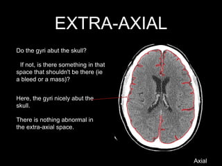
6.
EXTRA-AXIAL
Do the gyriabut the skull?
If not, is there something in that
space that shouldn't be there (ie
a bleed or a mass)?
Here, the gyri nicely abut the
skull.
There is nothing abnormal in
the extra-axial space.
Axial
7.
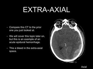
EXTRA-AXIAL
• Compare thisCT to the prior
one you just looked at.
• We will cover this topic later on,
but this is an example of an
acute epidural hemorrhage.
• This a bleed in the extra-axial
space.
Axial
8.
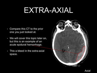
EXTRA-AXIAL
• Compare thisCT to the prior
one you just looked at.
• We will cover this topic later on,
but this is an example of an
acute epidural hemorrhage.
• This a bleed in the extra-axial
space.
Axial
9.
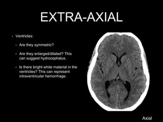
EXTRA-AXIAL
• Ventricles:
• Arethey symmetric?
• Are they enlarged/dilated? This
can suggest hydrocephalus.
• Is there bright white material in the
ventricles? This can represent
intraventricular hemorrhage.
Axial
10.
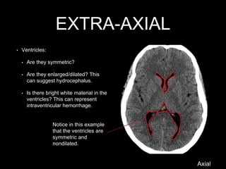
EXTRA-AXIAL
Notice in thisexample
that the ventricles are
symmetric and
nondilated.
Axial
• Ventricles:
• Are they symmetric?
• Are they enlarged/dilated? This
can suggest hydrocephalus.
• Is there bright white material in the
ventricles? This can represent
intraventricular hemorrhage.
11.
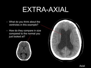
EXTRA-AXIAL
• What doyou think about the
ventricles in this example?
• How do they compare in size
compared to the normal you
just looked at?
Axial
12.
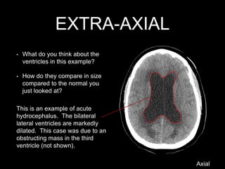
EXTRA-AXIAL
This is anexample of acute
hydrocephalus. The bilateral
lateral ventricles are markedly
dilated. This case was due to an
obstructing mass in the third
ventricle (not shown).
• What do you think about the
ventricles in this example?
• How do they compare in size
compared to the normal you
just looked at?
Axial
13.
EXTRA-AXIAL
Axial
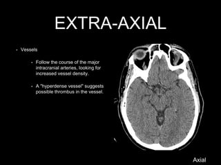
• Vessels
• Followthe course of the major
intracranial arteries, looking for
increased vessel density.
• A "hyperdense vessel" suggests
possible thrombus in the vessel.
14.
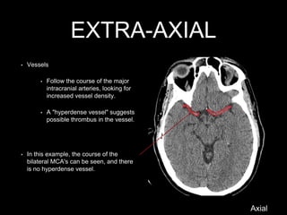
EXTRA-AXIAL
• Vessels
• Followthe course of the major
intracranial arteries, looking for
increased vessel density.
• A "hyperdense vessel" suggests
possible thrombus in the vessel.
• In this example, the course of the
bilateral MCA's can be seen, and there
is no hyperdense vessel.
Axial
15.
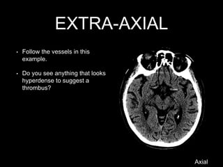
EXTRA-AXIAL
• Follow thevessels in this
example.
• Do you see anything that looks
hyperdense to suggest a
thrombus?
Axial
16.
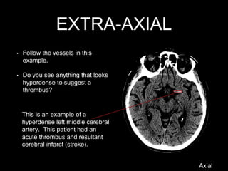
EXTRA-AXIAL
• Follow thevessels in this
example.
• Do you see anything that looks
hyperdense to suggest a
thrombus?
This is an example of a
hyperdense left middle cerebral
artery. This patient had an
acute thrombus and resultant
cerebral infarct (stroke).
Axial
17.
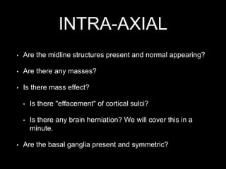
INTRA-AXIAL
• Are themidline structures present and normal appearing?
• Are there any masses?
• Is there mass effect?
• Is there "effacement" of cortical sulci?
• Is there any brain herniation? We will cover this in a
minute.
• Are the basal ganglia present and symmetric?
18.
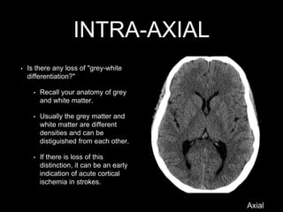
INTRA-AXIAL
• Is thereany loss of "grey-white
differentiation?"
• Recall your anatomy of grey
and white matter.
• Usually the grey matter and
white matter are different
densities and can be
distiguished from each other.
• If there is loss of this
distinction, it can be an early
indication of acute cortical
ischemia in strokes.
Axial
19.
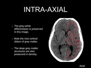
INTRA-AXIAL
• The grey-white
differentiaionis preserved
in this image.
• Note the nice cortical
ribbon of grey matter.
• The deep grey matter
structures are also
preseverd in density.
Axial
20.
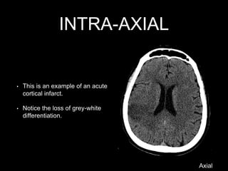
INTRA-AXIAL
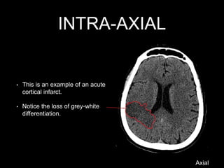
• This isan example of an acute
cortical infarct.
• Notice the loss of grey-white
differentiation.
Axial
21.
INTRA-AXIAL
• This isan example of an acute
cortical infarct.
• Notice the loss of grey-white
differentiation.
Axial
22.
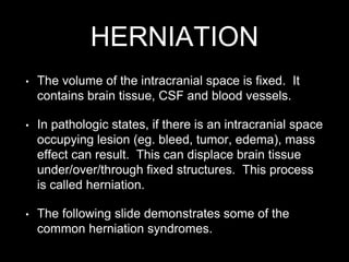
HERNIATION
• The volumeof the intracranial space is fixed. It
contains brain tissue, CSF and blood vessels.
• In pathologic states, if there is an intracranial space
occupying lesion (eg. bleed, tumor, edema), mass
effect can result. This can displace brain tissue
under/over/through fixed structures. This process
is called herniation.
• The following slide demonstrates some of the
common herniation syndromes.
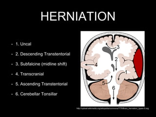
HERNIATION
Axial
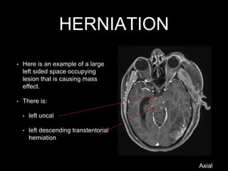
• Here isan example of a large
left sided space occupying
lesion that is causing mass
effect.
• There is:
• left uncal
• left descending transtentorial
herniation
25.
HERNIATION
• Here isan example of a large
left sided space occupying
lesion that is causing mass
effect.
• There is:
• left uncal
• left descending transtentorial
herniation
Axial
26.
HERNIATION
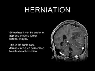
• Sometimes itcan be easier to
appreciate herniation on
coronal images.
• This is the same case,
demonstrating left descending
transtentorial herniation.
27.
HERNIATION
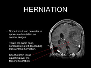
• Sometimes itcan be easier to
appreciate herniation on
coronal images.
• This is the same case,
demonstrating left descending
transtentorial herniation.
See the brain tissue
squishing over the
tentorium cerebelli.
28.
BONES
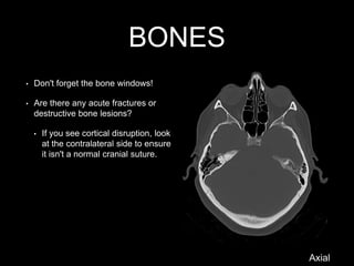
• Don't forgetthe bone windows!
• Are there any acute fractures or
destructive bone lesions?
• If you see cortical disruption, look
at the contralateral side to ensure
it isn't a normal cranial suture.
Axial
29.
BONES
• Don't forgetthe bone windows!
• Are there any acute fractures or
destructive bone lesions?
• If you see cortical disruption, look
at the contralateral side to ensure
it isn't a normal cranial suture.
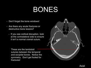
These are the lambdoid
sutures between the temporal
and occipital bones. Notice the
symmetry. Don't get fooled for
fractures!
Axial
30.
BONES
• Don't forgetthe bone windows!
• Are there any acute fractures or
destructive bone lesions?
• If you see cortical disruption, look
at the contralateral side to ensure
it isn't a normal cranial suture.
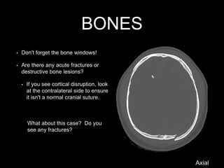
What about this case? Do you
see any fractures?
Axial
31.
BONES
• Don't forgetthe bone windows!
• Are there any acute fractures or
destructive bone lesions?
• If you see cortical disruption, look
at the contralateral side to ensure
it isn't a normal cranial suture.
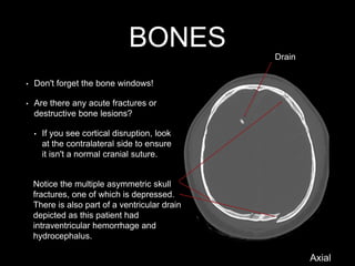
Notice the multiple asymmetric skull
fractures, one of which is depressed.
There is also part of a ventricular drain
depicted as this patient had
intraventricular hemorrhage and
hydrocephalus.
Drain
Axial
32.
• Introduction toNeuroimaging - DIL part 1
• Basic Brain Anatomy - DIL part 1
• Standardized Approach to Image Interpretation - DIL part 2
• Common Pathology
• Bleeds (Hemorrhages) - DIL part 3
• Strokes (Infarcts) DIL part 4
• Masses (Tumors) part 5
End of module 2


![Hypothalamus short ppt by Dr. Neha [PT].pptx](https://cdn.slidesharecdn.com/ss_thumbnails/hypothalamusbydr-260124145759-b9f94a93-thumbnail.jpg?width=640&height=640&fit=bounds)

