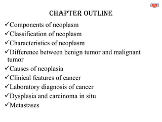
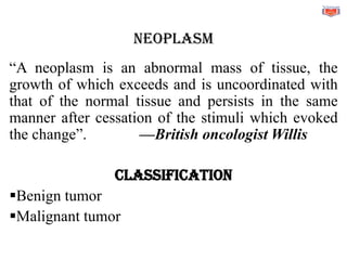
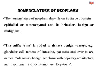
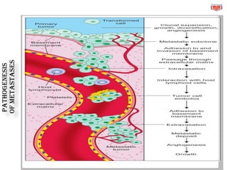
CHAPTER
4
CELL GROWTH
AND NEOPLASM
CHAPTER OUTLINE
✓Components of neoplasm
✓Classification of neoplasm
✓Characteristics of neoplasm
✓Difference between benign tumor and malignant
tumor
✓Causes of neoplasia
✓Clinical features of cancer
✓Laboratory diagnosis of cancer
✓Dysplasia and carcinoma in situ
✓Metastases
NEOPLASM
“A neoplasm is an abnormal mass of tissue, the
growth of which exceeds and is uncoordinated with
that of the normal tissue and persists in the same
manner after cessation of the stimuli which evoked
the change”. —British oncologist Willis
CLASSIFICATION
▪Benign tumor
▪Malignant tumor
Nomenclature of neoplasm
✓The nomenclature of neoplasm depends on its tissue of origin –
epithelial or mesenchymal and its behavior- benign or
malignant.
✓The suffix ‘oma’ is added to denote benign tumors, e.g.
glandular cell tumors of intestine, pancreas and ovaries are
named ‘Adenoma’, benign neoplasm with papillary architecture
are ‘papilloma’, liver cell tumor are ‘Hepatoma’.
✓‘Carcinoma’ suffix is used for malignant neoplasm
derived from epithelial cells, like squamous cell carcinoma,
and adenocarcinoma. Endocrine tumors are named as
follicular carcinoma of thyroid etc.
✓Sarcoma is malignant neoplasm derived from
mesenchymal cells (e.g., liposarcoma arises from fat cells,
leiomyosarcoma from smooth muscle cells).
CHARACTERISTICS
• Rate of Growth of Tumor
• Differentiation
• Grading of Malignant Tumor
✓Grading of malignant tumor is done based on differentiation, like
✓Well differentiated tumor is Grade I.
✓Moderately differentiated is Grade II.
✓Poorly differentiated is Grade III.
•Clinical Features of Tumor- may be asymptomatic.
Malignant tumors are associated with some
generalized symptoms like low grade fever, anorexia,
weight loss, anemia, weakness and fatigue.
•Morphologic Features
•Capsulation
•Mitotic activity
•Growth pattern
•Size of the tumor
•Invasion
•Metastases
Anaplasia or cellular atypia
This term refers to the lack of differentiation and is feature of
aggressiveness of the tumor. Cytologic evidence of anaplasia
includes:
• Variation in the size and shape of cells and cell nuclei
(pleomorphism).
• Enlarged and hyperchromatic nuclei with coarsely clumped
chromatin.
• Atypical mitoses.
• Bizarre cells, including tumor giant cells.
• Loss of polarity of nuclei.
• Presence of prominent nucleoli.
• High nuclear-cytoplasmic ratio (N/C ratio).
• Functional (cytoplasmic) changes which can be
qualitative and quantitative.
• Genetic abnormalities- All types of genetic
abnormalities like point mutations, frame shift
mutation, deletions, chromosomal translocations etc
can exist in malignant tumors.
Benign and malignant tumors
BENIGN TUMORS
❑Compress surrounding tissue
❑Do not metastasize
❑Are well circumscribed or encapsulated
❑Morphologically cells resemble tissue of origin very
closely.
❑Do not secrete hormones or other substances
❑Atypical mitosis is not seen
❑Necrosis is absent
❑May not require treatment if not health-threatening
❑Unlikely to recur if removed or require further
MALIGNANT TUMORS
❑Break the basement membrane and invade into
surrounding tissue
❑Metastases via bloodstream or lymphatic system is the
hallmark.
❑Capsule is incomplete or absent
❑Cells show anaplasia
❑Can secrete cytokines that cause fatigue, weight loss and
paraneoplastic syndrome.
❑Atypical mitosis arec haracteristically present
❑Necrosis -Often present
❑Require aggressive treatment, including surgery,
radiation, chemotherapy, and immunotherapy
medications
❑Recurrence and relapse is common
Environmental Causes
CHEMICALS
Include those that are man-made (such as aniline dyes in
bladder cancer), drugs (cigarette smoke in lung cancer),
and natural compounds (aflatoxins in liver cancer) which
are carcinogenic.
ONCOGENIC VIRUSES
The genomes of oncogenic DNA viruses integrate into and form
stable associations with the host cell genome and cause genetic
mutations resulting in formation of carcinoma. Some DNA
oncoviruses are human papillomavirus (HPV), implicated in most
squamous cell carcinomas of cervix and oral cavity, Epstein-Barr
virus (EBV) implicated in African Burkitt’s lymphoma, and hepatitis
B virus (HBV) implicated in development of hepatocellular
carcinomas.
RADIATION
Ultraviolet light induces pyrimidine dimers in DNA
and promotes skin cancers. Ionizing radiation (such
as gamma radiation) induces mutations in DNA and
promotes malignancies such as leukemia, thyroid,
lung, colon, and breast cancers.
CHEMICAL CARCINOGENESIS
Initiation: It is the induction of a mutation in a critical
gene involved in the control of cell proliferation. An
initiating carcinogenic agent irreversibly damages cell
DNA to start the process. Examples of carcinogenic
initiators include: alkylating agents like
cyclophosphamide, polycyclic aromatic hydrocarbons
found in smoked foods, aromatic amines or azo dyes
used in food coloring, aflatoxins in moldy peanuts, and
nitrosamines in pickled foods.
Promotion: A promoting agent (which may be
the same as the carcinogen) then acts to cause
proliferation of a neoplastic cell clone. It is a
reversible event. Examples of promoters include:
hormones such as estrogen, drugs such as
diethylstilbesterol, and chemicals.
CLINICAL FEATURES OF CANCER
Some general signs and symptoms associated with, but not specific to,
cancer, include:
• Fatigue
• Lump or area of thickening that can be felt under the skin·
• Unintended weight loss of more than 10 kgs in six months
• An existing mole which suddenly increases in size Changes in
bowel or bladder habits
•Persistent cough or dyspnoea
•Difficulty in swallowing
•Hoarseness of voice
•Persistent indigestion or discomfort after eating
•Persistent, unexplained muscle or joint pain
•Persistent, unexplained fevers or night sweats
•Unexplained bleeding or bruising
LABORATORY DIAGNOSIS OF CANCER
•Biopsy: Small tissue biopsy is the method that
samples small pieces of tissue from the affected site.
•Cytology: Cytological methods involve study of cells
on a stained smear.
•Radiographic technique:
Computed Tomography (CT), Magnetic Resonance
Imaging (MRI), Mammography, and Ultrasonography
(USG)
•Genetictesting: Genetic markers include chromosomal
alterations (translocations, deletions, duplication, etc.),
specific gene defects and gene rearrangements,
•Tumor marker: Tumor marker, are tumor-associated
enzymes, hormones and proteins found in blood and are
used for detection of a tumor.
DYSPLASIA AND CARCINOMA IN SITU
•premalignant changes
•Dysplasia means “disordered growth.” of epithelium.
•When dysplasia is severe and involves the entire thickness
of epithelial lining, it is called carcinoma-in-situ.
Cervical Intraepithelial Neoplasia
Dysplasia and carcinoma in situ (CIS) in squamous lining of uterine
cervix, is known as cervical intraepithelial neoplasia (CIN).
❑CIN–I: When the dysplastic cells involve lower one third of the
epithelium (Mild dysplasia)
❑CIN–II: When the dysplastic cells involves lower two thirds of the
epithelium (Moderate dysplasia)
❑CIN–III or Carcinoma in Situ (CIS): When the dysplastic
cells involves almost full thickness of the epithelium (severe
dysplasia).
METASTASIS
It is the spread of a tumor to sites that are discontinuous with the
primary tumor site
ROUTES OF METASTASIS
1. Lymphatic- It Is seen usually in carcinoma. The lymphatic spread
involves deposition of cancer cells in the draining lymph nodes.
2. Hematogenous- It is seen usually in sarcoma. It is also seen in
advanced stages of carcinomas. E.g. liver, the lungs, bone marrow
3. Direct seeding- Peritoneal, pleural and pericardial cavity and
subarachnoid spaces may be involved in metastases
Pathogenesis
of
metastases
Cancer cachexia (wasting
syndrome)
➢Progressive loss of body fat and lean body mass
➢Accompanied by profound weakness, anorexia, and
anemia.
➢Cachexia is caused by the action of soluble factors
such as cytokines produced by the tumor or host.
➢ It is the most common manifestation of the advanced
malignant disease, leading to death
PATHOLOGY-CHAPTER-4-CELL-GROWTH-AND-NEOPLASM.pdf

PATHOLOGY-CHAPTER-4-CELL-GROWTH-AND-NEOPLASM.pdf

  • 1.
  • 2.
    CHAPTER OUTLINE ✓Components ofneoplasm ✓Classification of neoplasm ✓Characteristics of neoplasm ✓Difference between benign tumor and malignant tumor ✓Causes of neoplasia ✓Clinical features of cancer ✓Laboratory diagnosis of cancer ✓Dysplasia and carcinoma in situ ✓Metastases
  • 3.
    NEOPLASM “A neoplasm isan abnormal mass of tissue, the growth of which exceeds and is uncoordinated with that of the normal tissue and persists in the same manner after cessation of the stimuli which evoked the change”. —British oncologist Willis CLASSIFICATION ▪Benign tumor ▪Malignant tumor
  • 4.
    Nomenclature of neoplasm ✓Thenomenclature of neoplasm depends on its tissue of origin – epithelial or mesenchymal and its behavior- benign or malignant. ✓The suffix ‘oma’ is added to denote benign tumors, e.g. glandular cell tumors of intestine, pancreas and ovaries are named ‘Adenoma’, benign neoplasm with papillary architecture are ‘papilloma’, liver cell tumor are ‘Hepatoma’.
  • 5.
    ✓‘Carcinoma’ suffix isused for malignant neoplasm derived from epithelial cells, like squamous cell carcinoma, and adenocarcinoma. Endocrine tumors are named as follicular carcinoma of thyroid etc. ✓Sarcoma is malignant neoplasm derived from mesenchymal cells (e.g., liposarcoma arises from fat cells, leiomyosarcoma from smooth muscle cells).
  • 6.
    CHARACTERISTICS • Rate ofGrowth of Tumor • Differentiation • Grading of Malignant Tumor ✓Grading of malignant tumor is done based on differentiation, like ✓Well differentiated tumor is Grade I. ✓Moderately differentiated is Grade II. ✓Poorly differentiated is Grade III.
  • 7.
    •Clinical Features ofTumor- may be asymptomatic. Malignant tumors are associated with some generalized symptoms like low grade fever, anorexia, weight loss, anemia, weakness and fatigue. •Morphologic Features •Capsulation •Mitotic activity •Growth pattern •Size of the tumor •Invasion •Metastases
  • 8.
    Anaplasia or cellularatypia This term refers to the lack of differentiation and is feature of aggressiveness of the tumor. Cytologic evidence of anaplasia includes: • Variation in the size and shape of cells and cell nuclei (pleomorphism). • Enlarged and hyperchromatic nuclei with coarsely clumped chromatin. • Atypical mitoses. • Bizarre cells, including tumor giant cells.
  • 9.
    • Loss ofpolarity of nuclei. • Presence of prominent nucleoli. • High nuclear-cytoplasmic ratio (N/C ratio). • Functional (cytoplasmic) changes which can be qualitative and quantitative. • Genetic abnormalities- All types of genetic abnormalities like point mutations, frame shift mutation, deletions, chromosomal translocations etc can exist in malignant tumors.
  • 10.
  • 11.
    BENIGN TUMORS ❑Compress surroundingtissue ❑Do not metastasize ❑Are well circumscribed or encapsulated ❑Morphologically cells resemble tissue of origin very closely. ❑Do not secrete hormones or other substances ❑Atypical mitosis is not seen ❑Necrosis is absent ❑May not require treatment if not health-threatening ❑Unlikely to recur if removed or require further
  • 12.
    MALIGNANT TUMORS ❑Break thebasement membrane and invade into surrounding tissue ❑Metastases via bloodstream or lymphatic system is the hallmark. ❑Capsule is incomplete or absent ❑Cells show anaplasia ❑Can secrete cytokines that cause fatigue, weight loss and paraneoplastic syndrome.
  • 13.
    ❑Atypical mitosis arecharacteristically present ❑Necrosis -Often present ❑Require aggressive treatment, including surgery, radiation, chemotherapy, and immunotherapy medications ❑Recurrence and relapse is common
  • 14.
    Environmental Causes CHEMICALS Include thosethat are man-made (such as aniline dyes in bladder cancer), drugs (cigarette smoke in lung cancer), and natural compounds (aflatoxins in liver cancer) which are carcinogenic.
  • 15.
    ONCOGENIC VIRUSES The genomesof oncogenic DNA viruses integrate into and form stable associations with the host cell genome and cause genetic mutations resulting in formation of carcinoma. Some DNA oncoviruses are human papillomavirus (HPV), implicated in most squamous cell carcinomas of cervix and oral cavity, Epstein-Barr virus (EBV) implicated in African Burkitt’s lymphoma, and hepatitis B virus (HBV) implicated in development of hepatocellular carcinomas.
  • 16.
    RADIATION Ultraviolet light inducespyrimidine dimers in DNA and promotes skin cancers. Ionizing radiation (such as gamma radiation) induces mutations in DNA and promotes malignancies such as leukemia, thyroid, lung, colon, and breast cancers.
  • 17.
    CHEMICAL CARCINOGENESIS Initiation: Itis the induction of a mutation in a critical gene involved in the control of cell proliferation. An initiating carcinogenic agent irreversibly damages cell DNA to start the process. Examples of carcinogenic initiators include: alkylating agents like cyclophosphamide, polycyclic aromatic hydrocarbons found in smoked foods, aromatic amines or azo dyes used in food coloring, aflatoxins in moldy peanuts, and nitrosamines in pickled foods.
  • 18.
    Promotion: A promotingagent (which may be the same as the carcinogen) then acts to cause proliferation of a neoplastic cell clone. It is a reversible event. Examples of promoters include: hormones such as estrogen, drugs such as diethylstilbesterol, and chemicals.
  • 19.
    CLINICAL FEATURES OFCANCER Some general signs and symptoms associated with, but not specific to, cancer, include: • Fatigue • Lump or area of thickening that can be felt under the skin· • Unintended weight loss of more than 10 kgs in six months • An existing mole which suddenly increases in size Changes in bowel or bladder habits
  • 20.
    •Persistent cough ordyspnoea •Difficulty in swallowing •Hoarseness of voice •Persistent indigestion or discomfort after eating •Persistent, unexplained muscle or joint pain •Persistent, unexplained fevers or night sweats •Unexplained bleeding or bruising
  • 21.
    LABORATORY DIAGNOSIS OFCANCER •Biopsy: Small tissue biopsy is the method that samples small pieces of tissue from the affected site. •Cytology: Cytological methods involve study of cells on a stained smear. •Radiographic technique: Computed Tomography (CT), Magnetic Resonance Imaging (MRI), Mammography, and Ultrasonography (USG)
  • 22.
    •Genetictesting: Genetic markersinclude chromosomal alterations (translocations, deletions, duplication, etc.), specific gene defects and gene rearrangements, •Tumor marker: Tumor marker, are tumor-associated enzymes, hormones and proteins found in blood and are used for detection of a tumor.
  • 23.
    DYSPLASIA AND CARCINOMAIN SITU •premalignant changes •Dysplasia means “disordered growth.” of epithelium. •When dysplasia is severe and involves the entire thickness of epithelial lining, it is called carcinoma-in-situ.
  • 24.
    Cervical Intraepithelial Neoplasia Dysplasiaand carcinoma in situ (CIS) in squamous lining of uterine cervix, is known as cervical intraepithelial neoplasia (CIN). ❑CIN–I: When the dysplastic cells involve lower one third of the epithelium (Mild dysplasia) ❑CIN–II: When the dysplastic cells involves lower two thirds of the epithelium (Moderate dysplasia) ❑CIN–III or Carcinoma in Situ (CIS): When the dysplastic cells involves almost full thickness of the epithelium (severe dysplasia).
  • 25.
    METASTASIS It is thespread of a tumor to sites that are discontinuous with the primary tumor site ROUTES OF METASTASIS 1. Lymphatic- It Is seen usually in carcinoma. The lymphatic spread involves deposition of cancer cells in the draining lymph nodes. 2. Hematogenous- It is seen usually in sarcoma. It is also seen in advanced stages of carcinomas. E.g. liver, the lungs, bone marrow 3. Direct seeding- Peritoneal, pleural and pericardial cavity and subarachnoid spaces may be involved in metastases
  • 26.
  • 27.
    Cancer cachexia (wasting syndrome) ➢Progressiveloss of body fat and lean body mass ➢Accompanied by profound weakness, anorexia, and anemia. ➢Cachexia is caused by the action of soluble factors such as cytokines produced by the tumor or host. ➢ It is the most common manifestation of the advanced malignant disease, leading to death