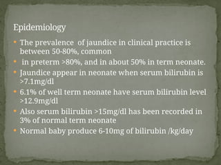
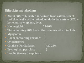
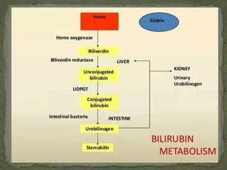
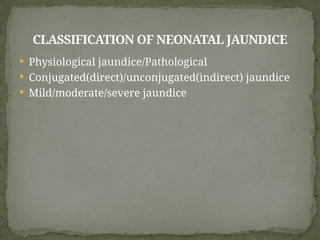
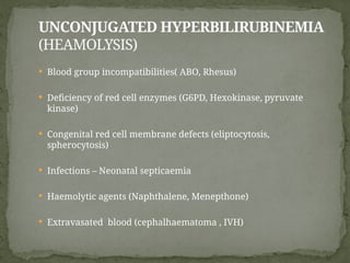
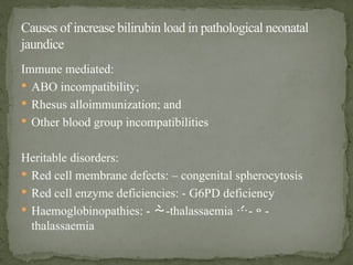
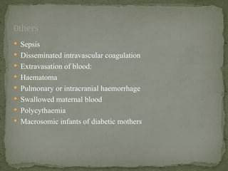
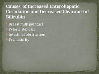
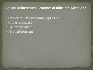
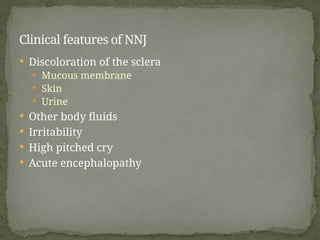
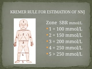
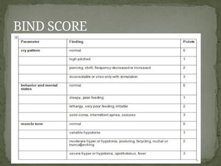
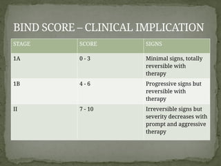
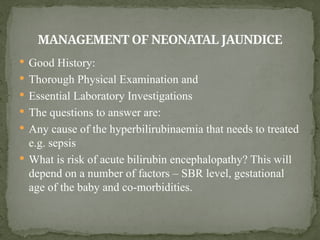
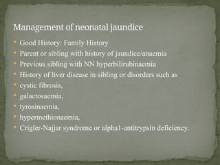
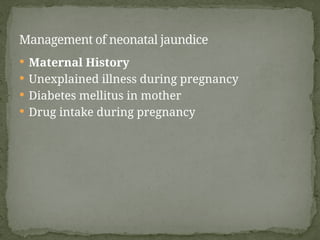
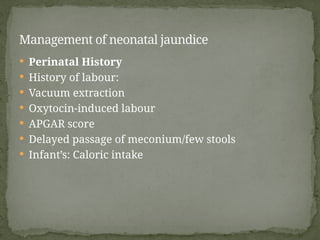
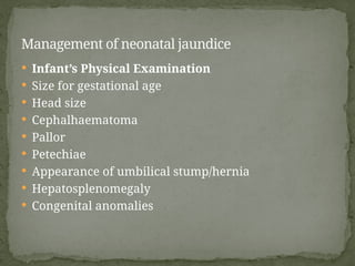
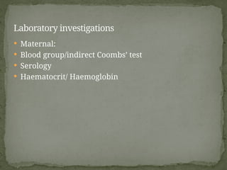
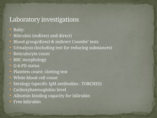
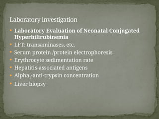
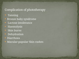
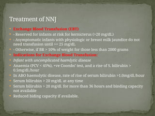
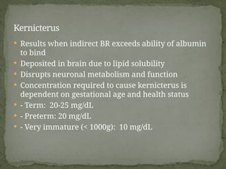
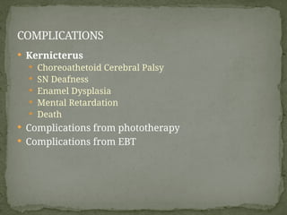
NEONATAL JAUNDICE
BY
DR MUHAMMAD HASSAN
25/04/24
GBMC
 OUTLINE:
 INTRODUCTION
 EPIDEMIOLOGY
 BILIRUBIN METABOLISM
 CLASSIFICATION
 CAUSES
 CLINICAL MANIFESTATION
 INVESTIGATIONS
 TREATMENT
 COMPLICATION
 PREVENTION
 DEFINATION: Jaundice is a clinical conditions that is
characterized by yellowish discoloration of : sclera,
mucous membrane, skin
 It occur due increased level of bilirubin, a pigment
derived mainly from HB
INTRODUCTION
 The prevalence of jaundice in clinical practice is
between 50-80%, common
 in preterm >80%, and in about 50% in term neonate.
 Jaundice appear in neonate when serum bilirubin is
>7.1mg/dl
 6.1% of well term neonate have serum bilirubin level
>12.9mg/dl
 Also serum bilirubin >15mg/dl has been recorded in
3% of normal term neonate
 Normal baby produce 6-10mg of bilirubin /kg/day
Epidemiology
 About 80% of bilirubin is derived from catabolism of
red blood cells in the reticulo-endothelial system (RES) –
bone marrow, spleen, liver;
 Haemoglobin 75-80%
 The remaining 20% from other sources which include:
 Myoglobin }
 Haem-containing enzymes }
 Cytochromes }
 Catalase /Peroxidases } 20-25%
 Tryptophan pyrrolase }
 In-effective erythropoiesis
Bilirubin metabolism
 Physiological jaundice/Pathological
 Conjugated(direct)/unconjugated(indirect) jaundice
 Mild/moderate/severe jaundice
CLASSIFICATION OF NEONATAL JAUNDICE
UNCONJUGATED HYPERBILIRUBINEMIA
(HEAMOLYSIS)
 Blood group incompatibilities( ABO, Rhesus)
 Deficiency of red cell enzymes (G6PD, Hexokinase, pyruvate
kinase)
 Congenital red cell membrane defects (eliptocytosis,
spherocytosis)
 Infections – Neonatal septicaemia
 Haemolytic agents (Naphthalene, Menepthone)
 Extravasated blood (cephalhaematoma , IVH)
UNCONJUGATED HYPERBILIRUBINEMIA
(DEFECTIVE CONJUGATION)
 Crigler Najjar syndrome
 Breast milk jaundice
 Familial neonatal hyperbilirubinemia
 Hypothyroidism
 Down’s syndrome
 Infants of diabetic mothers
 prematurity
CAUSES OF CONJUGATED
HYPERBILIRUBINEMIA
 INFECTION
 Toxoplasmosis, CMV, Hepatitis B, Rubella
 Giant cell hepatitis
 BIOCHEMICAL/METABOLIC
 Galactosemia, alfa-1-antitripsin deficiency, Roto’s & Dubin
Johnson syndrome
 Hypothyroidism, tyrosinosis
 CONGENITAL OBLITERATION
 Congenital biliary atresia, Choledochal cyst, Bile duct
stenosis
 Inspissated bile duct syndrome
 It is a diagnosis of exclusion. Only diagnosed by
excluding infection, haemolysis, and metabolic
diseases, etc.
 Causes are multifactorial: RBC mass, RBC life span,
decreased UDP-glucuronyl transferase and ligandin,
increased enterohepatic circulation
 Begins at >36 hrs (usually not earlier than 24 hours)
 Peaks to 5-6mg/dl on 4th
day
 Bilirubin levels not more than 12mg/dl
 Returns to below 2mg/dl on the 7- 10th
day
 Direct bilirubin < 1.5-2mg/dl or 15% of total SBR
PHYSIOLOGIC HYPERBILIRUBINAEMIA
 Onset before 24 hours of life;
 Rate of rise of SBR > 5mg/dl./24 hours or >0.5 mg/dL/hour;
usually signifying active haemolysis
 Term infant with SBR is above 12.9mg/dl.
 Preterm infant with SBR is above 15mg/dl.
 Direct bilirubin is above 1.5-2mg/dl. or 15%
 Jaundice persisting after 7 or 14 days in term and preterm
babies.
 There may be associated hepatosplenomegaly or anaemia
 Due to Increased bilirubin load or Impaired bilirubin
excretion or Mixed
Pathological jaundice
Immune mediated:
 ABO incompatibility;
 Rhesus alloimmunization; and
 Other blood group incompatibilities
Heritable disorders:
 Red cell membrane defects: – congenital spherocytosis
 Red cell enzyme deficiencies: - G6PD deficiency
 Haemoglobinopathies: - -thalassaemia --
thalassaemia
Causes of increase bilirubin load in pathological neonatal
jaundice
 Sepsis
 Disseminated intravascular coagulation
 Extravasation of blood:
 Haematoma
 Pulmonary or intracranial haemorrhage
 Swallowed maternal blood
 Polycythaemia
 Macrosomic infants of diabetic mothers
0thers
 Breast milk jaundice
 Pyloric stenosis
 Intestinal obstruction
 Prematurity
Causes of Increased Enterohepatic
Circulation and Decreased Clearance of
Bilirubin
 Crigler-Najjar Syndrome types I and II
 Gilbert’s disease
 Hypothyroidism
 Hypopituitarism
Causes Of increased Clearance of Bilirubin: Metabolic
 Extrahepatic biliary atresia
 Trisomy 18
 Intrahepatic biliary atresia (nonsyndromatic
paucity of bile ducts)
 Extrahepatic biliary atresia and choledochal cyst
 Bile plug syndrome
 Cystic disease
Causes of ductal disturbances in
pathological jaundice
 Major Aetiological Factors of Pathological Neonatal
Hyperbilirubinaemia in Nigeria
 i. ABO Incompatibility,
 ii. Bacterial Infections(sepsis)
 iii G-6-PD Deficiency
 iv. Low Birthweight
In a variable proportion of cases the cause is unknown – partly
due incomplete evaluation.
Major causes of pathological jaundice in Nigeria
COMMONLY USED ICTEROGENIC AGENTS
 Jaundice may appear any time during neonatal period
 Jaundice begins on the face and as the level increases,
progress to the abdomen and feet
 Unconjugated bilirubin appears bright yellow or orange
 Conjugated bilirubin appears greenish or muddy yellow
 Infants usually lethargic and feed poorly
 Signs of primary disease.(sepsis, etc)
CLINICAL PRESENTATION
 Discoloration of the sclera
 Mucous membrane
 Skin
 Urine
 Other body fluids
 Irritability
 High pitched cry
 Acute encephalopathy
Clinical features of NNJ
KREMER RULE FOR ESTIMATION OF NNJ
Zone SBR mmol/L
 1 = 100 mmol/L
 2 = 150 mmol/L
 3 = 200 mmol/L
 4 = 250 mmol/L
 5 > 250 mmol/L
BIND SCORE
BIND SCORE – CLINICAL IMPLICATION
STAGE SCORE SIGNS
1A 0 - 3 Minimal signs, totally
reversible with
therapy
1B 4 - 6 Progressive signs but
reversible with
therapy
II 7 - 10 Irreversible signs but
severity decreases with
prompt and aggressive
therapy
 Good History:
 Thorough Physical Examination and
 Essential Laboratory Investigations
 The questions to answer are:
 Any cause of the hyperbilirubinaemia that needs to treated
e.g. sepsis
 What is risk of acute bilirubin encephalopathy? This will
depend on a number of factors – SBR level, gestational
age of the baby and co-morbidities.
MANAGEMENT OF NEONATAL JAUNDICE
 Good History: Family History
 Parent or sibling with history of jaundice/anaemia
 Previous sibling with NN hyperbilirubinaemia
 History of liver disease in sibling or disorders such as
 cystic fibrosis,
 galactosaemia,
 tyrosinaemia,
 hypermethionaemia,
 Crigler-Najjar syndrome or alpha1-antitrypsin deficiency.
Management of neonatal jaundice
 Maternal History
 Unexplained illness during pregnancy
 Diabetes mellitus in mother
 Drug intake during pregnancy
Management of neonatal jaundice
 Perinatal History
 History of labour:
 Vacuum extraction
 Oxytocin-induced labour
 APGAR score
 Delayed passage of meconium/few stools
 Infant’s: Caloric intake
Management of neonatal jaundice
 Infant’s Physical Examination
 Size for gestational age
 Head size
 Cephalhaematoma
 Pallor
 Petechiae
 Appearance of umbilical stump/hernia
 Hepatosplenomegaly
 Congenital anomalies
Management of neonatal jaundice
 Maternal:
 Blood group/indirect Coombs’ test
 Serology
 Haematocrit/ Haemoglobin
Laboratory investigations
 Baby:
 Bilirubin (indirect and direct)
 Blood group/direct & indirect Coombs’ tests
 Urinalysis (including test for reducing substances)
 Reticulocyte count
 RBC morphology
 G-6-PD status
 Platelets count; clotting test
 White blood cell count
 Serology (specific IgM antibodies - TORCHES)
 Carboxyhaemoglobin level
 Albumin binding capacity for bilirubin
 Free bilirubin
Laboratory investigations
 Laboratory Evaluation of Neonatal Conjugated
Hyperbilirubinemia
 LFT: transaminases, etc.
 Serum protein /protein electrophoresis
 Erythrocyte sedimentation rate
 Hepatitis-associated antigens
 Alpha1-anti-trypsin concentration
 Liver biopsy
Laboratory investigation
 Urine Tests:
 Routine urinalysis,
 Urine culture
 Bilirubin and urobilinogen
 Amino acid screening
Laboratory investigation
PRINCIPLES
 Observation/monitoring
 Resucitation
 Reduce bilirubin load
 Phototherapy(unconjugated)
 Exchange blood transfusion
 Phenobarbital therapy
 Treat underlying cause
 Prevent/control complications
 Supportive therapy
Treatment of NNJ
Treatment of NNJ
 Phototherapy
 Effectiveness depends on:
 Amount of light energy emitted in effective wavelengths
 Distance from light source to the baby
 Amount of skin exposed
 Rate of underlying process generating excess bilirubin
 In vivo metabolism and excretion of bilirubin
 Indication: unconjugated bilirubin 16-18 mg/dl; lower in preterm
infants
 Contra indicated in obstructive jaundice
 Has significantly reduced the need for exchange blood transfusion
 Must simultaneously treat identifiable causes of
hyperbilirubinemia
 Prophylactically used in VLBW infants
Treatment of NNJ
 Tanning
 Bronze baby syndrome
 Lactose intolerance
 Haemolysis
 Skin burns
 Dehydration
 Diarrhoea
 Macular-papular Skin rashes
Complication of phototherapy
 Exchange Blood Transfusion (EBT)
 - Reserved for infants at risk for kernicterus (>20 mg/dL)
 - Asymptomatic infants with physiologic or breast milk jaundice do not
need transfusion until >= 25 mg/dL
 - Otherwise, if BR > 10% of weight for those less than 2000 grams
 Indications for Exchange Blood Transfusion:
 Infant with uncomplicated haemlytic disease
 Anaemia (PCV < 45%), +ve Coombs’ test, and a rise of S. bilirubin >
0.5mg/dl./hour
 In ABO haemolytic disease, rate of rise of serum bilirubin >1.0mg/dl./hour
 Serum bilirubin > 20 mg/dl. at any time
 Serum bilirubin > 20 mg/dl. for more than 36 hours and binding capacity
not available
 Reduced biding capacity if available.
Treatment of NNJ
 Results when indirect BR exceeds ability of albumin
to bind
 Deposited in brain due to lipid solubility
 Disrupts neuronal metabolism and function
 Concentration required to cause kernicterus is
dependent on gestational age and health status
 - Term: 20-25 mg/dL
 - Preterm: 20 mg/dL
 - Very immature (< 1000g): 10 mg/dL
Kernicterus
 Kernicterus
 Choreoathetoid Cerebral Palsy
 SN Deafness
 Enamel Dysplasia
 Mental Retardation
 Death
 Complications from phototherapy
 Complications from EBT
COMPLICATIONS
 Hyperbilirubinemia in neonates is a common
condition caused by multiple and variety of
factors which if identified can be treated
 Poor management of jaundice can lead to
severe neurological damage and death
CONCLUSION
 B.C Ibe. Neonatal jaundice. In: Paediatrics and Child
Health in the Tropical Region .Eds. Azubuike and
Nkanginieme 2nd
Edition African Pub.2007, 204-211
 Martins C.M and Cloherty J.P . Neonatal
hyperbilirubinaemia. In : MANUAL OF NEONATAL
CARE 6th
ED , 2008;181-212
 Kaplan M, Merlob P, Regev R. guidelines for the
management of neonatal hyperbilirubinaemia. J
Perinatol 2008;1-9
REFERENCES

NEONATAL JAUNDICE GBMC PRESENTATION.pptx

  • 1.
  • 2.
     OUTLINE:  INTRODUCTION EPIDEMIOLOGY  BILIRUBIN METABOLISM  CLASSIFICATION  CAUSES  CLINICAL MANIFESTATION  INVESTIGATIONS  TREATMENT  COMPLICATION  PREVENTION
  • 3.
     DEFINATION: Jaundiceis a clinical conditions that is characterized by yellowish discoloration of : sclera, mucous membrane, skin  It occur due increased level of bilirubin, a pigment derived mainly from HB INTRODUCTION
  • 4.
     The prevalenceof jaundice in clinical practice is between 50-80%, common  in preterm >80%, and in about 50% in term neonate.  Jaundice appear in neonate when serum bilirubin is >7.1mg/dl  6.1% of well term neonate have serum bilirubin level >12.9mg/dl  Also serum bilirubin >15mg/dl has been recorded in 3% of normal term neonate  Normal baby produce 6-10mg of bilirubin /kg/day Epidemiology
  • 5.
     About 80%of bilirubin is derived from catabolism of red blood cells in the reticulo-endothelial system (RES) – bone marrow, spleen, liver;  Haemoglobin 75-80%  The remaining 20% from other sources which include:  Myoglobin }  Haem-containing enzymes }  Cytochromes }  Catalase /Peroxidases } 20-25%  Tryptophan pyrrolase }  In-effective erythropoiesis Bilirubin metabolism
  • 7.
     Physiological jaundice/Pathological Conjugated(direct)/unconjugated(indirect) jaundice  Mild/moderate/severe jaundice CLASSIFICATION OF NEONATAL JAUNDICE
  • 8.
    UNCONJUGATED HYPERBILIRUBINEMIA (HEAMOLYSIS)  Bloodgroup incompatibilities( ABO, Rhesus)  Deficiency of red cell enzymes (G6PD, Hexokinase, pyruvate kinase)  Congenital red cell membrane defects (eliptocytosis, spherocytosis)  Infections – Neonatal septicaemia  Haemolytic agents (Naphthalene, Menepthone)  Extravasated blood (cephalhaematoma , IVH)
  • 9.
    UNCONJUGATED HYPERBILIRUBINEMIA (DEFECTIVE CONJUGATION) Crigler Najjar syndrome  Breast milk jaundice  Familial neonatal hyperbilirubinemia  Hypothyroidism  Down’s syndrome  Infants of diabetic mothers  prematurity
  • 10.
    CAUSES OF CONJUGATED HYPERBILIRUBINEMIA INFECTION  Toxoplasmosis, CMV, Hepatitis B, Rubella  Giant cell hepatitis  BIOCHEMICAL/METABOLIC  Galactosemia, alfa-1-antitripsin deficiency, Roto’s & Dubin Johnson syndrome  Hypothyroidism, tyrosinosis  CONGENITAL OBLITERATION  Congenital biliary atresia, Choledochal cyst, Bile duct stenosis  Inspissated bile duct syndrome
  • 11.
     It isa diagnosis of exclusion. Only diagnosed by excluding infection, haemolysis, and metabolic diseases, etc.  Causes are multifactorial: RBC mass, RBC life span, decreased UDP-glucuronyl transferase and ligandin, increased enterohepatic circulation  Begins at >36 hrs (usually not earlier than 24 hours)  Peaks to 5-6mg/dl on 4th day  Bilirubin levels not more than 12mg/dl  Returns to below 2mg/dl on the 7- 10th day  Direct bilirubin < 1.5-2mg/dl or 15% of total SBR PHYSIOLOGIC HYPERBILIRUBINAEMIA
  • 12.
     Onset before24 hours of life;  Rate of rise of SBR > 5mg/dl./24 hours or >0.5 mg/dL/hour; usually signifying active haemolysis  Term infant with SBR is above 12.9mg/dl.  Preterm infant with SBR is above 15mg/dl.  Direct bilirubin is above 1.5-2mg/dl. or 15%  Jaundice persisting after 7 or 14 days in term and preterm babies.  There may be associated hepatosplenomegaly or anaemia  Due to Increased bilirubin load or Impaired bilirubin excretion or Mixed Pathological jaundice
  • 13.
    Immune mediated:  ABOincompatibility;  Rhesus alloimmunization; and  Other blood group incompatibilities Heritable disorders:  Red cell membrane defects: – congenital spherocytosis  Red cell enzyme deficiencies: - G6PD deficiency  Haemoglobinopathies: - -thalassaemia -- thalassaemia Causes of increase bilirubin load in pathological neonatal jaundice
  • 14.
     Sepsis  Disseminatedintravascular coagulation  Extravasation of blood:  Haematoma  Pulmonary or intracranial haemorrhage  Swallowed maternal blood  Polycythaemia  Macrosomic infants of diabetic mothers 0thers
  • 15.
     Breast milkjaundice  Pyloric stenosis  Intestinal obstruction  Prematurity Causes of Increased Enterohepatic Circulation and Decreased Clearance of Bilirubin
  • 16.
     Crigler-Najjar Syndrometypes I and II  Gilbert’s disease  Hypothyroidism  Hypopituitarism Causes Of increased Clearance of Bilirubin: Metabolic
  • 17.
     Extrahepatic biliaryatresia  Trisomy 18  Intrahepatic biliary atresia (nonsyndromatic paucity of bile ducts)  Extrahepatic biliary atresia and choledochal cyst  Bile plug syndrome  Cystic disease Causes of ductal disturbances in pathological jaundice
  • 18.
     Major AetiologicalFactors of Pathological Neonatal Hyperbilirubinaemia in Nigeria  i. ABO Incompatibility,  ii. Bacterial Infections(sepsis)  iii G-6-PD Deficiency  iv. Low Birthweight In a variable proportion of cases the cause is unknown – partly due incomplete evaluation. Major causes of pathological jaundice in Nigeria
  • 19.
  • 20.
     Jaundice mayappear any time during neonatal period  Jaundice begins on the face and as the level increases, progress to the abdomen and feet  Unconjugated bilirubin appears bright yellow or orange  Conjugated bilirubin appears greenish or muddy yellow  Infants usually lethargic and feed poorly  Signs of primary disease.(sepsis, etc) CLINICAL PRESENTATION
  • 21.
     Discoloration ofthe sclera  Mucous membrane  Skin  Urine  Other body fluids  Irritability  High pitched cry  Acute encephalopathy Clinical features of NNJ
  • 22.
    KREMER RULE FORESTIMATION OF NNJ Zone SBR mmol/L  1 = 100 mmol/L  2 = 150 mmol/L  3 = 200 mmol/L  4 = 250 mmol/L  5 > 250 mmol/L
  • 23.
  • 24.
    BIND SCORE –CLINICAL IMPLICATION STAGE SCORE SIGNS 1A 0 - 3 Minimal signs, totally reversible with therapy 1B 4 - 6 Progressive signs but reversible with therapy II 7 - 10 Irreversible signs but severity decreases with prompt and aggressive therapy
  • 25.
     Good History: Thorough Physical Examination and  Essential Laboratory Investigations  The questions to answer are:  Any cause of the hyperbilirubinaemia that needs to treated e.g. sepsis  What is risk of acute bilirubin encephalopathy? This will depend on a number of factors – SBR level, gestational age of the baby and co-morbidities. MANAGEMENT OF NEONATAL JAUNDICE
  • 26.
     Good History:Family History  Parent or sibling with history of jaundice/anaemia  Previous sibling with NN hyperbilirubinaemia  History of liver disease in sibling or disorders such as  cystic fibrosis,  galactosaemia,  tyrosinaemia,  hypermethionaemia,  Crigler-Najjar syndrome or alpha1-antitrypsin deficiency. Management of neonatal jaundice
  • 27.
     Maternal History Unexplained illness during pregnancy  Diabetes mellitus in mother  Drug intake during pregnancy Management of neonatal jaundice
  • 28.
     Perinatal History History of labour:  Vacuum extraction  Oxytocin-induced labour  APGAR score  Delayed passage of meconium/few stools  Infant’s: Caloric intake Management of neonatal jaundice
  • 29.
     Infant’s PhysicalExamination  Size for gestational age  Head size  Cephalhaematoma  Pallor  Petechiae  Appearance of umbilical stump/hernia  Hepatosplenomegaly  Congenital anomalies Management of neonatal jaundice
  • 30.
     Maternal:  Bloodgroup/indirect Coombs’ test  Serology  Haematocrit/ Haemoglobin Laboratory investigations
  • 31.
     Baby:  Bilirubin(indirect and direct)  Blood group/direct & indirect Coombs’ tests  Urinalysis (including test for reducing substances)  Reticulocyte count  RBC morphology  G-6-PD status  Platelets count; clotting test  White blood cell count  Serology (specific IgM antibodies - TORCHES)  Carboxyhaemoglobin level  Albumin binding capacity for bilirubin  Free bilirubin Laboratory investigations
  • 32.
     Laboratory Evaluationof Neonatal Conjugated Hyperbilirubinemia  LFT: transaminases, etc.  Serum protein /protein electrophoresis  Erythrocyte sedimentation rate  Hepatitis-associated antigens  Alpha1-anti-trypsin concentration  Liver biopsy Laboratory investigation
  • 33.
     Urine Tests: Routine urinalysis,  Urine culture  Bilirubin and urobilinogen  Amino acid screening Laboratory investigation
  • 34.
    PRINCIPLES  Observation/monitoring  Resucitation Reduce bilirubin load  Phototherapy(unconjugated)  Exchange blood transfusion  Phenobarbital therapy  Treat underlying cause  Prevent/control complications  Supportive therapy Treatment of NNJ
  • 35.
  • 36.
     Phototherapy  Effectivenessdepends on:  Amount of light energy emitted in effective wavelengths  Distance from light source to the baby  Amount of skin exposed  Rate of underlying process generating excess bilirubin  In vivo metabolism and excretion of bilirubin  Indication: unconjugated bilirubin 16-18 mg/dl; lower in preterm infants  Contra indicated in obstructive jaundice  Has significantly reduced the need for exchange blood transfusion  Must simultaneously treat identifiable causes of hyperbilirubinemia  Prophylactically used in VLBW infants Treatment of NNJ
  • 37.
     Tanning  Bronzebaby syndrome  Lactose intolerance  Haemolysis  Skin burns  Dehydration  Diarrhoea  Macular-papular Skin rashes Complication of phototherapy
  • 38.
     Exchange BloodTransfusion (EBT)  - Reserved for infants at risk for kernicterus (>20 mg/dL)  - Asymptomatic infants with physiologic or breast milk jaundice do not need transfusion until >= 25 mg/dL  - Otherwise, if BR > 10% of weight for those less than 2000 grams  Indications for Exchange Blood Transfusion:  Infant with uncomplicated haemlytic disease  Anaemia (PCV < 45%), +ve Coombs’ test, and a rise of S. bilirubin > 0.5mg/dl./hour  In ABO haemolytic disease, rate of rise of serum bilirubin >1.0mg/dl./hour  Serum bilirubin > 20 mg/dl. at any time  Serum bilirubin > 20 mg/dl. for more than 36 hours and binding capacity not available  Reduced biding capacity if available. Treatment of NNJ
  • 39.
     Results whenindirect BR exceeds ability of albumin to bind  Deposited in brain due to lipid solubility  Disrupts neuronal metabolism and function  Concentration required to cause kernicterus is dependent on gestational age and health status  - Term: 20-25 mg/dL  - Preterm: 20 mg/dL  - Very immature (< 1000g): 10 mg/dL Kernicterus
  • 40.
     Kernicterus  ChoreoathetoidCerebral Palsy  SN Deafness  Enamel Dysplasia  Mental Retardation  Death  Complications from phototherapy  Complications from EBT COMPLICATIONS
  • 41.
     Hyperbilirubinemia inneonates is a common condition caused by multiple and variety of factors which if identified can be treated  Poor management of jaundice can lead to severe neurological damage and death CONCLUSION
  • 42.
     B.C Ibe.Neonatal jaundice. In: Paediatrics and Child Health in the Tropical Region .Eds. Azubuike and Nkanginieme 2nd Edition African Pub.2007, 204-211  Martins C.M and Cloherty J.P . Neonatal hyperbilirubinaemia. In : MANUAL OF NEONATAL CARE 6th ED , 2008;181-212  Kaplan M, Merlob P, Regev R. guidelines for the management of neonatal hyperbilirubinaemia. J Perinatol 2008;1-9 REFERENCES

Editor's Notes

  • #11 This usually appears 2-3rd day of life
  • #12 Pathogenesis:
  • #23 Unbound bilirubin crosses the blood brain barrier and causes toxic damage to brain cells of the basal ganglia,hippocampus, substantia nigra, geniculate body and cranial nerve nuclei. It is not known at what serum bilirubin concentration or under what circumstance significant brain damage occurs 1/3 of infants with HD and bilirubin >25-30 will develop acute bilirubin encephalopathy At autopsy 2 1% of hyperbirubinemic preterm babies have bilirubin encephalopathy
  • #34 Phenobarbitone – useful as prophylaxis
  • #36 PHOTOTHERAPY IS Contraindicated in porphyria PHAMACOTHERAPY Augments uptake of bilirubin by hepatic cells It increases the activity of UDPG-T enzyme Use of tin-mesoporphyrin to inhibit the production of haeme oxygenase has shown some promise but not yet in routine clinical use.
  • #38 COMPLICATIONS OF EBT Embolization with air or thrombi Thrombosis Arrhythmias/ arrest Volume overload Hyponatraemia, hypokalaemia Acidosis Hypocalcaemia, Hypoglycaemia Overheparinization Thrombocytopaenia Bacteria, malaria HIV/AIDES, Syphilis Hepatitis B virus etc Necrotising enterocolitis, Haemolysis of old donor blood or mechanical injury Hypothermia from overexposure Perforation of vessels or viscera Retrolental fibroplasia from substitution of adult haemoglobin for fetal haemoglobin in preterm infants
  • #39 - If other factors: sepsis, meningitis, haemolysis, asphyxia, hypoxia, hypothermia, hypoglycaemia, BR displacing drugs: 20 mg/dL Furusemide, benzyl penicillin, phenobarb