
Recommended
More Related Content
Similar to Nanofiber Based Drug Delivery technology
Similar to Nanofiber Based Drug Delivery technology (20)
Recently uploaded
Recently uploaded (20)
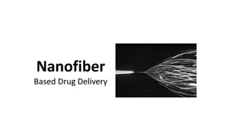
Nanofiber Based Drug Delivery technology
- 2. INTRODUCTION Electrospinning is a process that was originally developed in the early 1930s, but did not receive much attention until recent decades. the increased interest is due to the focus of research on nanotechnology.
- 3. A typical electrospinning process involves: Dissolving the drug of interest and a polymer in an appropriate solvent. The solution is then placed in a syringe, and a high voltage is applied. A small amount of the polymer solution is drawn out of the syringe, forming a Taylor cone. Increasing the applied voltage further results in the initiation of a charged fluid jet, which follows a chaotic trajectory of stretching and bending until it reaches the grounded target.
- 5. A stable jet is formed when 1. the charge is increased above a critical voltage 2. there is a balance between the surface tension of the fluid and the repulsive nature of the charge distribution on the surface of the fluid.
- 6. The presence of molecular entanglements in the polymer solution prevents the jet from breaking into droplets (electrospraying), and when combined with the electrical forces results in a whip-like motion of the jet, known as bending instability. This process typically results in the drawing of a virtually endless fiber with a nanometer-sized to micrometer-sized diameter.
- 7. Scanning electron microscopy (SEM) is a typical method to evaluate the nanofibers produced through electrospinning.
- 8. Nanofibers have a very large surface-area-to-volume ratio, as large as 1000 times that of a microfiber. This physical property useful for drug delivery of poorly soluble drug substances.
- 9. Currently, the pharmaceutical industry uses methods such as milling to reduce the particle size of a drug substance, but this is a high-energy method which can lead to stability issues and often cannot produce truly nanosized drug particles. In conventional dry milling, the limit of fineness is reached in the region of 100 μm. In wet bead milling produces further reduction in the particle size, but often not below the micrometer range.
- 10. Electrospinning process : 1. single-step creation of nanosized drug particles 2. low-energy process
- 11. The selection of the polymer also controls the drug-release properties. • immediate-release nanofibers can be created by water-soluble polymers. • enteric-release nanofibers can be created by enteric polymers such as methacrylic acid copolymers. • sustained-release nanofibers can be created by polylactic acid or polyvinyl acetate polymers.
- 12. Although the diameter of electrospun fibers is often characterized using SEM as proof that they are nanosized, it is important to note that the size of the drug particles embedded in the nanosized fiber is significantly smaller than the diameter of the fibers themselves.
- 13. Further utility may be found with electrospinning by embedding it at the end of the chemical synthesis of the drug substance, which would streamline the transfer of material from chemical development to pharmaceutical development. The last step in the chemical synthesis of a drug is usually a purification/precipitation step to create a drug powder. Then the powder is transferred to pharmaceutical development teams, which often mill the powder to a particular particle size and further granulate/process the material into a tablet. This powder handling and processing, which often requires safety controls
- 14. can be avoided if the last step in the chemical synthesis is transformed into a step where the drug and a preferred polymer are in an appropriate solution for electrospinning. Then the drug/polymer solution can be directly electrospun into a final product, thus creating a seamless process from chemical synthesis through creation of an appropriate final pharmaceutical product. This would eliminate powder handling of the drug substance, and possibly reduce variations in the final particle size of the drug substance during manufacturing.
- 15. DISSOLUTION ENHANCEMENT FOR IMMEDIATE- RELEASE DOSAGE FORMS • tablets or capsules which immediately release their drug when orally ingested are the most common dosage form in the pharmaceutical industry. • Creating a product which performs this task is challenging with low-solubility drugs. Inappropriate formulation of low-solubility drugs can result in * slow or limited drug dissolution in the gastrointestinal (GI) fluids * minimal drug being absorbed into systemic circulation.
- 16. Common technique to overcome this low bioavailability issue is to reduce the particle size of the drug substance to increase the dissolution of the drug into the GI fluids. The particle size of the drug is directly related to the rate that it dissolves. Nernst and Brunner diffusion layer model Q is the amount of the drug dissolved S the effectivesurface area of the drug particles surface area of the drug particle is a key physicochemical property which can be used to increase the rate of drug dissolution. The particle size of the drug substance is often used as marker for surface area.
- 17. Electrospinning presents a mechanism to allow further reduction of the particle size. Reduction of the particle size of the drug substance through electrospinning with a rapidly dissolving polymer causes the drug dissolution rate to be very rapid.
- 18. Another advantage of electrospinning as a dissolution-enhancement tool is the ability to control the morphology of the drug substance. By selecting a polymer such as hydroxypropylmethylcellulose acetate succinate (HPMC-AS), which has an amorphous character, one can create an amorphous drug substance through electrospinning. Amorphous drug substances are higher energy state higher solubility higher dissolution rate compared with crystalline materials.
- 19. • In addition to increasing the solubility and dissolution by creating an amorphous drug substance with HPMC-AS, it has also been shown to reduce in vivo precipitation of the drug by maintaining the drug as a supersaturated solution in the GI tract.
- 20. • Spray-drying with the drug substance and HPMC-AS has been performed to create amorphous particles to increase the drug exposure for rapid screening of drugs in preclinical models. • Electrospinning could be used in a similar fashion and it has two additional benefits: first, the ability to create smaller-diameter fibers than the typical particle size of spray-dried material. second, process collects the product onto a grounded surface, which results in very high efficiencies (99%+) and simplified recovery.
- 21. researchers used hydroxypropylmethylcellulose as the electrospinning polymer, and itraconazole as the model drug. On the basis of differential scanning calorimetry, they generated data supporting the conclusion that itraconazole was in the amorphous form, and performed dissolution studies to evaluate the rate of release.
- 22. SUSTAINED-RELEASE ELECTROSPUN FIBERS large range of drug-release profiles which can be obtained by careful selection of the polymer. For release of a drug substance for multiple days to months, a polymer in the biodegradable family, such as polyglycolide, polylactic acid, or polycaprolactone, can be used. A copolymer of ε-caprolactone and ethyl ethylene phosphate was used to sustain the release of human β-nerve growth factor for at least three months . However, selection of the polymer needs to be carefully screened because the polymer drug compatibility has been shown to play a critical role in the distribution of drug within the fiber.
- 23. Polymers which hydrate or swell but are insoluble can also be used to create sustained-release nanofibers. Electrospun segmented polyurethane itraconazole fibers to produce a sustained release of the drug substance. This type of release can be achieved through the use of generally regarded as safe (GRAS) polymers for oral drug delivery such as polyvinylacetate (PVAc) nanofibers. The intrinsic properties of the polymer form the basis for the type of drug release that occurs from the nanofibers.
- 24. LARGE-SCALE MANUFACTURING Although electrospinning is still at an early stage in the pharmaceutical industry, commercial use of nanofibers produced by electrospinning in other industries has already been established. Donaldson Company’s nanofiber filter media production has increased well beyond 10,000 m2/day during the last 20 years.
- 25. The large-scale manufacturing of electrospun nanofibers for pharmaceutical applications can progress at a rapid rate, including the adoption and implementation of appropriate quality control tools for nanofiber production such as automated fiber sizing and mechanical integrity testing.
- 26. Automated Fiber Sizing Full process control of the nanofiber production requires measurement and control of fiber size, fiber size distribution, and quantity of fibers. Therefore, there is a need for a tool that can directly measure mean fiber size and fiber size distribution. Routine sampling and measurement of these properties are achieved using an SEM and a developed analysis methodology.
- 27. Measuring nanofiber diameters usually consists of manually comparing the diameter of fibers in a photomicrograph to a known scale. The process is very time-consuming, and operator consistency and fatigue can reduce the accuracy.
- 28. For drug-delivery applications, fiber diameter and fiber diameter distribution will be important parameters to measure. fiber diameter is one of the variables that can be controlled to tune the rate of fiber dissolution and control drug release.
- 29. An algorithm has been developed at Donaldson Company to overcome the limitations of commercially available software. 1) In the automated fiber sizing process, the SEM image is first cropped to a desired size, and unwanted details are eliminated. 2) The calibration bar from the SEM image is used to set the number of pixels per micrometer. 3) Next, the program converts the image to black and white using a gray scale function. 4) The black (nonfiber) areas are sorted according to size. Starting with the largest black area, a straight line is defined on the border pixels and a diameter is drawn from one pixel across the white area at 90°. 5) The diameter drawing stops when another black pixel is intersected. 6) The process is repeated around the black shape at an interval of approximately every five pixels, and then around each black area in order of size
- 31. All fiber diameter lengths are recorded and a histogram is generated Characterization of fiber size, fiber size distribution, and quantity of fibers is critical to ensure a quality product.
- 32. The mechanical integrity of a nanofiber structure can be important for sustainedrelease drug-delivery applications such as wound dressings, tissue engineering, and regenerative medicine. Knowledge of the strain–stress characteristics is important in understanding the performance of nanofiber composites under dynamic stress such as the gastric compression and peristaltic action of the GI tract. Owing to the small size of fibers and extremely low weight of the layer, traditional measurement methods do not give useful results.
- 33. Mechanical Integrity Testing • The DL Bending Tester was developed to study the strain properties of nanofibers. • First, a sample is secured in the tester and positioned in the optical microscope. • A motor then bends and extends the sample around a cylinder with known diameter. The strain condition approximates plane strain because the sample is thin compared with the relatively large radius of the cylinder. Thus, differences in the strain condition between the upper and lower surfaces of the sample are minimal.
- 37. • A camera mounted on the microscope sends a dynamic image to a monitor that is used to observe the sample throughout the test. • The first sign of relative movement between the components of the composite structure is an indicator of critical strain. • An operator records the relative movements and angles of the first destruction in the nanofiber layer and full destruction of nanofiber layer. Comparison of the angles for different composites gives a measure of integrity of the material.