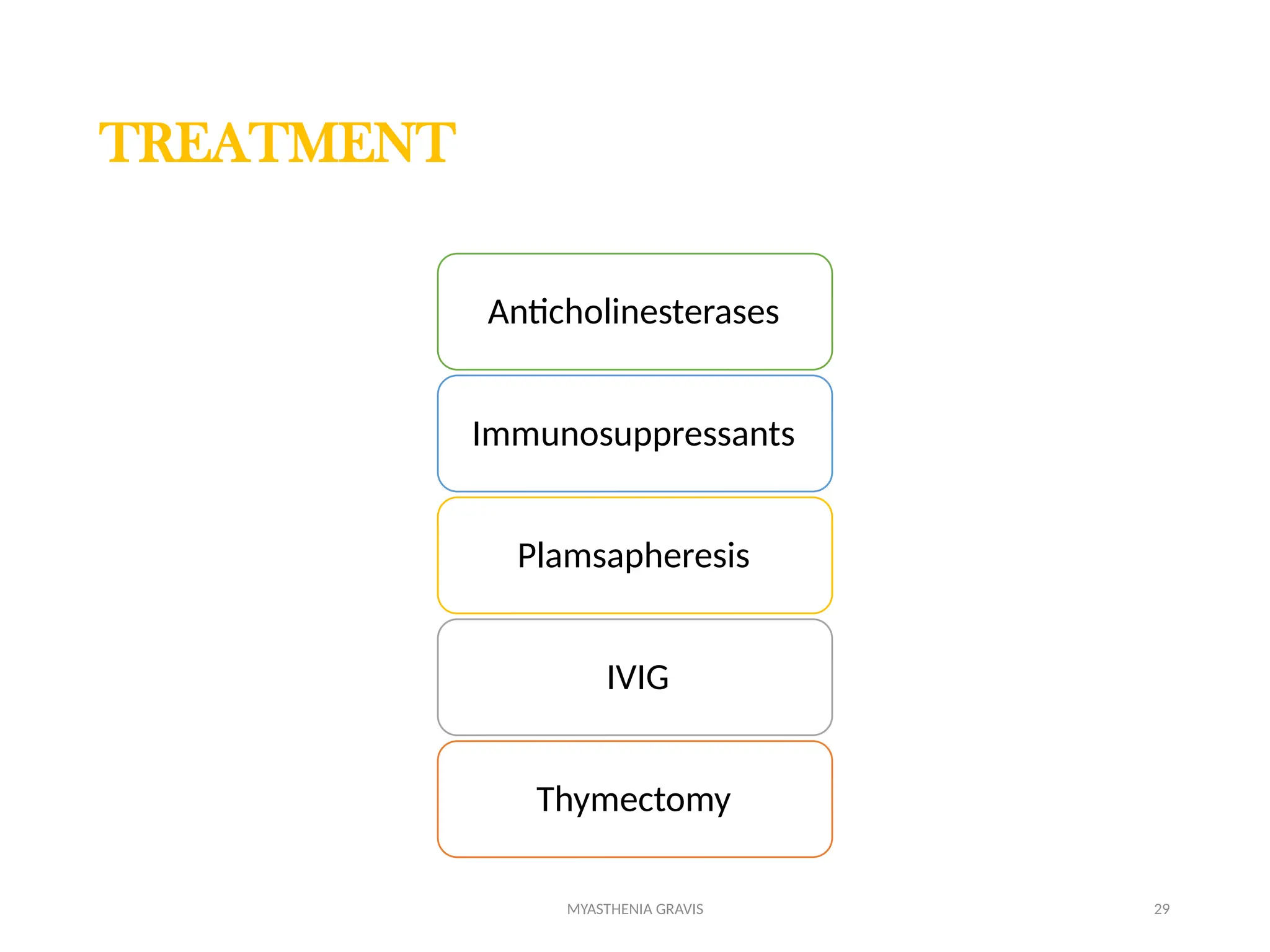
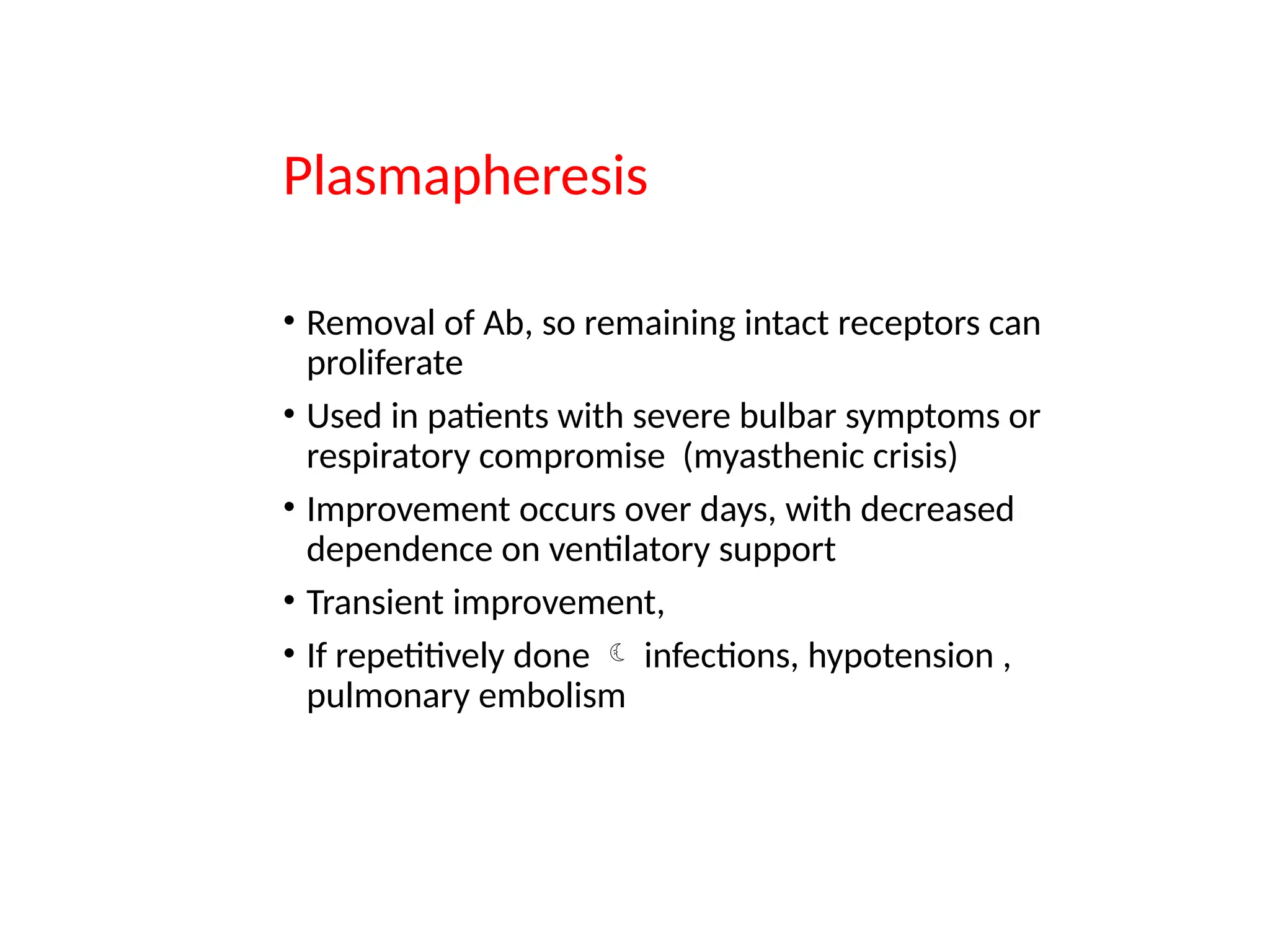
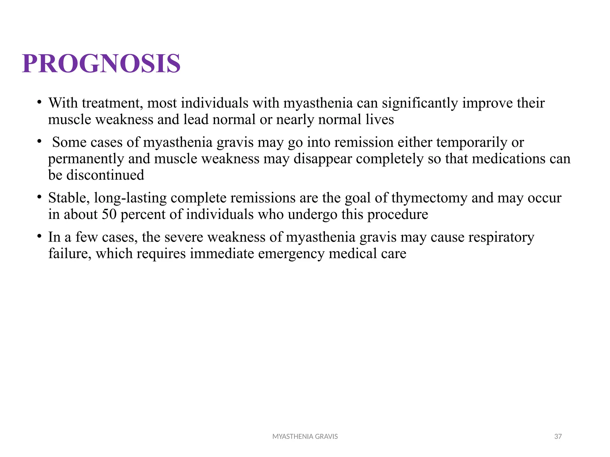
1
Case Discussion
History
A 55 yrs old male presented to ER with
Left eye ptosis for 3 days
He had noticed double vision, which was somewhat corrected by his
glasses, but he had not seen ophthalmologist .He continued his normal
activity, but noticed loss of balance
K/C/0 T2DM/SHTN for 5 yrs. on medication
3
Primary Survey
Airway Patent
Breathing Regular
SpO2: 98%(RA)
RR - 20/min
Circulation
BP:90/60 mmHg
PR: 90/min
IV line secured- IV Fluids started
Sample sent for Routine Investigation
Disability
GCS:E4V5M6
GRBS:90mg/dl
Pupils – 2.5mm reactive to light
Exposure
TEMP:98.3’F
4
Primary Adjuncts
ABG on 6L of O2
pH -7.32 mmHg, PCO2 - 48.5 mmHg, HCO3 - 23.5 mmHg, PO2 – 76
mmHg,
ECG :Sinus tachycardia
Bedside 2D echo
•IVC collapsible.
•No RA/RV dilatation.
•Ejection fraction of 60%
•No Left ventricular Hypertrophy.
•No regional wall motion abnormality
Chest X - Ray
5
G/E: Conscious /oriented afebrile
No pallor/Icterus/Clubbing/Pedal Edema
S/E
CVS : S1S2 heard, no murmurs.
Respiratory system :
Bilateral equal air entry
Abdomen : Soft, Bowel sounds present.
CNS: GCS :15/15
Neurological examination
1)Higher mental status Examination: Patient oriented (Time, Place & Person), Alert,
good memory ,
speaking with understable language , No emotional disturbances noted
2)Cranial Nerve Examination
Ptosis and dysphagia (+)
All other cranial nerve intact
3)Motor Function :
Power: Right and Left upper and lower limb no weakness elicited
Tone: normal
Muscle coordination : Normal
4)Sensory Function :Intact
5)Cerebellar Function : No abnormality noted
6)Reflex - Normal
7
MYASTHENIA GRAVIS
8
Overview
Pathophysiology
Clinical presentation
Signs and symptoms
Diagnosis
Treatment and management
Prognosis
9
OVERVIEW
• The name myasthenia gravis, which is Latin and Greek in origin, literally
means "grave muscle weakness“
• Antibody-mediated autoimmune disease of the neuromuscular
junction
• The hallmark of myasthenia gravis is muscle weakness that increases
during periods of activity and improves after periods of rest
10
• Certain muscles such as those that control eye and eyelid movement, facial
expression, chewing, talking, and swallowing are often, but not always,
involved in the disorder
• The muscles that control breathing and neck and limb movements may also be
affected
• Myasthenia gravis occurs in both sexes
• It most commonly affects women under 40 and Men from the age of 50 to 70
years of age, but it has been known to occur at any age .
• Myasthenic crisis a severe generalized quadriparesis or life-threatening
respiratory muscle weakness, occurs in about 15 to 20% of patients at least once
in their life. Once respiratory insufficiency begins, respiratory failure may occur
rapidly
11
The Thymus in Myasthenia
Gravis
10% of patients with myasthenia gravis have a thymic
tumor and 70% have hyperplastic changes that indicate an
active immune response
The thymus contains myoid cells helps in developing
mature T lymphocytes
In thymic abnormalities these cells produces the immune-
mediated T lymphocytes (antibodies) which attacks the
AChR in myasthenia gravis.
12
However, it is still uncertain whether the role of the thymus in the
pathogenesis of myasthenia gravis is primary or secondary.
Most thymic tumors in patients with myasthenia gravis are benign,
well-differentiated and encapsulated, and can be removed completely at
surgery.
Pathophysiology
1
3
14
15
At a Glance
16
Eyes
• In about two-thirds of individuals, the initial symptom of MG is related to
the weakness of muscles around the eye
• There may be eyelid drooping (ptosis due to weakness of levator palpebrae
superioris) and double vision (diplopia due to weakness of the extraocular
muscles)
• The term "ocular myasthenia gravis" describes a subtype of MG where
muscle weakness is confined to the eyes, i.e. extra ocular muscles, levator
palpebrae superioris and orbicularis oculi. Typically, this subtype evolves
into generalized MG, usually after a few years.
17
Eating
• Weakness of the muscles involved in swallowing may lead to swallowing
difficulty (dysphagia).
• Weakness of the muscles that move the jaw (muscles of mastication) may
cause difficulty chewing.
• In individuals with MG, chewing tends to become more tiring when chewing
tough, fibrous foods. Difficulty in swallowing, chewing and speaking is the
first symptom in about one-sixth of individuals.
Voice
• Weakness of the muscles involved in speaking may lead to dysarthria
and hypophonia. Speech may be slow and slurred or have a nasal quality.
In some cases a singing hobby or profession must be abandoned.
18
Head and neck
• Due to weakness of the muscles of facial expression and muscles of
mastication, there may be facial weakness and a snarling appearance when
attempting to smile.
• Together with drooping eyelids, facial weakness may make the individual
appear sleepy or sad. There may be difficulty in holding the head upright.
19
Respiratory muscles
• The muscles that control breathing (dyspnea) and limb movements can also be
affected, but rarely do these present as the first symptoms of MG, and they develop
over months to years.
• In a myasthenic crisis, a paralysis of the respiratory muscles occurs, necessitating
assisted ventilation to sustain life.
• Crisis may be triggered by various biological stressors such as infection, fever, an
adverse reaction to medication, or emotional stress.
MYASTHENIA GRAVIS 20
MYASTHENIA GRAVIS FOUNDATION OF AMERICA
CLINICAL CLASSIFICATION
Class Description
I
Any eye muscle weakness, possible ptosis, no other evidence of muscle
weakness elsewhere
II Eye muscle weakness of any severity, mild weakness of other muscles
IIa Predominantly limb or axial muscles
IIb Predominantly bulbar and/or respiratory muscles
III Eye muscle weakness of any severity, moderate weakness of other muscles
IIIa Predominantly limb or axial muscles
IIIb Predominantly bulbar and/or respiratory muscles
IV Eye muscle weakness of any severity, severe weakness of other muscles
IVa Predominantly limb or axial muscles
IVb Predominantly bulbar and/or respiratory muscles
V Intubation needed to maintain airway
MYASTHENIA GRAVIS 21
DIAGNOSIS
History
Physical exam
Clinical investigations
Tensilon test
Serum antibody
(x-AChR & X-MUSK)
Ice test
EMG
(RNS & SFEMG)
Imaging
( X ray,CT,MRI)
Pulmonary function test
MYASTHENIA GRAVIS 22
History
• The first steps of diagnosing myasthenia gravis include a review of the
individual's medical history, and physical and neurological examinations.
• A characteristic of MG is that patients have weakness that comes on with
activity and improves following rest.
Physical examination
• During a physical examination to check for MG, a doctor might ask the
potentially affected person to look at a fixed point for 30 seconds and to relax
the muscles of their forehead.
• This is done because a person with MG and ptosis of their eyes might be
involuntarily using their forehead muscles to compensate for the weakness in
their eyelids.
• The clinical examiner might also try to elicit the "curtain sign" in a patient by
holding one of the person's eyes open, which in the case of MG will lead the
other eye to close.
MYASTHENIA GRAVIS 23
The Edrophonium Chloride (Tensilon®) Test
• Weakness caused by abnormal neuromuscular transmission characteristically
improves after intravenous administration of edrophonium chloride, commonly
referred to as the Tensilon®
Test
• Edrophonium chloride, is injected intravenously, 2 mg at a time to a total
of 10 mg. 30 seconds after injection, facial muscle weakness and
ptosis should resolve for about 5 minutes. This immediate
improvement in muscle strength after administration of this agent
represents a positive test and usually confirms the diagnosis
MYASTHENIA GRAVIS 24
Ice test
• Applying ice for two to five minutes to the muscles reportedly has a sensitivity
and specificity of 76.9% and 98.3%, respectively, for the identification of MG.
• Acetylcholinesterase is thought to be inhibited at the lower temperature, and
this is the basis for this diagnostic test.
• This generally is performed on the eyelids when a ptosis is present and is
deemed positive if there is a ≥2mm raise in the eyelid after the ice is removed.
MYASTHENIA GRAVIS 25
Serum antibody testing
• A special blood test can detect the presence of acetylcholine receptor antibodies.
Most patients with myasthenia gravis have abnormally elevated levels of these
antibodies.
• Recently, a second antibody—called the anti-MuSK antibody—has been found in
about 30 to 40 percent of individuals with myasthenia gravis who do not have
acetylcholine receptor antibodies.
• Acetylcholine Receptor Antibody— a blood test for the abnormal antibodies
can be performed to see if they are present. Approximately 85% of MG
patients have this antibody and, when detected with an elevated concentration
the AChR antibody test is strongly indicative of MG.
• Anti-MuSK Antibody testing----a blood test for the remaining 15% of MG
patients who have tested negative for the acetylcholine antibody. These
patients have seronegative (SN) MG. About 40% of patients with SNMG test
positive for the anti-MuSK antibody.
MYASTHENIA GRAVIS 26
Electromyography
Single fibre electromyography(SFEMG)
• SFEMG measures the electrical potential of muscle cells when single
muscle fibre are stimulated by electrical impulse. Muscle fibres in
myasthenia gravis, as well as other neuromuscular disorders, do not
respond to electrical stimulation compared to muscles from normal
individuals.
Repetitive Nerve Stimulation (RNS)
• This test records weakening muscle responses when the nerves are
repetitively stimulated by small pulses of electricity.
• The amplitude of the compound muscle action potential (CMAP) elicited
by repetitive nerve stimulation is normal or only slightly reduced in
normal ones.
• The amplitude of the fourth or fifth response to a train of low frequency
nerve stimuli falls at least 10% from the initial value in myasthenic
patients. This decrementing response to RNS is seen more often in
proximal muscles, such as the facial muscles, biceps, deltoid, and
trapezius than in hand muscles. A significant decrement to RNS in either
a hand or shoulder muscle is found in about 60% of patients with
myasthenia gravis.
MYASTHENIA GRAVIS 27
Diagnostic imaging
• A chest X-ray may identify widening of the mediastinum suggestive of
thymoma.
• Diagnostic imaging of the chest, using computed tomography (CT) or
magnetic resonance imaging (MRI), may be used to identify the presence of a
thymoma.
• MRI Brain /orbit may also be performed to exclude compressive lesions of
the cranial nerves and ocular muscles
2/19/2017
MYASTHENIA GRAVIS 28
Pulmonary function test
• Pulmonary function testing, which measures breathing strength, helps
to predict whether respiration may fail and lead to a myasthenic crisis.
• The forced vital capacity may be monitored at intervals to detect
increasing muscular weakness.
• Acutely, negative inspiratory force may be used to determine
adequacy of ventilation; it is performed on those individuals with MG
MYASTHENIA GRAVIS 29
TREATMENT
Anticholinesterases
Immunosuppressants
Plamsapheresis
IVIG
Thymectomy
MYASTHENIA GRAVIS 30
Cholinesterase Inhibitors
• ChE inhibitors retard the enzymatic hydrolysis of ACh at cholinergic synapses,
so that ACh accumulates at the neuromuscular junction and its effect is
prolonged.
• ChE inhibitors cause considerable improvement in some patients and little to
none in others. Strength rarely returns to normal. Pyridostigmine bromide
(Mestinon) and neostigmine bromide (Prostigmin) are the most commonly used
ChE inhibitors
• Neostigmine: 15 mg qid every 3 hrs up to 60 mg p/o
• Pyridostigmine:30 mg tds every 3 hrs upto 120 mg p/o
MYASTHENIA GRAVIS 31
• Adverse effects of ChE inhibitors may result from ACh accumulation at
muscarinic receptors on muscle fibre,Therefore Gastrointestinal complaints
are common; loose stools, nausea, vomiting, abdominal cramps, and diarrhea.
Increased bronchial and oral secretions are a serious problem in patients with
swallowing or respiratory insufficiency.
• Cholinergic crisis is muscular weakness that can result when the dose of
anticholinesterase drugs (eg, neostigmine, pyridostigmine) is too high. A mild
crisis may be difficult to differentiate from worsening myasthenia. Severe
cholinergic crisis can usually be differentiated because it, unlike myasthenia
gravis, results in increased lacrimation and salivation, tachycardia, and
diarrhoea.
Immunosuppression
• Prevents or attenuates the destruction of Ach receptors at
motor end-plate
• Corticosteroids improve condition of most myasthenics
{ most commonly used but a/w various side effects)
• Prednisone 15-25mg/kg  increased slowly to 50-60mg/kg
• Azathioprine(2.5-3.5mg/kg)- if resistant to steroids (a/w
BM suppression, hepatotoxicity, nephrotoxicity)
• Cyclosporine(4-5mg/kg/day) was used in MG patients and
had less SE than Azathioprine( nephrotoxic)
• Mycophenolate Mofetil(1-1.5mg/kg).
Plasmapheresis
• Removal of Ab, so remaining intact receptors can
proliferate
• Used in patients with severe bulbar symptoms or
respiratory compromise (myasthenic crisis)
• Improvement occurs over days, with decreased
dependence on ventilatory support
• Transient improvement,
• If repetitively done  infections, hypotension ,
pulmonary embolism
MYASTHENIA GRAVIS 34
Intravenous Immune Globulin (IVIG)
• Several groups have reported a favorable response to high-dose (2 grams/kg infused
over 2 to 5 days) IVIG. Possible mechanisms of action include down-regulation of
antibodies directed against AChR and the introduction of anti-idiotypic antibodies
• Improvement occurs in 50 to 100% of patients, usually beginning within 1 week and
lasting for several weeks or months.
• The mechanism of action is not known but is probably non-specific down regulation of
antibody production.
MYASTHENIA GRAVIS 35
Thymectomy
• Thymectomy is recommended by many physicians for most patients with
myasthenia gravis. Most reports do not correlate the severity of weakness
before surgery and the timing or degree of improvement after thymectomy.
• The maximal favorable response generally occurs 2 to 5 years after surgery.
However, the response is relatively unpredictable and significant impairment
may continue for months or years after surgery.
• Sometimes, improvement is only appreciated in retrospect. The best
responses to thymectomy are in young people early in the course of their
disease, but improvement can occur even after 30 years of symptoms.
Patients with disease onset after the age of 60 rarely show substantial
improvement from thymectomy.
MYASTHENIA GRAVIS 36
Myasthenia
weakness
exaggerated
Quinine
Magnesium
Pencillamine
Erythromycin
Azithromycin
Ciprofolxacin
Levofloxacin
Ofloxacin
Gatifloxacin
Non-depolarising
muscle relaxants
(pancuronium)
Propanol
Atenolol
Local anaesthetic
(lignocaine)
Streptomycin
MYASTHENIA GRAVIS 37
PROGNOSIS
• With treatment, most individuals with myasthenia can significantly improve their
muscle weakness and lead normal or nearly normal lives
• Some cases of myasthenia gravis may go into remission either temporarily or
permanently and muscle weakness may disappear completely so that medications can
be discontinued
• Stable, long-lasting complete remissions are the goal of thymectomy and may occur
in about 50 percent of individuals who undergo this procedure
• In a few cases, the severe weakness of myasthenia gravis may cause respiratory
failure, which requires immediate emergency medical care
MYASTHENIA GRAVIS 38
Myasthenic Crisis Cholinergic Crisis
Under medication Overmedication
Increased HR/BP/RR Decreased BP
Bowel and bladder incontinence Abd cramps,N/V, Diarrhea
Decreased urine output Blurred vision
Absent cough and swallow reflex Pallor, Facial muscle twitching
May need mechanical ventilation Constriction of pupils
Temporary improvement of symptoms with
administration of Tensilon
Tensilon has no effect Symptoms improve with
administration of anticholinergics (Atropine)
MYASTHENIA GRAVIS 39
MYASTHENIA GRAVIS 40
References:
1.Werner Hoch, John McConville,Sigrun Helms,John Newsom,Arthur Melms &
Angela Vincent(2001): Auto-antibodies to the receptor tyrosine kinase MuSK in
patients with myasthenia gravis without acetylcholine receptor antibodies.
2.Curtis Wells Dewey, Cleta Sue Bailey, G. Diane Shelton, Philip H. Kass, and G. H.
Cardinet(1995):Clinical Forms of Acquired Myasthenia Gravis in Dogs.
3.Jon M. Linderstrom, PhD, Marjorie E. Seybold, MD,(2002):Antibody to
acetylcholine receptor in myasthenia gravis
4.The Merck Veterinary Manual(9th
edition)
41
THANK YOU

Myasthenia Gravis case presentation pptn

  • 1.
  • 2.
    History A 55 yrsold male presented to ER with Left eye ptosis for 3 days He had noticed double vision, which was somewhat corrected by his glasses, but he had not seen ophthalmologist .He continued his normal activity, but noticed loss of balance K/C/0 T2DM/SHTN for 5 yrs. on medication
  • 3.
    3 Primary Survey Airway Patent BreathingRegular SpO2: 98%(RA) RR - 20/min Circulation BP:90/60 mmHg PR: 90/min IV line secured- IV Fluids started Sample sent for Routine Investigation Disability GCS:E4V5M6 GRBS:90mg/dl Pupils – 2.5mm reactive to light Exposure TEMP:98.3’F
  • 4.
    4 Primary Adjuncts ABG on6L of O2 pH -7.32 mmHg, PCO2 - 48.5 mmHg, HCO3 - 23.5 mmHg, PO2 – 76 mmHg, ECG :Sinus tachycardia Bedside 2D echo •IVC collapsible. •No RA/RV dilatation. •Ejection fraction of 60% •No Left ventricular Hypertrophy. •No regional wall motion abnormality Chest X - Ray
  • 5.
    5 G/E: Conscious /orientedafebrile No pallor/Icterus/Clubbing/Pedal Edema S/E CVS : S1S2 heard, no murmurs. Respiratory system : Bilateral equal air entry Abdomen : Soft, Bowel sounds present. CNS: GCS :15/15
  • 6.
    Neurological examination 1)Higher mentalstatus Examination: Patient oriented (Time, Place & Person), Alert, good memory , speaking with understable language , No emotional disturbances noted 2)Cranial Nerve Examination Ptosis and dysphagia (+) All other cranial nerve intact 3)Motor Function : Power: Right and Left upper and lower limb no weakness elicited Tone: normal Muscle coordination : Normal 4)Sensory Function :Intact 5)Cerebellar Function : No abnormality noted 6)Reflex - Normal
  • 7.
  • 8.
    8 Overview Pathophysiology Clinical presentation Signs andsymptoms Diagnosis Treatment and management Prognosis
  • 9.
    9 OVERVIEW • The namemyasthenia gravis, which is Latin and Greek in origin, literally means "grave muscle weakness“ • Antibody-mediated autoimmune disease of the neuromuscular junction • The hallmark of myasthenia gravis is muscle weakness that increases during periods of activity and improves after periods of rest
  • 10.
    10 • Certain musclessuch as those that control eye and eyelid movement, facial expression, chewing, talking, and swallowing are often, but not always, involved in the disorder • The muscles that control breathing and neck and limb movements may also be affected • Myasthenia gravis occurs in both sexes • It most commonly affects women under 40 and Men from the age of 50 to 70 years of age, but it has been known to occur at any age . • Myasthenic crisis a severe generalized quadriparesis or life-threatening respiratory muscle weakness, occurs in about 15 to 20% of patients at least once in their life. Once respiratory insufficiency begins, respiratory failure may occur rapidly
  • 11.
    11 The Thymus inMyasthenia Gravis 10% of patients with myasthenia gravis have a thymic tumor and 70% have hyperplastic changes that indicate an active immune response The thymus contains myoid cells helps in developing mature T lymphocytes In thymic abnormalities these cells produces the immune- mediated T lymphocytes (antibodies) which attacks the AChR in myasthenia gravis.
  • 12.
    12 However, it isstill uncertain whether the role of the thymus in the pathogenesis of myasthenia gravis is primary or secondary. Most thymic tumors in patients with myasthenia gravis are benign, well-differentiated and encapsulated, and can be removed completely at surgery.
  • 13.
  • 14.
  • 15.
  • 16.
    16 Eyes • In abouttwo-thirds of individuals, the initial symptom of MG is related to the weakness of muscles around the eye • There may be eyelid drooping (ptosis due to weakness of levator palpebrae superioris) and double vision (diplopia due to weakness of the extraocular muscles) • The term "ocular myasthenia gravis" describes a subtype of MG where muscle weakness is confined to the eyes, i.e. extra ocular muscles, levator palpebrae superioris and orbicularis oculi. Typically, this subtype evolves into generalized MG, usually after a few years.
  • 17.
    17 Eating • Weakness ofthe muscles involved in swallowing may lead to swallowing difficulty (dysphagia). • Weakness of the muscles that move the jaw (muscles of mastication) may cause difficulty chewing. • In individuals with MG, chewing tends to become more tiring when chewing tough, fibrous foods. Difficulty in swallowing, chewing and speaking is the first symptom in about one-sixth of individuals. Voice • Weakness of the muscles involved in speaking may lead to dysarthria and hypophonia. Speech may be slow and slurred or have a nasal quality. In some cases a singing hobby or profession must be abandoned.
  • 18.
    18 Head and neck •Due to weakness of the muscles of facial expression and muscles of mastication, there may be facial weakness and a snarling appearance when attempting to smile. • Together with drooping eyelids, facial weakness may make the individual appear sleepy or sad. There may be difficulty in holding the head upright.
  • 19.
    19 Respiratory muscles • Themuscles that control breathing (dyspnea) and limb movements can also be affected, but rarely do these present as the first symptoms of MG, and they develop over months to years. • In a myasthenic crisis, a paralysis of the respiratory muscles occurs, necessitating assisted ventilation to sustain life. • Crisis may be triggered by various biological stressors such as infection, fever, an adverse reaction to medication, or emotional stress.
  • 20.
    MYASTHENIA GRAVIS 20 MYASTHENIAGRAVIS FOUNDATION OF AMERICA CLINICAL CLASSIFICATION Class Description I Any eye muscle weakness, possible ptosis, no other evidence of muscle weakness elsewhere II Eye muscle weakness of any severity, mild weakness of other muscles IIa Predominantly limb or axial muscles IIb Predominantly bulbar and/or respiratory muscles III Eye muscle weakness of any severity, moderate weakness of other muscles IIIa Predominantly limb or axial muscles IIIb Predominantly bulbar and/or respiratory muscles IV Eye muscle weakness of any severity, severe weakness of other muscles IVa Predominantly limb or axial muscles IVb Predominantly bulbar and/or respiratory muscles V Intubation needed to maintain airway
  • 21.
    MYASTHENIA GRAVIS 21 DIAGNOSIS History Physicalexam Clinical investigations Tensilon test Serum antibody (x-AChR & X-MUSK) Ice test EMG (RNS & SFEMG) Imaging ( X ray,CT,MRI) Pulmonary function test
  • 22.
    MYASTHENIA GRAVIS 22 History •The first steps of diagnosing myasthenia gravis include a review of the individual's medical history, and physical and neurological examinations. • A characteristic of MG is that patients have weakness that comes on with activity and improves following rest. Physical examination • During a physical examination to check for MG, a doctor might ask the potentially affected person to look at a fixed point for 30 seconds and to relax the muscles of their forehead. • This is done because a person with MG and ptosis of their eyes might be involuntarily using their forehead muscles to compensate for the weakness in their eyelids. • The clinical examiner might also try to elicit the "curtain sign" in a patient by holding one of the person's eyes open, which in the case of MG will lead the other eye to close.
  • 23.
    MYASTHENIA GRAVIS 23 TheEdrophonium Chloride (Tensilon®) Test • Weakness caused by abnormal neuromuscular transmission characteristically improves after intravenous administration of edrophonium chloride, commonly referred to as the Tensilon® Test • Edrophonium chloride, is injected intravenously, 2 mg at a time to a total of 10 mg. 30 seconds after injection, facial muscle weakness and ptosis should resolve for about 5 minutes. This immediate improvement in muscle strength after administration of this agent represents a positive test and usually confirms the diagnosis
  • 24.
    MYASTHENIA GRAVIS 24 Icetest • Applying ice for two to five minutes to the muscles reportedly has a sensitivity and specificity of 76.9% and 98.3%, respectively, for the identification of MG. • Acetylcholinesterase is thought to be inhibited at the lower temperature, and this is the basis for this diagnostic test. • This generally is performed on the eyelids when a ptosis is present and is deemed positive if there is a ≥2mm raise in the eyelid after the ice is removed.
  • 25.
    MYASTHENIA GRAVIS 25 Serumantibody testing • A special blood test can detect the presence of acetylcholine receptor antibodies. Most patients with myasthenia gravis have abnormally elevated levels of these antibodies. • Recently, a second antibody—called the anti-MuSK antibody—has been found in about 30 to 40 percent of individuals with myasthenia gravis who do not have acetylcholine receptor antibodies. • Acetylcholine Receptor Antibody— a blood test for the abnormal antibodies can be performed to see if they are present. Approximately 85% of MG patients have this antibody and, when detected with an elevated concentration the AChR antibody test is strongly indicative of MG. • Anti-MuSK Antibody testing----a blood test for the remaining 15% of MG patients who have tested negative for the acetylcholine antibody. These patients have seronegative (SN) MG. About 40% of patients with SNMG test positive for the anti-MuSK antibody.
  • 26.
    MYASTHENIA GRAVIS 26 Electromyography Singlefibre electromyography(SFEMG) • SFEMG measures the electrical potential of muscle cells when single muscle fibre are stimulated by electrical impulse. Muscle fibres in myasthenia gravis, as well as other neuromuscular disorders, do not respond to electrical stimulation compared to muscles from normal individuals. Repetitive Nerve Stimulation (RNS) • This test records weakening muscle responses when the nerves are repetitively stimulated by small pulses of electricity. • The amplitude of the compound muscle action potential (CMAP) elicited by repetitive nerve stimulation is normal or only slightly reduced in normal ones. • The amplitude of the fourth or fifth response to a train of low frequency nerve stimuli falls at least 10% from the initial value in myasthenic patients. This decrementing response to RNS is seen more often in proximal muscles, such as the facial muscles, biceps, deltoid, and trapezius than in hand muscles. A significant decrement to RNS in either a hand or shoulder muscle is found in about 60% of patients with myasthenia gravis.
  • 27.
    MYASTHENIA GRAVIS 27 Diagnosticimaging • A chest X-ray may identify widening of the mediastinum suggestive of thymoma. • Diagnostic imaging of the chest, using computed tomography (CT) or magnetic resonance imaging (MRI), may be used to identify the presence of a thymoma. • MRI Brain /orbit may also be performed to exclude compressive lesions of the cranial nerves and ocular muscles 2/19/2017
  • 28.
    MYASTHENIA GRAVIS 28 Pulmonaryfunction test • Pulmonary function testing, which measures breathing strength, helps to predict whether respiration may fail and lead to a myasthenic crisis. • The forced vital capacity may be monitored at intervals to detect increasing muscular weakness. • Acutely, negative inspiratory force may be used to determine adequacy of ventilation; it is performed on those individuals with MG
  • 29.
  • 30.
    MYASTHENIA GRAVIS 30 CholinesteraseInhibitors • ChE inhibitors retard the enzymatic hydrolysis of ACh at cholinergic synapses, so that ACh accumulates at the neuromuscular junction and its effect is prolonged. • ChE inhibitors cause considerable improvement in some patients and little to none in others. Strength rarely returns to normal. Pyridostigmine bromide (Mestinon) and neostigmine bromide (Prostigmin) are the most commonly used ChE inhibitors • Neostigmine: 15 mg qid every 3 hrs up to 60 mg p/o • Pyridostigmine:30 mg tds every 3 hrs upto 120 mg p/o
  • 31.
    MYASTHENIA GRAVIS 31 •Adverse effects of ChE inhibitors may result from ACh accumulation at muscarinic receptors on muscle fibre,Therefore Gastrointestinal complaints are common; loose stools, nausea, vomiting, abdominal cramps, and diarrhea. Increased bronchial and oral secretions are a serious problem in patients with swallowing or respiratory insufficiency. • Cholinergic crisis is muscular weakness that can result when the dose of anticholinesterase drugs (eg, neostigmine, pyridostigmine) is too high. A mild crisis may be difficult to differentiate from worsening myasthenia. Severe cholinergic crisis can usually be differentiated because it, unlike myasthenia gravis, results in increased lacrimation and salivation, tachycardia, and diarrhoea.
  • 32.
    Immunosuppression • Prevents orattenuates the destruction of Ach receptors at motor end-plate • Corticosteroids improve condition of most myasthenics { most commonly used but a/w various side effects) • Prednisone 15-25mg/kg  increased slowly to 50-60mg/kg • Azathioprine(2.5-3.5mg/kg)- if resistant to steroids (a/w BM suppression, hepatotoxicity, nephrotoxicity) • Cyclosporine(4-5mg/kg/day) was used in MG patients and had less SE than Azathioprine( nephrotoxic) • Mycophenolate Mofetil(1-1.5mg/kg).
  • 33.
    Plasmapheresis • Removal ofAb, so remaining intact receptors can proliferate • Used in patients with severe bulbar symptoms or respiratory compromise (myasthenic crisis) • Improvement occurs over days, with decreased dependence on ventilatory support • Transient improvement, • If repetitively done  infections, hypotension , pulmonary embolism
  • 34.
    MYASTHENIA GRAVIS 34 IntravenousImmune Globulin (IVIG) • Several groups have reported a favorable response to high-dose (2 grams/kg infused over 2 to 5 days) IVIG. Possible mechanisms of action include down-regulation of antibodies directed against AChR and the introduction of anti-idiotypic antibodies • Improvement occurs in 50 to 100% of patients, usually beginning within 1 week and lasting for several weeks or months. • The mechanism of action is not known but is probably non-specific down regulation of antibody production.
  • 35.
    MYASTHENIA GRAVIS 35 Thymectomy •Thymectomy is recommended by many physicians for most patients with myasthenia gravis. Most reports do not correlate the severity of weakness before surgery and the timing or degree of improvement after thymectomy. • The maximal favorable response generally occurs 2 to 5 years after surgery. However, the response is relatively unpredictable and significant impairment may continue for months or years after surgery. • Sometimes, improvement is only appreciated in retrospect. The best responses to thymectomy are in young people early in the course of their disease, but improvement can occur even after 30 years of symptoms. Patients with disease onset after the age of 60 rarely show substantial improvement from thymectomy.
  • 36.
  • 37.
    MYASTHENIA GRAVIS 37 PROGNOSIS •With treatment, most individuals with myasthenia can significantly improve their muscle weakness and lead normal or nearly normal lives • Some cases of myasthenia gravis may go into remission either temporarily or permanently and muscle weakness may disappear completely so that medications can be discontinued • Stable, long-lasting complete remissions are the goal of thymectomy and may occur in about 50 percent of individuals who undergo this procedure • In a few cases, the severe weakness of myasthenia gravis may cause respiratory failure, which requires immediate emergency medical care
  • 38.
    MYASTHENIA GRAVIS 38 MyasthenicCrisis Cholinergic Crisis Under medication Overmedication Increased HR/BP/RR Decreased BP Bowel and bladder incontinence Abd cramps,N/V, Diarrhea Decreased urine output Blurred vision Absent cough and swallow reflex Pallor, Facial muscle twitching May need mechanical ventilation Constriction of pupils Temporary improvement of symptoms with administration of Tensilon Tensilon has no effect Symptoms improve with administration of anticholinergics (Atropine)
  • 39.
  • 40.
    MYASTHENIA GRAVIS 40 References: 1.WernerHoch, John McConville,Sigrun Helms,John Newsom,Arthur Melms & Angela Vincent(2001): Auto-antibodies to the receptor tyrosine kinase MuSK in patients with myasthenia gravis without acetylcholine receptor antibodies. 2.Curtis Wells Dewey, Cleta Sue Bailey, G. Diane Shelton, Philip H. Kass, and G. H. Cardinet(1995):Clinical Forms of Acquired Myasthenia Gravis in Dogs. 3.Jon M. Linderstrom, PhD, Marjorie E. Seybold, MD,(2002):Antibody to acetylcholine receptor in myasthenia gravis 4.The Merck Veterinary Manual(9th edition)
  • 41.

Editor's Notes

  • #11  Thymic abnormalities are clearly associated with myasthenia gravis but the nature of the association is uncertain. 10% of patients with myasthenia gravis have a thymic tumor and 70% have hyperplastic changes that indicate an active immune response. The thymus is the central organ for immunological self-tolerance, it is reasonable to suspect that thymic abnormalities cause the breakdown in tolerance that causes an immune-mediated attack on AChR in myasthenia gravis. The thymus contains all the necessary elements for the pathogenesis of myasthenia gravis: myoid cells that express the AChR antigen, antigen presenting cells, and immunocompetent T-cells.