1. Mutation Detection
Using the AdvanCE™ FS CE System
a d v a n c e d a n a ly t i c a l
A high throughput cApill Ary electrophoresis system And reAgent kit for AutomAted,
rApid And sensitive detection of induced And nAturAl point mutAtions.
Accurately detecting natural or induced point mutation can be a time consuming task when using
the traditional slab gel method, usually requiring specialized know-how. A process developed for
use with the AdvanCE™ FS instrument platform offers significant advantages over the traditional
methods by streamlining and automating the process. Unique features of this process include
no clean up step, no need for labeled primers, simple sample handling and rapid turn-around
times to results.
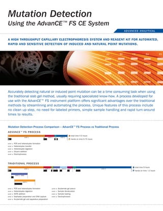
Mutation detection Process comparison – AdvanCE™ FS Process vs traditional Process
advance™ FS ProceS S
total time 4.5 hours
hands on time 0.75 hours
SteP 1: PCR and heteroduplex formation
SteP 2: Heteroduplex transfer
SteP 3: Heteroduplex digestion
SteP 4: Diluent addition
SteP 5: Electrophoresis
traditional ProceSS
total time 9 hours
hands on time >2 hours
SteP 1: PCR and heteroduplex formation SteP 6: Acrylamide gel prerun
SteP 2: Heteroduplex digestion SteP 7: Sample denaturation
SteP 3: EDTA addition SteP 8: Sample loading
SteP 4: Sephadex preparation and DNA capture SteP 9: Electrophoresis
SteP 5: Acrylamide gel and apparatus preparation
2. Mutation Detection Using the AdvanCE™ FS CE System continued
a d v a n c e d a n a ly t i c a l
The streamlined process shaves off a significant amount of time from each step. Compared to
the traditional method, which requires numerous manual steps including manual gel preparations,
plate handling and set up, heteroduplex clean up steps and manual gel loading and subsequent
analysis, the Mutation Detection kit and subsequent separation on the AdvanCE™ FS96 reduces
hands on time of both the sample handling and the analysis.
A comparison of the Post PCR process steps are listed below for each instrument platform.
P o S t P c r P r o c e S S c o M Pa r i S o n
M U tat i o n d e t e c t i o n K i t P r o c e S S traditional ProceSS
1. Add 2µL PCR product to 2µL enzyme 1. Add 20µL Cel I cocktail to PCR products
solution 2. Spin 1 min
2. Spin 10 sec 3. Incubate @45ºC for 15 minutes
3. Incubate @45ºC for 45 minutes 4. Add 5µL of EDTA
4. Add 24µL diluent buffer 5. Purify on Sephadex or EtOH precipitate
5. Place on ice until CE 6. Run gel
F e at U r e S / B e n e F i t S
> no clean up step needed: > Abilityto look at larger fragments 10,000bp:
Eliminates several steps of the traditional process, Exceed size limitation over traditional slab gel methods.
reduces overall time and potential sample loss Improve primer design
> potential to reduce gdnA input amount: > eliminate use of labeled primer sets:
Smaller PCR setup and high sensitivity means less input Saves time and cost for expensive labels. No signal loss
gDNA is required — saves precious DNA over time
> fast electrophoresis run times (30 minutes): > Ability to identify multiple cuts in one gene:
Get more separations done per instrument per day Sensitive intercalating dye allows easy detection of
> minimal labor (no pouring gels or cleaning plates): multiple fragment cut sites
The automated process significantly reduces hands-on > Analytical software for fragment sizing and
time handling fragile glass plates and toxic chemicals concentration:
> Analyze up to 16 gene copies: Easy to use data analysis software eliminates manual
Maximize throughput by maximizing organism pooling screening of gel pictures. Aids in displaying and sizing
cut fragments
Advanced Analytical Technologies, Inc.
2711 South Loop Drive, Suite 4150 Phone: +1-515-296-6600
Ames, IA 50010 USA Fax: +1-515-294-7141
www.aati-us.com E-mail: sales-fs@aati-us.com