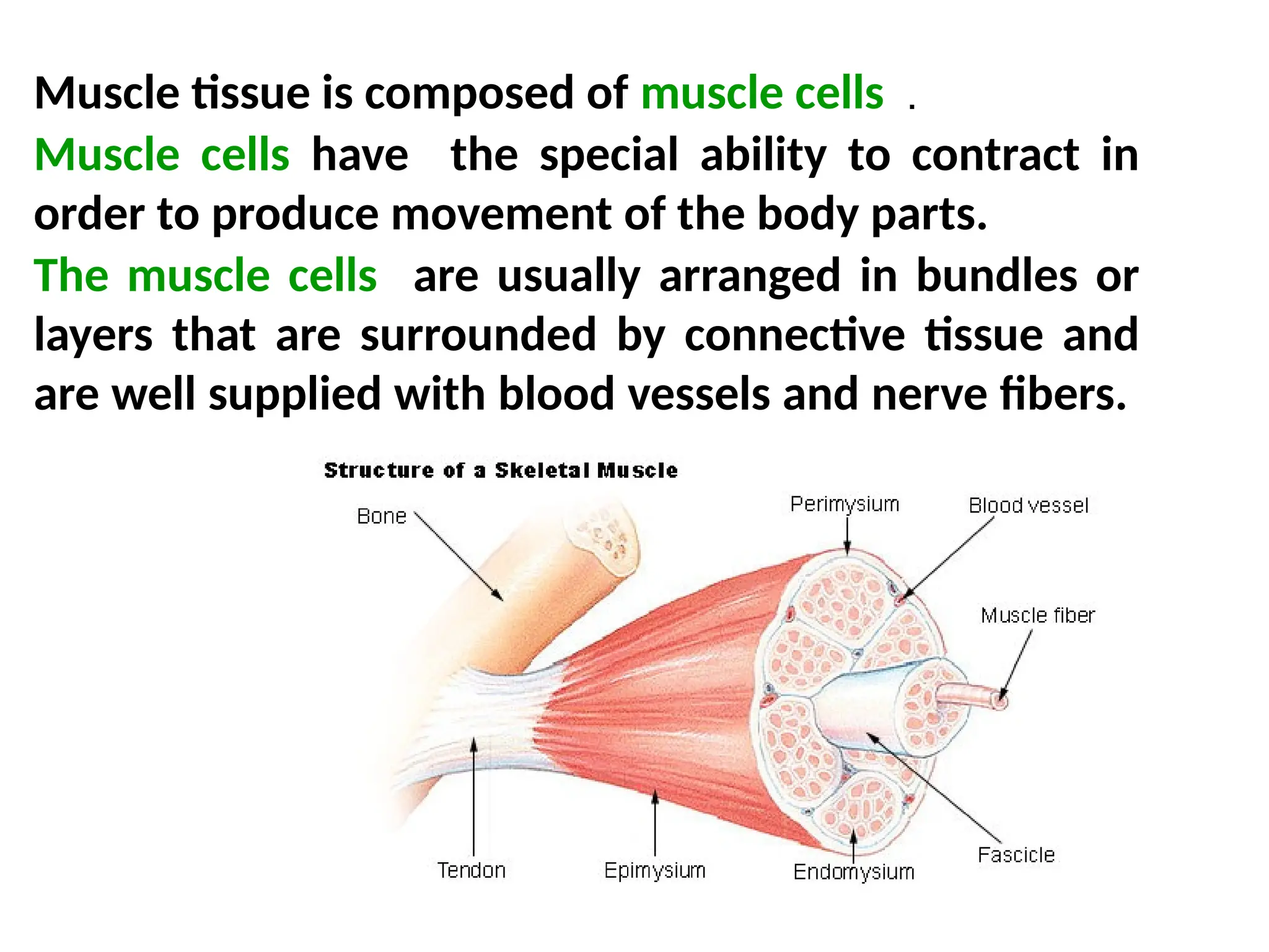
Muscle tissue iscomposed of muscle cells .
Muscle cells have the special ability to contract in
order to produce movement of the body parts.
The muscle cells are usually arranged in bundles or
layers that are surrounded by connective tissue and
are well supplied with blood vessels and nerve fibers.
4.
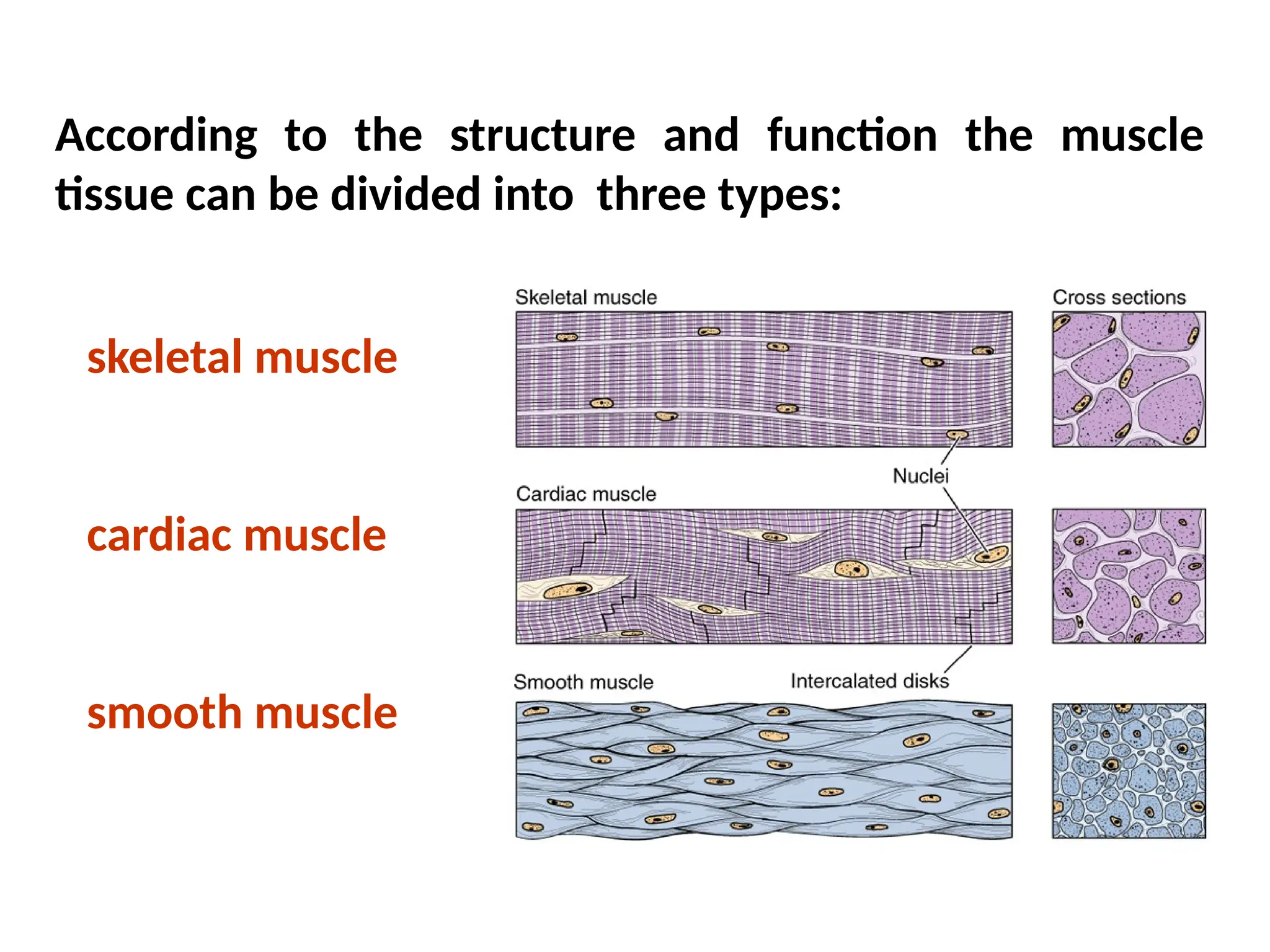
According to thestructure and function the muscle
tissue can be divided into three types:
skeletal muscle
cardiac muscle
smooth muscle
5.
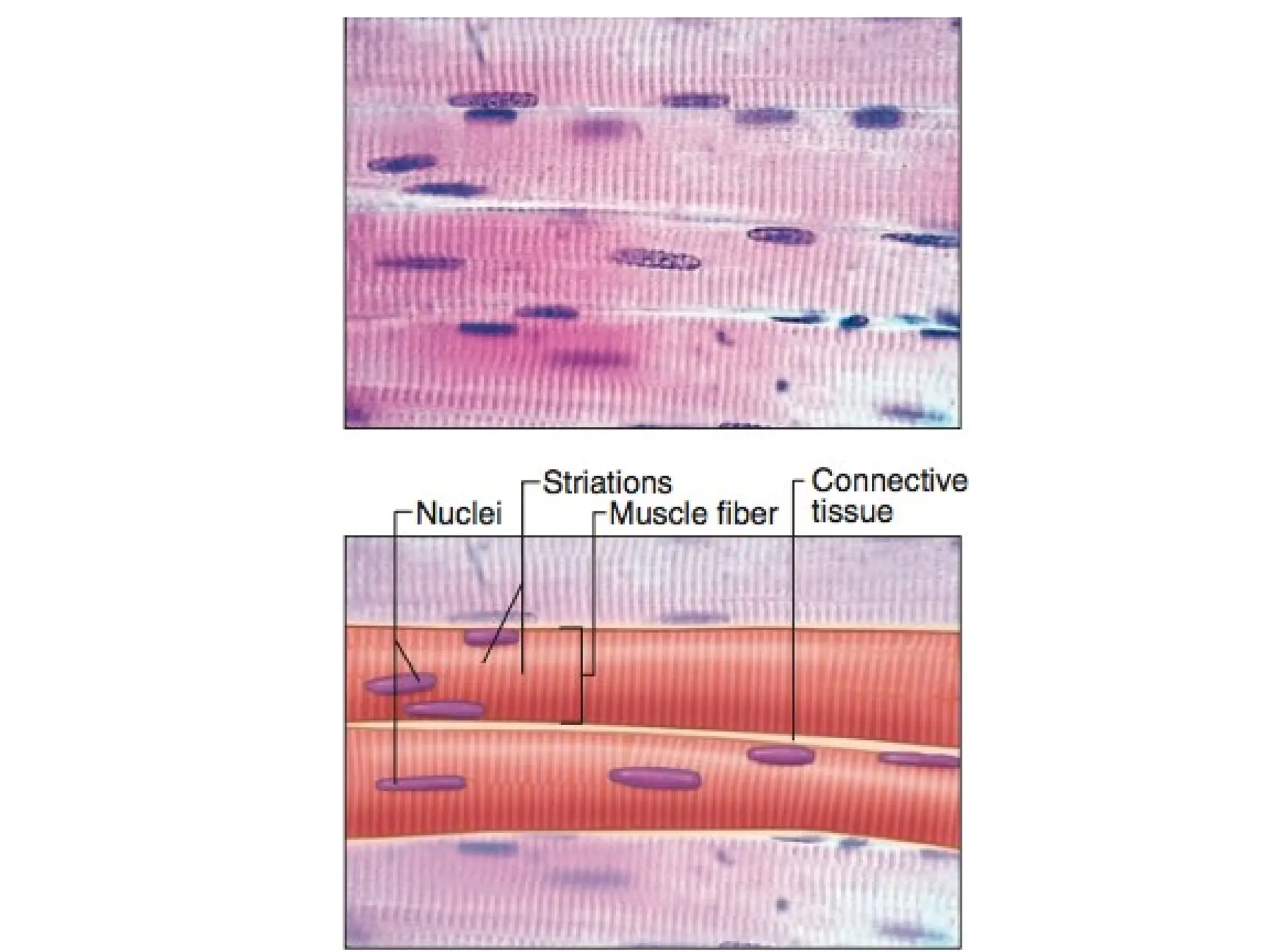
Muscle cells arelong and slender so they are
sometimes called muscle fibers or myofibers.
The membrane of muscle cell is called sarcolemma.
The cytoplasm of muscle cell is called sarcoplasm.
The smooth endoplasmic reticulum in the muscle
cell is called sarcoplasmic reticulum.
6.
• Two typesof myofilaments are associated
with cell contraction.
1. Thin filaments are composed primarily of
the protein actin.
2. Thick filaments are composed of the
protein myosin II.
7.
SKELETAL MUSCLE
• Inskeletal muscle, each muscle cell is
commonly called a muscle fiber, is actually a
multinucleated syncytium.
• The nuclei of a skeletal muscle fiber are
located in the cytoplasm immediately beneath
the plasma membrane, also called the
sarcolemma.
8.
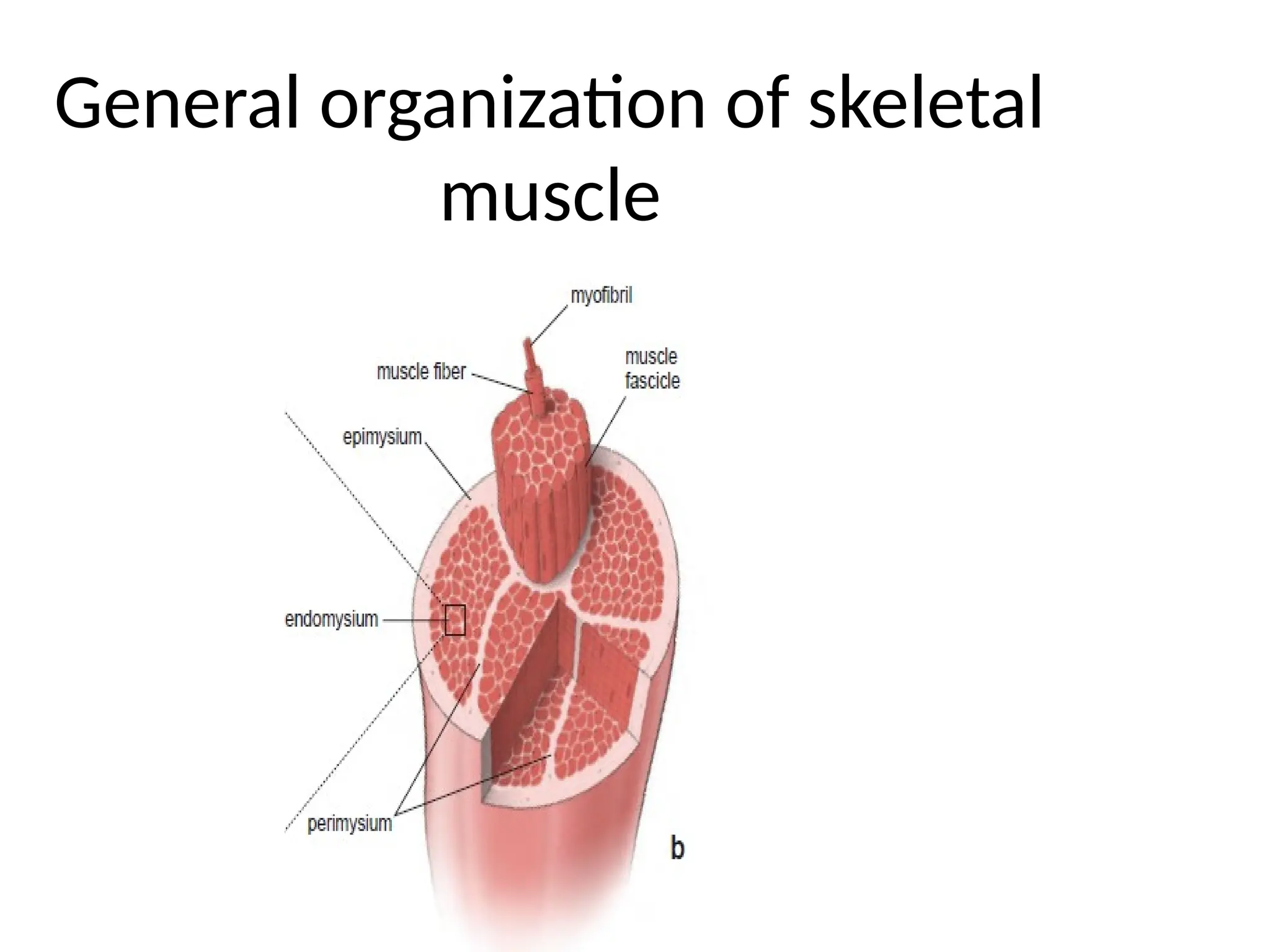
• A skeletalmuscle consists of striated muscle
fibers held together by connective tissue.
• The connective tissue associated with muscle
is named according to its relationship with the
muscle fibers:
• Endomysium is the delicate layer of reticular
fibers that immediately surrounds individual
muscle fibers.
9.
• Perimysium isa thicker connective tissue layer
that surrounds a group of fibers to form a
bundle or fascicle.
• Epimysium is the sheath of dense connective
tissue that surrounds a collection of fascicles
that constitutes the muscle.
Three types ofskeletal muscle
fiber
• Type I fibers or slow oxidative fibers are small
fibers that appear red in fresh specimens and
contain many mitochondria and large amounts
of myoglobin and cytochrome complexes.
• Type IIa fibers or fast oxidative glycolytic
fibers are of medium size with many
mitochondria and a high myoglobin content
and large amounts of glycogen and are
capable of anaerobic glycolysis.
12.
• Type IIbfibers or fast glycolytic fibers are
large fibers that appear light pink in fresh
specimens and contain less myoglobin and
fewer mitochondria than type I and type IIa
fibers.
• They have a low level of oxidative enzymes
and store a considerable amount of glycogen.
13.
Myofibrils and Myofilaments
•A muscle fiber is filled with longitudinally
arrayed structural subunits called
myofibrils.
• The structural and functional subunit of
the muscle fiber is the myofibril.
• Myofibrils are visible in favorable
histologic preparations and are best seen
in cross-sections of muscle fibers.
14.
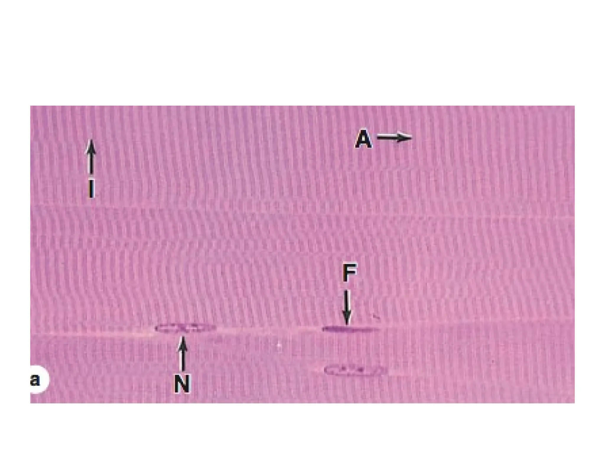
• Cross-striations areevident in H&E–stained
preparations of longitudinal sections of
muscle fibers in which they appear as
alternating light and dark bands.
• These bands are termed the A band and the I
band.
17.
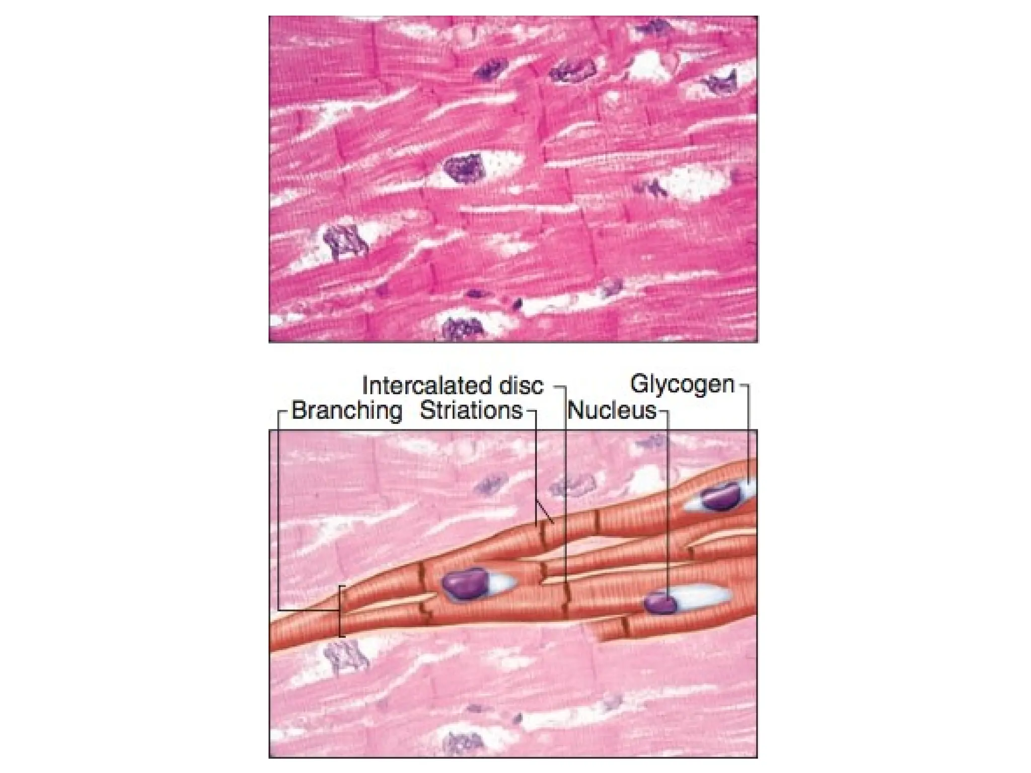
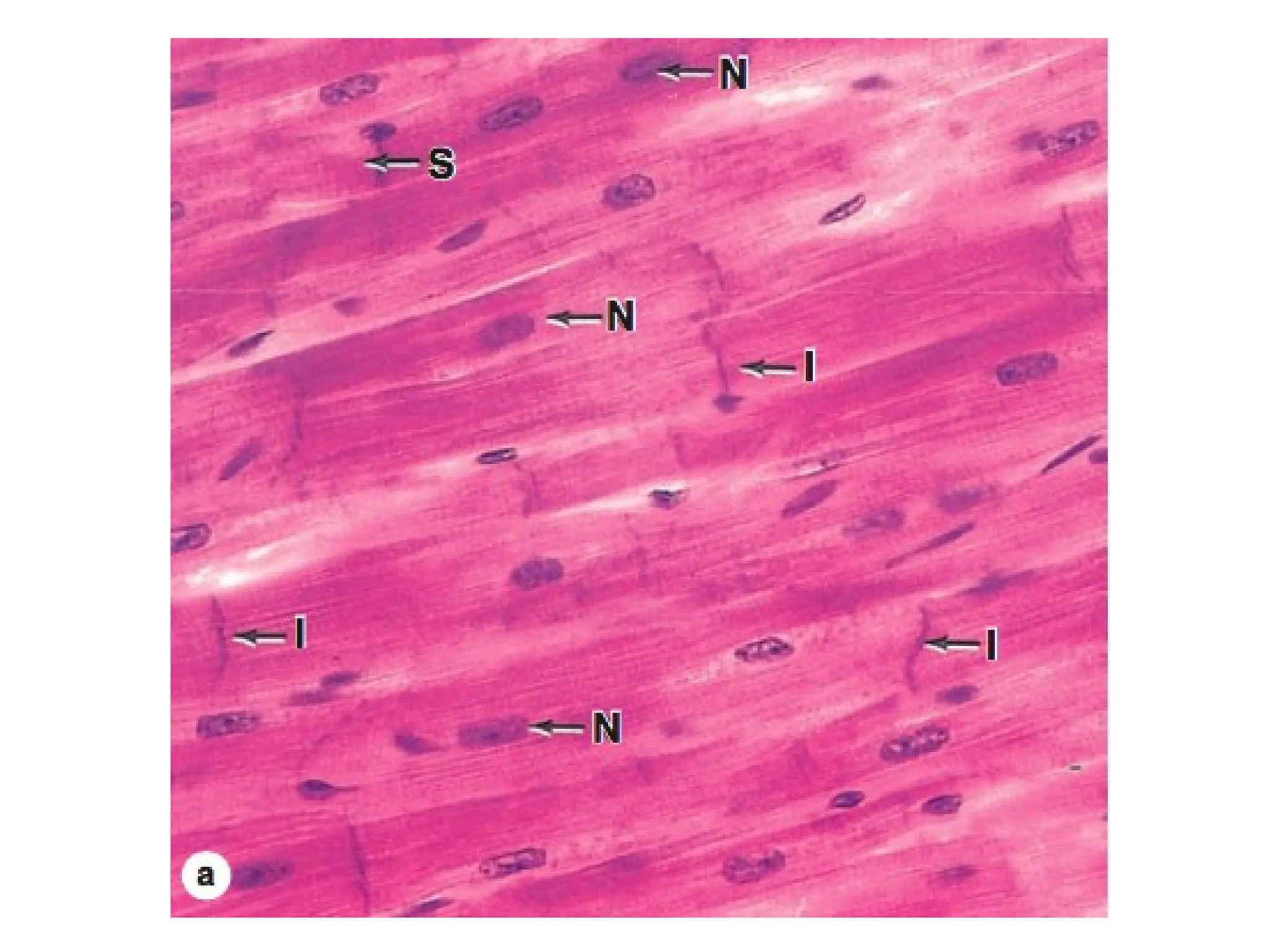
CARDIAC MUSCLE
• Cardiacmuscle has the same types and arrangement
of contractile filaments as skeletal muscle.
• Cardiac muscle cells and the fibers they exhibit cross-
striations evident in routine histologic sections.
• In addition, cardiac muscle fibers exhibit densely
staining cross-bands, called intercalated discs, that
cross the fibers in a linear fashion.
• The intercalated discs represent highly specialized
attachment sites between adjacent cells.
18.
Structure of CardiacMuscle
• The cardiac muscle nucleus lies in the center
of the cell.
• The central location of the nucleus in cardiac
muscle cells is one feature that helps
distinguish them from multinucleated skeletal
muscle fibers, whose nuclei lie immediately
under the plasma membrane.
21.
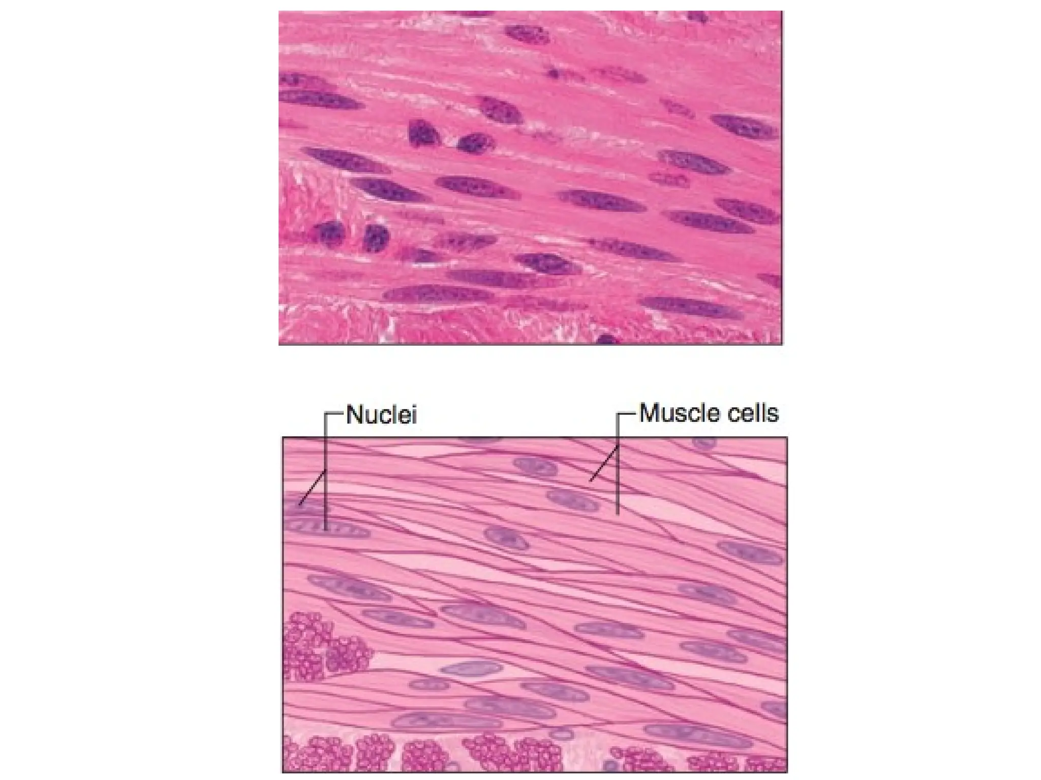
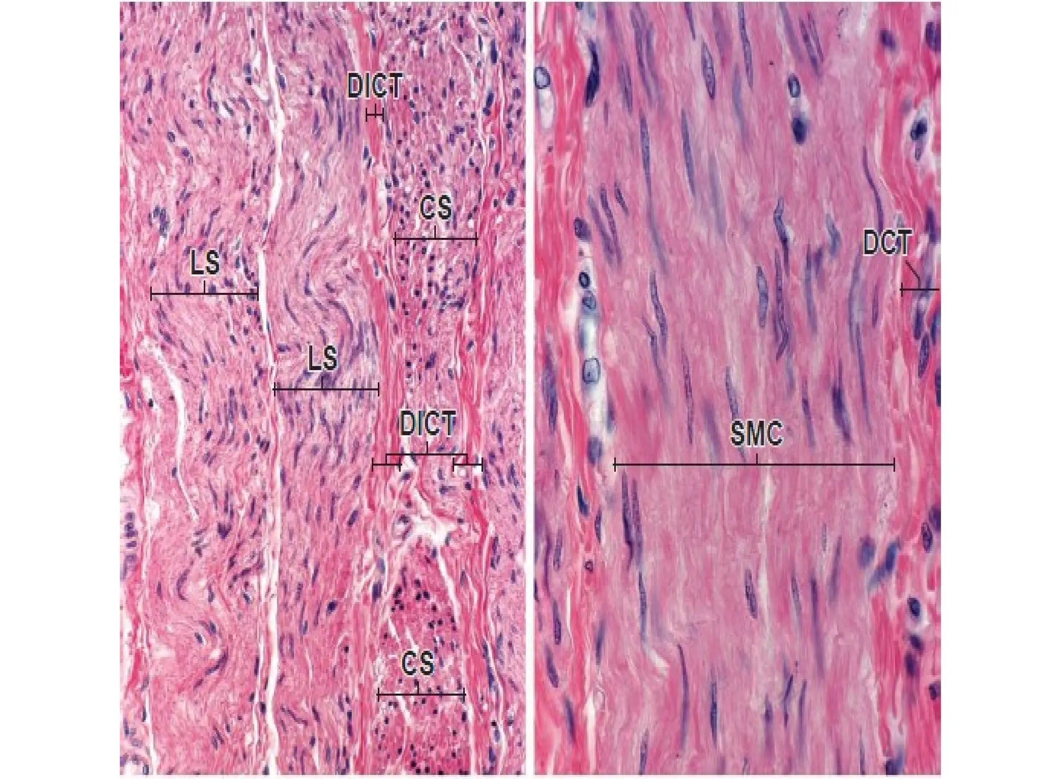
SMOOTH MUSCLE
• Smoothmuscle generally occurs as bundles of
elongated fusiform cells with finely tapered
ends.
• Range in length from 20 m in the walls of small
blood vessels to about 200 m in the wall of the
intestine; they may be as large as 500 m in the
wall of the uterus during pregnancy.
• The nuclei of smooth muscle cells are located in
the center of the cell and often have a
corkscrew appearance in longitudinal section.
22.
Structure of SmoothMuscle
• The sarcoplasm is filled with thin filaments
that form a part of the contractile apparatus.
• Thick myosin filaments are scattered
throughout the sarcoplasm of a smooth
muscle cell.
• They are extremely labile and tend to be lost
during tissue preparation.

![Histology of Muscles of the human body - Copy [A].pptx](https://cdn.slidesharecdn.com/ss_thumbnails/histologyofmuscle-copya-240705131152-8a1bfd61-thumbnail.jpg?width=640&height=640&fit=bounds)


![Muscular 1[2]](https://cdn.slidesharecdn.com/ss_thumbnails/fsyp7qstsuanexrpfthh-signature-4c28f0f13a30c4ea316a9d58353990586de4897ab085203d01a9b7b7228e72f9-poli-180213061217-thumbnail.jpg?width=640&height=640&fit=bounds)


