Download to read offline
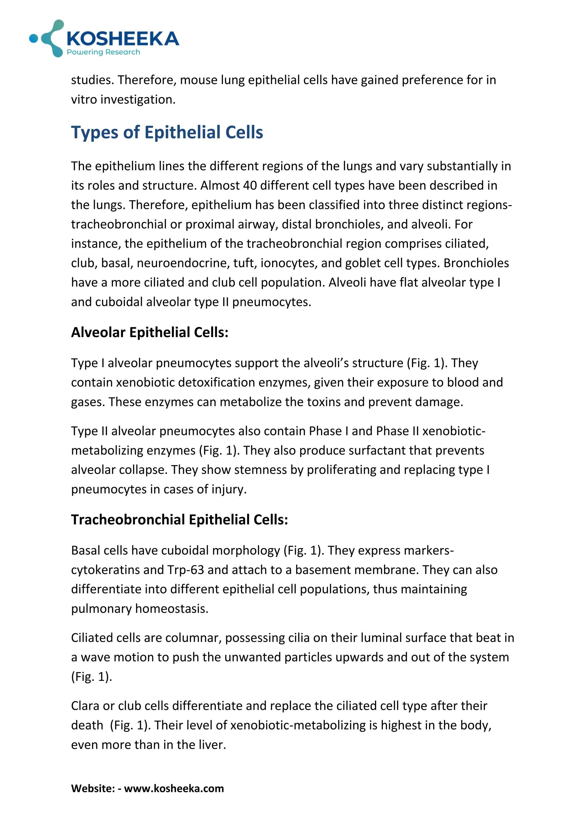


Lungs are the vital part of the respiratory system for oxygenating blood and removing carbon dioxide. Their cellular composition includes immune system, epithelium and endothelium. The epithelium has direct contact with the external environment, thus driving the immune response. The ability of genetic engineering in mice has also elevated its utilization as a replacement for humans in scientific studies. Therefore, mouse lung epithelial cells have gained preference for in vitro investigation.


![Lungsmarlenereyescorrection[1]final](https://cdn.slidesharecdn.com/ss_thumbnails/lungsmarlenereyescorrection1final-110210122726-phpapp01-thumbnail.jpg?width=640&height=640&fit=bounds)


