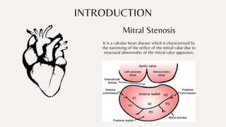
Mitral Stenosis
Presented by - Nikhil Raj Singh
Roll no. - 72
INTRODUCTION
Mitral Stenosis
It is a valvular heart disease which is characterized by

the narrowing of the orifice of the mitral value due to

structural abnormality of the mitral valve apparatus.
In Mitral stenosis, the mitral valve orifice is slowly diminished by progressive fibrosis, calcification

of the valve leaflets, and fusion of the cusps and subvalvular apparatus. The mitral valve orifice is

normally about 5 cm² in diastole but can be reduced to <1 cm² in severe mitral stenosis. The

patient is usually asymptomatic until the orifice is <2 cm². As stenosis progresses, left ventricular

filling becomes more dependent on left atrial contraction.
Pathogenesis
There is dilatation and hypertrophy of the LA and left atrial pressure rises, leading to pulmonary

venous congestion and breathlessness. Any increase in heart rate shortens diastole when the mitral

valve is open and produces a further rise in left atrial pressure. Situations that demand an increase in

cardiac output, such as pregnancy and exercise, also increase left atrial pressure and are poorly

tolerated. Atrial fibrillation is very common due to progressive dilatation of the LA. Its onset often

precipitates pulmonary oedema because the accompanying tachycardia and loss of atrial contraction

lead to marked haemodynamic deterioration and a rapid rise in left atrial pressure.
In the absence of AF, a more gradual rise in left atrial pressure may occur. In the presence or absence of

AF, pulmonary hypertension may occur, which can protect the patient from pulmonary oedema.
Pulmonary hypertension leads to right ventricular hypertrophy and dilatation, tricuspid regurgitation

and right heart failure.
Rheumatic fever is the most common cause of mitral stenosis. It develops

secondary to previous rheumatic fever due to infection with group A B-

hemolytic Streptococcus.
Mitral stenosis is more common in females.


The latent period from the first attack of rheumatic fever and the
development of onset of symptoms due to mitral stenosis is usually as short
as 1-2 years in India, compared to long in Western countries. This may be
due to repeated attacks of severe carditis in India.
Clinical manifestation in juvenile mitral stenosis/malig nant mitral stenosis
develops below the age of 19 years and is common in India. Pediatric mitral
stenosis manifests below the age of 12 years. Pin-point mitral valve is seen.
Atrial fibril lation is rare. Valve calcification is uncommon.
In rheumatic MS, chronic inflammation produces;
• Diffuse thickening of the mitral valve leaflets due to fibrosis and/or calcification
• Fusion of commissures and cusp •Fusion and shortening of the chordae tendineae.
The above morphological changes progress and cause rigidity of mitral valvular cusps which in turn leads to narrowing at the

apex of the funnel-shaped ("fish-mouth") mitral valve →severe narrowing (stenotic) of valve orifice and progressive immobility

of the valve cusps
•Rheumatic fever (leading cause)
•Congenital mitral stenosis (parachute mitral valve/Shone complex)
•Metastatic carcinoid tumor to the lung, or primary bronchial carcinoid
•Severe calcification of mitral valve apparatus, e.g., in elderly
•Systemic lupus erythematosus
•Rheumatoid arthritis (extremely rare)
•Endomyocardial fibrosis
•Gout
•Methysergide treatment
Causes
•Mucopolysaccharidosis - Hurler syndrome and Hunter syndrome
•Whipple's disease
•Infective endocarditis with large vegetations Mimics of MS
•Ball valve thrombus
•Myxoma
Salient features of mitral stenosis (MS):
•First chamber to fail in MS: Left atrium
•Ventricle to fail in MS: Right ventricle
• Atria that fibrillates in MS: Affects both right and left atria .
•Left ventricle in MS: Left ventricular end-diastolic volume (LVEDV) is reduced in

15%, while it is normal in the rest
• The most common complication of MS: Atrial fibrillation (AF)
Symptoms & Signs
Grading
Old-school Specs
Fashion eyeglasses from hippie
to preppy styles
Presentations are communication tools that can be used as
demonstrations, lectures, speeches, reports, and more. It is mostly
presented before an audience.
The
Mintmade
Fashion
Philosophy
My commitment to you
Unique product range
Carefully curated pieces
Diverse size range
Ethically-made
Easy return policy

Mitral stenosis.pdf

  • 1.
    Mitral Stenosis Presented by- Nikhil Raj Singh Roll no. - 72
  • 2.
    INTRODUCTION Mitral Stenosis It isa valvular heart disease which is characterized by the narrowing of the orifice of the mitral value due to structural abnormality of the mitral valve apparatus.
  • 3.
    In Mitral stenosis,the mitral valve orifice is slowly diminished by progressive fibrosis, calcification of the valve leaflets, and fusion of the cusps and subvalvular apparatus. The mitral valve orifice is normally about 5 cm² in diastole but can be reduced to <1 cm² in severe mitral stenosis. The patient is usually asymptomatic until the orifice is <2 cm². As stenosis progresses, left ventricular filling becomes more dependent on left atrial contraction. Pathogenesis There is dilatation and hypertrophy of the LA and left atrial pressure rises, leading to pulmonary venous congestion and breathlessness. Any increase in heart rate shortens diastole when the mitral valve is open and produces a further rise in left atrial pressure. Situations that demand an increase in cardiac output, such as pregnancy and exercise, also increase left atrial pressure and are poorly tolerated. Atrial fibrillation is very common due to progressive dilatation of the LA. Its onset often precipitates pulmonary oedema because the accompanying tachycardia and loss of atrial contraction lead to marked haemodynamic deterioration and a rapid rise in left atrial pressure.
  • 4.
    In the absenceof AF, a more gradual rise in left atrial pressure may occur. In the presence or absence of AF, pulmonary hypertension may occur, which can protect the patient from pulmonary oedema. Pulmonary hypertension leads to right ventricular hypertrophy and dilatation, tricuspid regurgitation and right heart failure.
  • 5.
    Rheumatic fever isthe most common cause of mitral stenosis. It develops secondary to previous rheumatic fever due to infection with group A B- hemolytic Streptococcus. Mitral stenosis is more common in females. The latent period from the first attack of rheumatic fever and the development of onset of symptoms due to mitral stenosis is usually as short as 1-2 years in India, compared to long in Western countries. This may be due to repeated attacks of severe carditis in India. Clinical manifestation in juvenile mitral stenosis/malig nant mitral stenosis develops below the age of 19 years and is common in India. Pediatric mitral stenosis manifests below the age of 12 years. Pin-point mitral valve is seen. Atrial fibril lation is rare. Valve calcification is uncommon. In rheumatic MS, chronic inflammation produces; • Diffuse thickening of the mitral valve leaflets due to fibrosis and/or calcification • Fusion of commissures and cusp •Fusion and shortening of the chordae tendineae. The above morphological changes progress and cause rigidity of mitral valvular cusps which in turn leads to narrowing at the apex of the funnel-shaped ("fish-mouth") mitral valve →severe narrowing (stenotic) of valve orifice and progressive immobility of the valve cusps
  • 6.
    •Rheumatic fever (leadingcause) •Congenital mitral stenosis (parachute mitral valve/Shone complex) •Metastatic carcinoid tumor to the lung, or primary bronchial carcinoid •Severe calcification of mitral valve apparatus, e.g., in elderly •Systemic lupus erythematosus •Rheumatoid arthritis (extremely rare) •Endomyocardial fibrosis •Gout •Methysergide treatment Causes •Mucopolysaccharidosis - Hurler syndrome and Hunter syndrome •Whipple's disease •Infective endocarditis with large vegetations Mimics of MS •Ball valve thrombus •Myxoma
  • 7.
    Salient features ofmitral stenosis (MS): •First chamber to fail in MS: Left atrium •Ventricle to fail in MS: Right ventricle • Atria that fibrillates in MS: Affects both right and left atria . •Left ventricle in MS: Left ventricular end-diastolic volume (LVEDV) is reduced in 15%, while it is normal in the rest • The most common complication of MS: Atrial fibrillation (AF)
  • 8.
  • 9.
  • 10.
    Old-school Specs Fashion eyeglassesfrom hippie to preppy styles Presentations are communication tools that can be used as demonstrations, lectures, speeches, reports, and more. It is mostly presented before an audience.
  • 11.
    The Mintmade Fashion Philosophy My commitment toyou Unique product range Carefully curated pieces Diverse size range Ethically-made Easy return policy