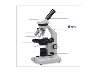
Call Girls Siliguri Just Call 9907093804 Top Class Call Girl Service Available
Microscopy and staining techniques details
1. What is light?
Light is electromagnetic radiation.
What we usually describe as light is
only the visible spectrum of this
radiation with wavelengths between
400nm and 700nm.
The elementary particle that defines
light is the photon.
There are 3 basic dimensions of light
a) Intensity (amplitude) which is related to the perception of brightness
b) Frequency (wavelength), perceived as colour
c) Polarization (angle of vibration) which is not or weakly perceptible to humans
a)
b)
3. Starting with use of a simple lens in ancient
times, to the first compound microscope
around 1590, and up to the microscopes you
are using in 7th grade life science, the
microscope has allowed scientists to make
discoveries about the “invisible world.”
The microscope has
become one of the most
recognizable symbols of
science.
4. Micro- = “small”; -scope = “to look at”
Photographs of cells are taken using a microscope, and these pictures
are called micrographs.
5. What is a microscope?
Theoretically a microscope is an array of two lenses.
Objective
lens
Tube lens Eyepiece
lens
Focal plane Image plane
Classic compound microscope
Image plane
7. What is magnification?
Magnification is a measure of how much larger a
microscope (or set of lenses within a microscope)
causes an object to appear.
Magnification is defined by the
magnification by the objective
x
the magnification by eyepiece
BUT maximum magnification does not mean maximum
resolution!
8. What is resolution?
Resolution describes the minimal distance of two points that can be distinguished.
The resolution of a microscope or lens is the smallest distance by which two points can
be separated and still be distinguished as separate objects.
The smaller this value, the higher the resolving power of the microscope and the better
the clarity and detail of the image.
Picture taken from http://microscopy.fsu.edu/primer/anatomy/numaperture.html
9. NA = n sin
What is the numerical aperture?
NA is an estimate of how much light from the sample is collected by the objective
α1
α2
Coverslip (n = 1.5)
Glass slide (n = 1.5)
Oil (n = 1.5)
Air (n = 1.0)
n = refractive index
α = angle of incident illumination
Objective lens
10. • The higher the numerical aperture of a
lens, the better the resolution of a
specimen will be which can be obtained
with that lens.
• d=0.5 λ/n sin Ɵ
– Where d= resolution
– Λ = wavelength of light used
11. Numerical aperture, NOT
magnification determines resolution!
Increasing NA
A lens with a larger NA will be able to visualize finer details and will also
collect more light and give a brighter image than a lens with lower NA.
13. Light (Optical) Microscopy
• Visible light is uses.
• Glass lens are used
• Advantage: It can often be performed on
living cells, so it’s possible to watch cells
carrying out their normal behaviors (e.g.,
migrating or dividing) under the
microscope.
14. Principle
• When a ray of light passes from one
medium to another it bends by
phenomena called refraction.
• Bending of light slows the speed.
• The bending of light is determined by
refractive index of the medium.
15. Types of Light Microscopes
1.Bright field Light Microscope
2.Phase Contrast Light Microscope
3.Dark-Field Light Microscope
4.Fluorescence Light Microscope
17. Brightfield
Piece of artificially grown skin
(www.igb.fhg.de/.../dt/PI_BioTechnica2001.dt.html )
Cross section of sunflower root
(http://www.zum.de/Faecher/Materialien/beck/12/bs12-5.htm)
Principle: Light is transmitted through the sample and absorbed by it.
Application: Only useful for specimens that can be contrasted via dyes.
Very little contrast in unstained specimens.
With a bright background, the human eye requires local intensity fluctuations
of at least 10 to 20% to be able to recognize objects.
20. Eyepiece
• Also known as the ocular
• Contains the first lens you look through
- usually a magnification of 10x
• Located on the top of the body tube
22. Objective Lenses
• Used in combination with the eyepiece
to provide a range of magnification
• Magnification ranges from 40x to 400x
• Located on the nose-piece at the
bottom of the body tube
26. Arm
• Supports the upper parts of the
microscope
• Used to carry the microscope
• When carrying a microscope, always
have one hand on the arm and one
hand on the base. Use two hands!!
28. Base
• Supports the whole microscope
• Used to carry the microscope
• When carrying a microscope, always
have one hand on the arm and one
hand on the base. Use two hands!!
32. Stage Clip
• Helps to hold the slide in place
• Usually one on each side of the hole
(stage opening) = 2 stage clips
• The stage opening allows light to pass
from the light source to the lenses.
34. Light Source
• Provides light necessary for viewing the
specimen
• Usually either a mirror or illuminator
• Sends light through the stage opening
to the diaphragm
36. Diaphragm
• Wheel or lever located below the stage
opening
• Regulates the amount of light that can
enter the lenses
• May need to be adjusted based on the
thickness of the specimen being
studied
40. Fine Adjustment Knob
• Raises and lowers the stage or
objective lenses a small distance for
exact focusing
• Used when focusing the medium
power (10x) and high power (40x)
objective lenses
42. Phase contrast
Principle: Incident light [Io] is out of phase with transmitted light [I] as it was slowed down while
passing through different parts of the sample and when the phases of the light are synchronized
by an interference lens, a new image with greater contrast is seen.
I
I0
Phase ring
aligned
not aligned
Phase stops
https://www.youtube.com/watch?v=fC
Zw4X7V5Pw
43. Phase contrast
Application: Phase contrast is the most commonly used contrasting technique All
tissue culture microscopes and the time-lapse microscopes are set up for phase.
wrong phase stop
brightfield right phase stop
44. Applications
• Determine morphologies of living
cells such as plant and animal cells
• Studying microbial motility and
structures of locomotion
• To detect certain microbial elements
such as the bacterial endospores
46. Applications
• Used in the visualization of bacterial agents such
as Mycobacterium tuberculosis.
• Used to identify specific antibodies produced
against bacterial antigens/pathogens in
immunofluorescence techniques by labeling the
antibodies with fluorochromes.
• Used in ecological studies to identify and observe
microorganisms labeled by the fluorochromes
• It can also be used to differentiate between dead
and live bacteria by the color they emit when
treated with special stains
48. Darkfield
Principle: The illuminating rays of light are directed through the sample from the side by putting a
dark disk into the condenser that hinders the main light beam to enter the objective. Only light that
is scattered by structures in the sample enters the objective.
Application: People use it a lot to look at Diatoms and other unstained/colourless specimens
Brightfield
Darkfield
Symbiotic Diatom colony
(www1.tip.nl/~t936927/making.html)
49. Applications
• It is used to visualize the internal
organs of larger cells such as the
eukaryotic cells
• Identification of bacterial cells with
distinctive shapes such
as Treponema pallidum, a causative
agent of syphilis.
https://www.youtube.com/watch?v=W9Mhk
myfMLQ