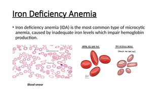
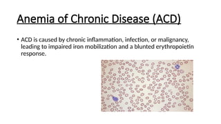
Microcytic Anemias and Its
Types
A Genetic and Clinical Overview
Presented by Amal Ishtiaq
2024-Mpil-1102
Introduction to Microcytic Anemias
• Microcytic anemias are characterized by small red blood cells (MCV <
80 fL). These conditions are typically caused by impaired hemoglobin
synthesis.
Overview of Hemoglobin Synthesis
• Hemoglobin consists of alpha and beta globin chains. Iron plays a
critical role in its synthesis. Defects in globin chains or iron
metabolism can lead to microcytic anemia.
Classification of Microcytic Anemias
Microcytic anemias can be classified into several types:
1. Iron Deficiency Anemia
2. Thalassemia
3. Anemia of Chronic Disease
4. Sideroblastic Anemia
5. Lead Poisoning
Iron Deficiency Anemia
• Iron deficiency anemia (IDA) is the most common type of microcytic
anemia, caused by inadequate iron levels which impair hemoglobin
production.
Blood smear
Causes of Iron Deficiency Anemia
1. Inadequate dietary intake
2. Chronic blood loss (e.g., gastrointestinal bleeding)
3. Malabsorption disorders (e.g., celiac disease)
4. Increased demand (e.g., pregnancy, rapid growth)
Genetic Considerations in Iron
Deficiency Anemia
• While iron deficiency is typically acquired, rare genetic mutations
(such as TMPRSS6) can impair iron metabolism (by reducing amount
of functional matriptase-2 preventing it from controlling hepcidin
levels, resulting in block of absorption of iron via the intestines and
release of iron from storage), leading to a hereditary form of iron
deficiency anemia.
Diagnosis of Iron Deficiency Anemia
• Laboratory tests include serum ferritin, transferrin saturation, and
serum iron. In severe cases, a bone marrow biopsy may be performed
to assess iron stores.
Treatment of Iron Deficiency Anemia
 Oral iron supplementation
IV iron therapy in severe or refractory cases
 Treatment of underlying causes (e.g., GI bleeding)
Thalassemia
• Thalassemia are genetic disorders that affect globin chain production,
leading to an imbalance in alpha or beta chains, hemolysis, and
microcytic anemia.
Genetics of Thalassemia
1. Alpha-thalassemia: Deletions in HBA1 and HBA2 genes
2. Beta-thalassemia: Mutations in the HBB gene
Severity depends on the number of affected genes.
Types of Alpha-thalassemia
1. Silent carrier: One gene deletion
2. Alpha-thalassemia trait: Two gene deletions
3. Hemoglobin H disease: Three gene deletions
4. Hydrops fetalis: Four gene deletions (lethal)
Clinical Manifestations of Alpha-
thalassemia
• Carriers are typically asymptomatic, but Hemoglobin H disease causes
moderate to severe anemia, and hydrops fetalis is non-survivable.
Genetics of Beta-thalassemia
• Mutations in the HBB gene lead to reduced beta chain production.
Over 200 different mutations are identified, with varying effects on
clinical severity.
Types of Beta-thalassemia
1. Thalassemia minor: Mild anemia, asymptomatic
2. Thalassemia intermedia: Moderate anemia, some transfusion
dependence
3. Thalassemia major: Severe anemia, lifelong transfusions required
Genetic Heterogeneity in Beta-
thalassemia
• Beta-thalassemia shows genetic heterogeneity, with a wide range of
mutations including missense, nonsense, frameshift, and splicing
mutations that affect the severity of the disease.
Diagnosis of Thalassemia
• Thalassemias are diagnosed through hemoglobin electrophoresis and
genetic testing for mutations in the HBB, HBA1, and HBA2 genes.
Management of Thalassemia
• Management includes regular blood transfusions (for major forms),
iron chelation therapy to prevent iron overload, and bone marrow
transplant (BMT) in severe cases.
Anemia of Chronic Disease (ACD)
• ACD is caused by chronic inflammation, infection, or malignancy,
leading to impaired iron mobilization and a blunted erythropoietin
response.
Pathophysiology of ACD
• Inflammation induces the overproduction of hepcidin, blocking iron
release from macrophages, leading to reduced red blood cell
production and anemia.
Genetic Aspects of ACD
• Polymorphisms in inflammatory cytokine genes (e.g., IL-6, TNF-alpha)
can influence the severity of ACD. Genetic variability in hepcidin
expression (HAMP gene) also affects disease presentation.
Diagnosis of ACD
• ACD diagnosis is based on laboratory findings such as normal to
increased ferritin, low serum iron, and transferrin saturation. ACD is a
diagnosis of exclusion.
Sideroblastic Anemia
• Sideroblastic anemia is caused by defective iron incorporation into
hemoglobin, leading to iron accumulation in mitochondria and the
formation of ringed sideroblasts.
Genetics of Sideroblastic Anemia
• X-linked sideroblastic anemia is caused by mutations in the ALAS2
gene, while autosomal recessive forms are linked to SLC25A38
mutations. Mitochondrial defects also play a role in iron-sulfur cluster
biogenesis.
Clinical Features of Sideroblastic
Anemia
• Clinical features include microcytic anemia, ringed sideroblasts in the
bone marrow, and iron overload, despite the presence of anemia.
Diagnosis of Sideroblastic Anemia
• Bone marrow biopsy reveals ringed sideroblasts, while serum iron and
ferritin levels are elevated due to iron accumulation. Genetic testing
can confirm specific mutations like ALAS2 and SLC25A38.
Treatment of Sideroblastic Anemia
Treatment options include Vitamin B6 supplementation for ALAS2
mutations, regular blood transfusions for severe cases, and iron
chelation therapy to manage iron overload.
Lead Poisoning and Microcytic
Anemia
• Lead poisoning interferes with heme synthesis and causes microcytic
anemia. Symptoms include anemia, neurological deficits, and
abdominal pain.
Diagnosis and Management of Lead
Poisoning
• Diagnosis involves measuring blood lead levels. Management includes
chelation therapy and removal from exposure to lead.
Summary
• Microcytic anemias are diverse conditions with both genetic and
acquired causes. Genetic understanding helps guide diagnosis and
treatment. Proper management depends on the specific type and
severity of the anemia.

Microcytic Anemias and Its Types .pptx

  • 1.
    Microcytic Anemias andIts Types A Genetic and Clinical Overview Presented by Amal Ishtiaq 2024-Mpil-1102
  • 2.
    Introduction to MicrocyticAnemias • Microcytic anemias are characterized by small red blood cells (MCV < 80 fL). These conditions are typically caused by impaired hemoglobin synthesis.
  • 3.
    Overview of HemoglobinSynthesis • Hemoglobin consists of alpha and beta globin chains. Iron plays a critical role in its synthesis. Defects in globin chains or iron metabolism can lead to microcytic anemia.
  • 4.
    Classification of MicrocyticAnemias Microcytic anemias can be classified into several types: 1. Iron Deficiency Anemia 2. Thalassemia 3. Anemia of Chronic Disease 4. Sideroblastic Anemia 5. Lead Poisoning
  • 5.
    Iron Deficiency Anemia •Iron deficiency anemia (IDA) is the most common type of microcytic anemia, caused by inadequate iron levels which impair hemoglobin production. Blood smear
  • 6.
    Causes of IronDeficiency Anemia 1. Inadequate dietary intake 2. Chronic blood loss (e.g., gastrointestinal bleeding) 3. Malabsorption disorders (e.g., celiac disease) 4. Increased demand (e.g., pregnancy, rapid growth)
  • 7.
    Genetic Considerations inIron Deficiency Anemia • While iron deficiency is typically acquired, rare genetic mutations (such as TMPRSS6) can impair iron metabolism (by reducing amount of functional matriptase-2 preventing it from controlling hepcidin levels, resulting in block of absorption of iron via the intestines and release of iron from storage), leading to a hereditary form of iron deficiency anemia.
  • 8.
    Diagnosis of IronDeficiency Anemia • Laboratory tests include serum ferritin, transferrin saturation, and serum iron. In severe cases, a bone marrow biopsy may be performed to assess iron stores.
  • 9.
    Treatment of IronDeficiency Anemia  Oral iron supplementation IV iron therapy in severe or refractory cases  Treatment of underlying causes (e.g., GI bleeding)
  • 10.
    Thalassemia • Thalassemia aregenetic disorders that affect globin chain production, leading to an imbalance in alpha or beta chains, hemolysis, and microcytic anemia.
  • 11.
    Genetics of Thalassemia 1.Alpha-thalassemia: Deletions in HBA1 and HBA2 genes 2. Beta-thalassemia: Mutations in the HBB gene Severity depends on the number of affected genes.
  • 12.
    Types of Alpha-thalassemia 1.Silent carrier: One gene deletion 2. Alpha-thalassemia trait: Two gene deletions 3. Hemoglobin H disease: Three gene deletions 4. Hydrops fetalis: Four gene deletions (lethal)
  • 13.
    Clinical Manifestations ofAlpha- thalassemia • Carriers are typically asymptomatic, but Hemoglobin H disease causes moderate to severe anemia, and hydrops fetalis is non-survivable.
  • 14.
    Genetics of Beta-thalassemia •Mutations in the HBB gene lead to reduced beta chain production. Over 200 different mutations are identified, with varying effects on clinical severity.
  • 15.
    Types of Beta-thalassemia 1.Thalassemia minor: Mild anemia, asymptomatic 2. Thalassemia intermedia: Moderate anemia, some transfusion dependence 3. Thalassemia major: Severe anemia, lifelong transfusions required
  • 16.
    Genetic Heterogeneity inBeta- thalassemia • Beta-thalassemia shows genetic heterogeneity, with a wide range of mutations including missense, nonsense, frameshift, and splicing mutations that affect the severity of the disease.
  • 17.
    Diagnosis of Thalassemia •Thalassemias are diagnosed through hemoglobin electrophoresis and genetic testing for mutations in the HBB, HBA1, and HBA2 genes.
  • 18.
    Management of Thalassemia •Management includes regular blood transfusions (for major forms), iron chelation therapy to prevent iron overload, and bone marrow transplant (BMT) in severe cases.
  • 19.
    Anemia of ChronicDisease (ACD) • ACD is caused by chronic inflammation, infection, or malignancy, leading to impaired iron mobilization and a blunted erythropoietin response.
  • 20.
    Pathophysiology of ACD •Inflammation induces the overproduction of hepcidin, blocking iron release from macrophages, leading to reduced red blood cell production and anemia.
  • 21.
    Genetic Aspects ofACD • Polymorphisms in inflammatory cytokine genes (e.g., IL-6, TNF-alpha) can influence the severity of ACD. Genetic variability in hepcidin expression (HAMP gene) also affects disease presentation.
  • 22.
    Diagnosis of ACD •ACD diagnosis is based on laboratory findings such as normal to increased ferritin, low serum iron, and transferrin saturation. ACD is a diagnosis of exclusion.
  • 23.
    Sideroblastic Anemia • Sideroblasticanemia is caused by defective iron incorporation into hemoglobin, leading to iron accumulation in mitochondria and the formation of ringed sideroblasts.
  • 24.
    Genetics of SideroblasticAnemia • X-linked sideroblastic anemia is caused by mutations in the ALAS2 gene, while autosomal recessive forms are linked to SLC25A38 mutations. Mitochondrial defects also play a role in iron-sulfur cluster biogenesis.
  • 25.
    Clinical Features ofSideroblastic Anemia • Clinical features include microcytic anemia, ringed sideroblasts in the bone marrow, and iron overload, despite the presence of anemia.
  • 26.
    Diagnosis of SideroblasticAnemia • Bone marrow biopsy reveals ringed sideroblasts, while serum iron and ferritin levels are elevated due to iron accumulation. Genetic testing can confirm specific mutations like ALAS2 and SLC25A38.
  • 27.
    Treatment of SideroblasticAnemia Treatment options include Vitamin B6 supplementation for ALAS2 mutations, regular blood transfusions for severe cases, and iron chelation therapy to manage iron overload.
  • 28.
    Lead Poisoning andMicrocytic Anemia • Lead poisoning interferes with heme synthesis and causes microcytic anemia. Symptoms include anemia, neurological deficits, and abdominal pain.
  • 29.
    Diagnosis and Managementof Lead Poisoning • Diagnosis involves measuring blood lead levels. Management includes chelation therapy and removal from exposure to lead.
  • 30.
    Summary • Microcytic anemiasare diverse conditions with both genetic and acquired causes. Genetic understanding helps guide diagnosis and treatment. Proper management depends on the specific type and severity of the anemia.