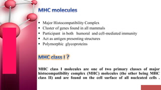
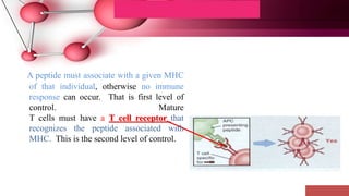
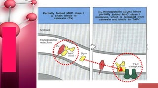
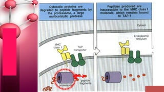
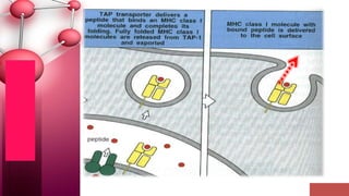
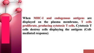
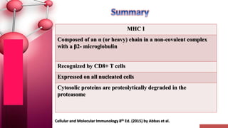
 Major Histocompatibility Complex
 Cluster of genes found in all mammals
 Participant in both humoral and cell-mediated immunity
 Act as antigen presenting structures
 Polymorphic glycoproteins
MHC class I molecules are one of two primary classes of major
histocompatibility complex (MHC) molecules (the other being MHC
class II) and are found on the cell surface of all nucleated cells .
1. MHC molecules are membrane-bound. Recognition by T cells
requires cell-cell contact.
2. Peptide from cytosol associates with class I MHC and is
recognized by Tc cells.
3. Although there is a high degree of polymorphism for a species,
an individual has maximum of six different class I MHC
products.
4. A peptide must associate with a given MHC
of that individual, otherwise no immune
response can occur. That is first level of
control. Mature
T cells must have a T cell receptor that
recognizes the peptide associated with
MHC. This is the second level of control.
• Their function is to display peptide fragments of non-
self proteins from within the cell to cytotoxic T cells;
• This will trigger an immediate response from the
immune system against a particular non-self antigen
displayed with the help of an MHC class I protein.
1
3
2
MHC-encoded -chain of 43kDa ,Made Up Of 3
Domains (1, 2 and 3)
2m
2-microglobulin, 12kDa, non-MHC encoded, non-
transmembrane, non covalently bound to 3 domain
The a3 segment of the MHC I , is highly conserved
among MHC1 & serves as a binding site for CD8
Peptide antigen in a groove formed from a pair of a-
helicies on a floor of anti-parallel b strands
-chain anchored to the cell membrane
• Conversion of native antigen (large globular protein) into peptides capable of
binding to MHC molecules
• Occurs in cellular compartments where MHC molecules are synthesized and
assembled (ER)
– Determines how antigen in different cellular compartments generates peptides
that are displayed by class I or class II MHC molecules
1. Only fragments of proteins (peptides) associated with MHC
molecules on surface of cells
• Helper T cells CD4+(Th) recognize peptide associated with
MHC class II molecules
• Cytotoxic T cells CD8+ (Tcyt) recognize peptide associated
with MHC class I molecules.
Dendritic cells
The fate of the virus-infected cell is almost
always induction of apoptosis through cell-
mediated immunity , reducing the risk of
infecting neighboring cells.
Therefore, in the absence of MHC I molecules,
NK cells are activated and recognize the cell as
aberrant, suggesting that it may be infected by
viruses attempting to evade immune destruction.
Cellular and Molecular Immunology 8th Ed. (2015) by Abbas et al.
MHC I
Composed of an α (or heavy) chain in a non-covalent complex
with a β2- microglobulin
Recognized by CD8+ T cells
Expressed on all nucleated cells
Cytosolic proteins are proteolytically degraded in the
proteasome
MHC I  molecules
MHC I  molecules

MHC I molecules

  • 2.
     Major HistocompatibilityComplex  Cluster of genes found in all mammals  Participant in both humoral and cell-mediated immunity  Act as antigen presenting structures  Polymorphic glycoproteins MHC class I molecules are one of two primary classes of major histocompatibility complex (MHC) molecules (the other being MHC class II) and are found on the cell surface of all nucleated cells .
  • 3.
    1. MHC moleculesare membrane-bound. Recognition by T cells requires cell-cell contact. 2. Peptide from cytosol associates with class I MHC and is recognized by Tc cells. 3. Although there is a high degree of polymorphism for a species, an individual has maximum of six different class I MHC products.
  • 4.
    4. A peptidemust associate with a given MHC of that individual, otherwise no immune response can occur. That is first level of control. Mature T cells must have a T cell receptor that recognizes the peptide associated with MHC. This is the second level of control.
  • 5.
    • Their functionis to display peptide fragments of non- self proteins from within the cell to cytotoxic T cells; • This will trigger an immediate response from the immune system against a particular non-self antigen displayed with the help of an MHC class I protein.
  • 6.
    1 3 2 MHC-encoded -chain of43kDa ,Made Up Of 3 Domains (1, 2 and 3) 2m 2-microglobulin, 12kDa, non-MHC encoded, non- transmembrane, non covalently bound to 3 domain The a3 segment of the MHC I , is highly conserved among MHC1 & serves as a binding site for CD8 Peptide antigen in a groove formed from a pair of a- helicies on a floor of anti-parallel b strands -chain anchored to the cell membrane
  • 8.
    • Conversion ofnative antigen (large globular protein) into peptides capable of binding to MHC molecules • Occurs in cellular compartments where MHC molecules are synthesized and assembled (ER) – Determines how antigen in different cellular compartments generates peptides that are displayed by class I or class II MHC molecules
  • 9.
    1. Only fragmentsof proteins (peptides) associated with MHC molecules on surface of cells • Helper T cells CD4+(Th) recognize peptide associated with MHC class II molecules • Cytotoxic T cells CD8+ (Tcyt) recognize peptide associated with MHC class I molecules.
  • 10.
  • 18.
    The fate ofthe virus-infected cell is almost always induction of apoptosis through cell- mediated immunity , reducing the risk of infecting neighboring cells. Therefore, in the absence of MHC I molecules, NK cells are activated and recognize the cell as aberrant, suggesting that it may be infected by viruses attempting to evade immune destruction.
  • 19.
    Cellular and MolecularImmunology 8th Ed. (2015) by Abbas et al. MHC I Composed of an α (or heavy) chain in a non-covalent complex with a β2- microglobulin Recognized by CD8+ T cells Expressed on all nucleated cells Cytosolic proteins are proteolytically degraded in the proteasome

Editor's Notes

  • #4 Aspects = visual expression
  • #13 Calnexin (CNX) is a 67kDa integral protein (that appears variously as a 90kDa, 80kDa, or 75kDa band on western blotting depending on the source of the antibody) of the endoplasmic reticulum (ER). Transporter associated with antigen processing (TAP) is a member of the ATP-binding-cassette transporter family.