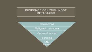
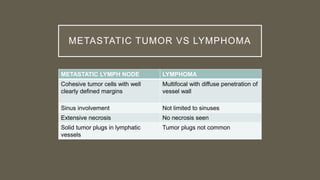
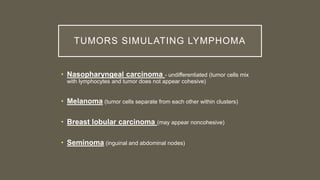
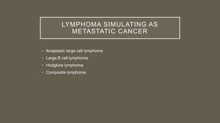
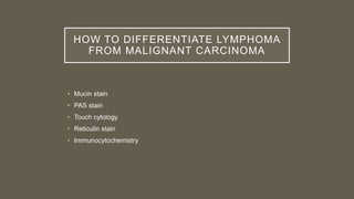
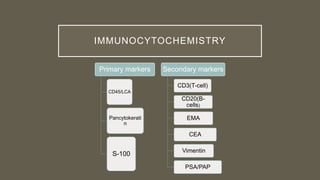
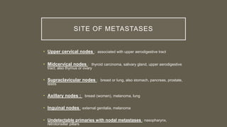
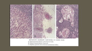
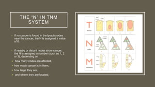
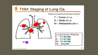
METASTATIC TUMORS TO LYMPH
NODE
Dr.Dur-e-Zahra
M.Phil Haematology
LEARNING OBJECTIVES
• Introduction to metastasis to lymph nodes
• Incidence of metastasis of Ca to lymph nodes
• Differentiate Lymphoma from Metastasis to lymph
node
• TNM staging
LYMPH NODE METASTASIS
• Most common site of metastatic malignancy
• Presence of metastasis is important for TNM staging
• Usually first clinical manifestation of disease
• Every lymph node enlargement calls for assessment by
pathologist whether its benign or malignant
MUST DETERMINE WHILE EXAMINING
A LYMPH NODE
Tumor Present or Not
Tumor Primary or
Metastatic
Possible sites of
Primary Tumor
Number of Metastatic
nodes involved
Presence of Extranodal or
Vascular involvement
INCIDENCE OF LYMPH NODE
METASTASIS
Carcinomas
Malignant melanoma
Germ cell tumors
Sarcoma
CNS
tumors
METASTATIC TUMOR VS LYMPHOMA
METASTATIC LYMPH NODE LYMPHOMA
Cohesive tumor cells with well
clearly defined margins
Multifocal with diffuse penetration of
vessel wall
Sinus involvement Not limited to sinuses
Extensive necrosis No necrosis seen
Solid tumor plugs in lymphatic
vessels
Tumor plugs not common
TUMORS SIMULATING LYMPHOMA
• Nasopharyngeal carcinoma - undifferentiated (tumor cells mix
with lymphocytes and tumor does not appear cohesive)
• Melanoma (tumor cells separate from each other within clusters)
• Breast lobular carcinoma (may appear noncohesive)
• Seminoma (inguinal and abdominal nodes)
LYMPHOMA SIMULATING AS
METASTATIC CANCER
• Anaplastic large cell lymphoma
• Large B cell lymphoma
• Hodgkins lymphoma
• Composite lymphoma
HOW TO DIFFERENTIATE LYMPHOMA
FROM MALIGNANT CARCINOMA
• Mucin stain
• PAS stain
• Touch cytology
• Reticulin stain
• Immunocytochemistry
IMMUNOCYTOCHEMISTRY
Primary markers
CD45/LCA
Pancytokerati
n
S-100
Secondary markers
CD3(T-cell)
CD20(B-
cells)
EMA
CEA
Vimentin
PSA/PAP
SITE OF METASTASES
• Upper cervical nodes : associated with upper aerodigestive tract
• Midcervical nodes : thyroid carcinoma, salivary gland, upper aerodigestive
tract; also thymus or ovary
• Supraclavicular nodes : breast or lung, also stomach, pancreas, prostate,
testis
• Axillary nodes : breast (women), melanoma, lung
• Inguinal nodes : external genitalia, melanoma
• Undetectable primaries with nodal metastases : nasopharynx,
retrotonsillar pillars
RESECTED
LYMPH NODE
SPECIMEN.
• A) 45mm yellowish
lobular structure can be
seen in the resected
lymph node
• B) The typical trabecular
structure pattern of
hepatocellular carcinoma
was confirmed
microscopically
GOOD TO
KNOW!
• Supraclavicular nodes
involved by intra-
abdominal carcinomas
are sometimes referred
to as Virchow or
Troisier nodes
SYSTEM FOR STAGING CANCER
• Lymph nodes play an important role in cancer staging,
which determines the extent of cancer in the body.
• The most commonly used systems for staging cancer is
the TNM system.
• It is based on the extent of the tumor (T), the extent of
spread to the lymph nodes (N), and the presence of
metastasis (M).
THE “N” IN TNM
SYSTEM
• If no cancer is found in the lymph nodes
near the cancer, the N is assigned a value
of 0.
• If nearby or distant nodes show cancer,
the N is assigned a number (such as 1, 2
or 3), depending on
 how many nodes are affected,
 how much cancer is in them,
 how large they are,
 and where they are located.
Metastatic tumors to lymph node

Metastatic tumors to lymph node

  • 1.
    METASTATIC TUMORS TOLYMPH NODE Dr.Dur-e-Zahra M.Phil Haematology
  • 2.
    LEARNING OBJECTIVES • Introductionto metastasis to lymph nodes • Incidence of metastasis of Ca to lymph nodes • Differentiate Lymphoma from Metastasis to lymph node • TNM staging
  • 3.
    LYMPH NODE METASTASIS •Most common site of metastatic malignancy • Presence of metastasis is important for TNM staging • Usually first clinical manifestation of disease • Every lymph node enlargement calls for assessment by pathologist whether its benign or malignant
  • 4.
    MUST DETERMINE WHILEEXAMINING A LYMPH NODE Tumor Present or Not Tumor Primary or Metastatic Possible sites of Primary Tumor Number of Metastatic nodes involved Presence of Extranodal or Vascular involvement
  • 5.
    INCIDENCE OF LYMPHNODE METASTASIS Carcinomas Malignant melanoma Germ cell tumors Sarcoma CNS tumors
  • 6.
    METASTATIC TUMOR VSLYMPHOMA METASTATIC LYMPH NODE LYMPHOMA Cohesive tumor cells with well clearly defined margins Multifocal with diffuse penetration of vessel wall Sinus involvement Not limited to sinuses Extensive necrosis No necrosis seen Solid tumor plugs in lymphatic vessels Tumor plugs not common
  • 7.
    TUMORS SIMULATING LYMPHOMA •Nasopharyngeal carcinoma - undifferentiated (tumor cells mix with lymphocytes and tumor does not appear cohesive) • Melanoma (tumor cells separate from each other within clusters) • Breast lobular carcinoma (may appear noncohesive) • Seminoma (inguinal and abdominal nodes)
  • 8.
    LYMPHOMA SIMULATING AS METASTATICCANCER • Anaplastic large cell lymphoma • Large B cell lymphoma • Hodgkins lymphoma • Composite lymphoma
  • 9.
    HOW TO DIFFERENTIATELYMPHOMA FROM MALIGNANT CARCINOMA • Mucin stain • PAS stain • Touch cytology • Reticulin stain • Immunocytochemistry
  • 10.
  • 11.
    SITE OF METASTASES •Upper cervical nodes : associated with upper aerodigestive tract • Midcervical nodes : thyroid carcinoma, salivary gland, upper aerodigestive tract; also thymus or ovary • Supraclavicular nodes : breast or lung, also stomach, pancreas, prostate, testis • Axillary nodes : breast (women), melanoma, lung • Inguinal nodes : external genitalia, melanoma • Undetectable primaries with nodal metastases : nasopharynx, retrotonsillar pillars
  • 12.
    RESECTED LYMPH NODE SPECIMEN. • A)45mm yellowish lobular structure can be seen in the resected lymph node • B) The typical trabecular structure pattern of hepatocellular carcinoma was confirmed microscopically
  • 15.
    GOOD TO KNOW! • Supraclavicularnodes involved by intra- abdominal carcinomas are sometimes referred to as Virchow or Troisier nodes
  • 16.
    SYSTEM FOR STAGINGCANCER • Lymph nodes play an important role in cancer staging, which determines the extent of cancer in the body. • The most commonly used systems for staging cancer is the TNM system. • It is based on the extent of the tumor (T), the extent of spread to the lymph nodes (N), and the presence of metastasis (M).
  • 17.
    THE “N” INTNM SYSTEM • If no cancer is found in the lymph nodes near the cancer, the N is assigned a value of 0. • If nearby or distant nodes show cancer, the N is assigned a number (such as 1, 2 or 3), depending on  how many nodes are affected,  how much cancer is in them,  how large they are,  and where they are located.