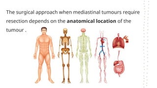
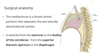
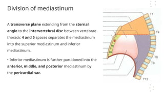
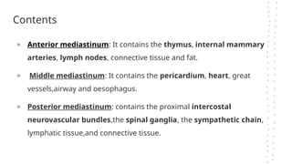
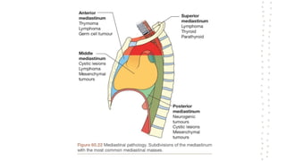
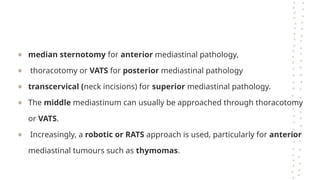
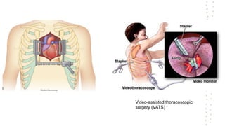
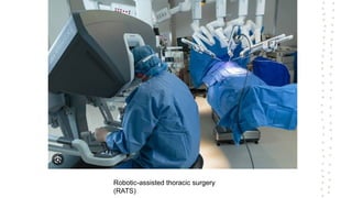
The Mediastinum
K U B U B A S H E E R
R O L L N O - 1 2 5
The surgical approach when mediastinal tumours require
resection depends on the anatomical location of the
tumour .
Surgical anatomy
● The mediastinum is a broad central
partition that separates the two laterally
placed pleural cavities.
● It extends from the sternum to the bodies
of the vertebrae; from the superior
thoracic aperture to the diaphragm.
Division of mediastinum
A transverse plane extending from the sternal
angle to the intervertebral disc between vertebrae
thoracic 4 and 5 spaces separates the mediastinum
into the superior mediastinum and inferior
mediastinum.
• Inferior mediastinum is further partitioned into the
anterior, middle, and posterior mediastinum by
the pericardial sac.
Contents
● Anterior mediastinum: It contains the thymus, internal mammary
arteries, lymph nodes, connective tissue and fat.
● Middle mediastinum: It contains the pericardium, heart, great
vessels,airway and oesophagus.
● Posterior mediastinum: contains the proximal intercostal
neurovascular bundles,the spinal ganglia, the sympathetic chain,
lymphatic tissue,and connective tissue.
Mediastinal Masses
● Various surgical procedures to approach structures in the
mediastinum are performed, usually as diagnostic procedures.
● The surgical approach when mediastinal tumours require
resection depends on the anatomical location of the tumour .
● median sternotomy for anterior mediastinal pathology,
● thoracotomy or VATS for posterior mediastinal pathology
● transcervical (neck incisions) for superior mediastinal pathology.
● The middle mediastinum can usually be approached through thoracotomy
or VATS.
● Increasingly, a robotic or RATS approach is used, particularly for anterior
mediastinal tumours such as thymomas.
Video-assisted thoracoscopic
surgery (VATS)
Robotic-assisted thoracic surgery
(RATS)

mediastinum................ Mmmmmmmmmmmmmmm

  • 1.
    The Mediastinum K UB U B A S H E E R R O L L N O - 1 2 5
  • 2.
    The surgical approachwhen mediastinal tumours require resection depends on the anatomical location of the tumour .
  • 3.
    Surgical anatomy ● Themediastinum is a broad central partition that separates the two laterally placed pleural cavities. ● It extends from the sternum to the bodies of the vertebrae; from the superior thoracic aperture to the diaphragm.
  • 4.
    Division of mediastinum Atransverse plane extending from the sternal angle to the intervertebral disc between vertebrae thoracic 4 and 5 spaces separates the mediastinum into the superior mediastinum and inferior mediastinum. • Inferior mediastinum is further partitioned into the anterior, middle, and posterior mediastinum by the pericardial sac.
  • 5.
    Contents ● Anterior mediastinum:It contains the thymus, internal mammary arteries, lymph nodes, connective tissue and fat. ● Middle mediastinum: It contains the pericardium, heart, great vessels,airway and oesophagus. ● Posterior mediastinum: contains the proximal intercostal neurovascular bundles,the spinal ganglia, the sympathetic chain, lymphatic tissue,and connective tissue.
  • 6.
  • 8.
    ● Various surgicalprocedures to approach structures in the mediastinum are performed, usually as diagnostic procedures. ● The surgical approach when mediastinal tumours require resection depends on the anatomical location of the tumour .
  • 9.
    ● median sternotomyfor anterior mediastinal pathology, ● thoracotomy or VATS for posterior mediastinal pathology ● transcervical (neck incisions) for superior mediastinal pathology. ● The middle mediastinum can usually be approached through thoracotomy or VATS. ● Increasingly, a robotic or RATS approach is used, particularly for anterior mediastinal tumours such as thymomas.
  • 10.
  • 11.