x Perform an ABCDEF primary survey, assessing the airway, breathing, circulation, neurological status, exposure and providing fluids.
x Take a thorough history to determine burn mechanism and rule out inhalational injuries or other trauma.
x Intubate if the airway is compromised or at risk of compromise from swelling. Monitor for signs of smoke inhalation like stridor.
x Ensure adequate oxygenation, treat for carbon monoxide poisoning if indicated, and consider escharotomies if burns are restricting breathing.
x Refer major burns covering over 10% of total body surface area or with complicating factors to a specialist burns unit.
pediatric emergency its from important topics in pediatric and in this show we will discuss the most important things like shock and its types with special overview on hypovomlemic shock and its treatment and anaphylactic shock also with its treatment then transfered to other important one which is status asthmaticus and its treatment and then status epiliptucs and poisning
pediatric emergency its from important topics in pediatric and in this show we will discuss the most important things like shock and its types with special overview on hypovomlemic shock and its treatment and anaphylactic shock also with its treatment then transfered to other important one which is status asthmaticus and its treatment and then status epiliptucs and poisning
GEMC: Hypertensive Urgency and Emergency: Resident TrainingOpen.Michigan
This is a lecture by Dr. Keith Kocher from the Ghana Emergency Medicine Collaborative. To download the editable version (in PPT), to access additional learning modules, or to learn more about the project, see http://openmi.ch/em-gemc. Unless otherwise noted, this material is made available under the terms of the Creative Commons Attribution Share Alike-3.0 License: http://creativecommons.org/licenses/by-sa/3.0/.
Acute management in burns
Types of burns
Admission criteria in burns
Fluid management in burns
Early surgical management in burns
Facial burn
Chemical burn
Eye burn
Ear burn
Hand burn
Electrical burn
Burn Management is complex topic to learn and master during MS General Surgery courses.It is pet favorite questions during viva voice/practical exams.With this ppt it will help you to gain basic knowledge related to this Topic.If you like my work then do share your response by mailing me on officialdrrishi@outlook.com
Burns management presentation by 2nd yr MSC nursing studentSigymol John
this ppt deals with the management part of burns, mainly divided as pre-hospital care, emergent phase,acute phase and rehabilitation phase along with nursing management,nursing diagnosis and interventions.
Burns are tissue damage that results from heat, overexposure to the sun or other radiation, or chemical or electrical contact. Burns can be minor medical problems or life-threatening emergencies. The treatment of burns depends on the location and severity of the damage.
Title: Sense of Smell
Presenter: Dr. Faiza, Assistant Professor of Physiology
Qualifications:
MBBS (Best Graduate, AIMC Lahore)
FCPS Physiology
ICMT, CHPE, DHPE (STMU)
MPH (GC University, Faisalabad)
MBA (Virtual University of Pakistan)
Learning Objectives:
Describe the primary categories of smells and the concept of odor blindness.
Explain the structure and location of the olfactory membrane and mucosa, including the types and roles of cells involved in olfaction.
Describe the pathway and mechanisms of olfactory signal transmission from the olfactory receptors to the brain.
Illustrate the biochemical cascade triggered by odorant binding to olfactory receptors, including the role of G-proteins and second messengers in generating an action potential.
Identify different types of olfactory disorders such as anosmia, hyposmia, hyperosmia, and dysosmia, including their potential causes.
Key Topics:
Olfactory Genes:
3% of the human genome accounts for olfactory genes.
400 genes for odorant receptors.
Olfactory Membrane:
Located in the superior part of the nasal cavity.
Medially: Folds downward along the superior septum.
Laterally: Folds over the superior turbinate and upper surface of the middle turbinate.
Total surface area: 5-10 square centimeters.
Olfactory Mucosa:
Olfactory Cells: Bipolar nerve cells derived from the CNS (100 million), with 4-25 olfactory cilia per cell.
Sustentacular Cells: Produce mucus and maintain ionic and molecular environment.
Basal Cells: Replace worn-out olfactory cells with an average lifespan of 1-2 months.
Bowman’s Gland: Secretes mucus.
Stimulation of Olfactory Cells:
Odorant dissolves in mucus and attaches to receptors on olfactory cilia.
Involves a cascade effect through G-proteins and second messengers, leading to depolarization and action potential generation in the olfactory nerve.
Quality of a Good Odorant:
Small (3-20 Carbon atoms), volatile, water-soluble, and lipid-soluble.
Facilitated by odorant-binding proteins in mucus.
Membrane Potential and Action Potential:
Resting membrane potential: -55mV.
Action potential frequency in the olfactory nerve increases with odorant strength.
Adaptation Towards the Sense of Smell:
Rapid adaptation within the first second, with further slow adaptation.
Psychological adaptation greater than receptor adaptation, involving feedback inhibition from the central nervous system.
Primary Sensations of Smell:
Camphoraceous, Musky, Floral, Pepperminty, Ethereal, Pungent, Putrid.
Odor Detection Threshold:
Examples: Hydrogen sulfide (0.0005 ppm), Methyl-mercaptan (0.002 ppm).
Some toxic substances are odorless at lethal concentrations.
Characteristics of Smell:
Odor blindness for single substances due to lack of appropriate receptor protein.
Behavioral and emotional influences of smell.
Transmission of Olfactory Signals:
From olfactory cells to glomeruli in the olfactory bulb, involving lateral inhibition.
Primitive, less old, and new olfactory systems with different path
Pulmonary Thromboembolism - etilogy, types, medical- Surgical and nursing man...VarunMahajani
Disruption of blood supply to lung alveoli due to blockage of one or more pulmonary blood vessels is called as Pulmonary thromboembolism. In this presentation we will discuss its causes, types and its management in depth.
Ozempic: Preoperative Management of Patients on GLP-1 Receptor Agonists Saeid Safari
Preoperative Management of Patients on GLP-1 Receptor Agonists like Ozempic and Semiglutide
ASA GUIDELINE
NYSORA Guideline
2 Case Reports of Gastric Ultrasound
Tom Selleck Health: A Comprehensive Look at the Iconic Actor’s Wellness Journeygreendigital
Tom Selleck, an enduring figure in Hollywood. has captivated audiences for decades with his rugged charm, iconic moustache. and memorable roles in television and film. From his breakout role as Thomas Magnum in Magnum P.I. to his current portrayal of Frank Reagan in Blue Bloods. Selleck's career has spanned over 50 years. But beyond his professional achievements. fans have often been curious about Tom Selleck Health. especially as he has aged in the public eye.
Follow us on: Pinterest
Introduction
Many have been interested in Tom Selleck health. not only because of his enduring presence on screen but also because of the challenges. and lifestyle choices he has faced and made over the years. This article delves into the various aspects of Tom Selleck health. exploring his fitness regimen, diet, mental health. and the challenges he has encountered as he ages. We'll look at how he maintains his well-being. the health issues he has faced, and his approach to ageing .
Early Life and Career
Childhood and Athletic Beginnings
Tom Selleck was born on January 29, 1945, in Detroit, Michigan, and grew up in Sherman Oaks, California. From an early age, he was involved in sports, particularly basketball. which played a significant role in his physical development. His athletic pursuits continued into college. where he attended the University of Southern California (USC) on a basketball scholarship. This early involvement in sports laid a strong foundation for his physical health and disciplined lifestyle.
Transition to Acting
Selleck's transition from an athlete to an actor came with its physical demands. His first significant role in "Magnum P.I." required him to perform various stunts and maintain a fit appearance. This role, which he played from 1980 to 1988. necessitated a rigorous fitness routine to meet the show's demands. setting the stage for his long-term commitment to health and wellness.
Fitness Regimen
Workout Routine
Tom Selleck health and fitness regimen has evolved. adapting to his changing roles and age. During his "Magnum, P.I." days. Selleck's workouts were intense and focused on building and maintaining muscle mass. His routine included weightlifting, cardiovascular exercises. and specific training for the stunts he performed on the show.
Selleck adjusted his fitness routine as he aged to suit his body's needs. Today, his workouts focus on maintaining flexibility, strength, and cardiovascular health. He incorporates low-impact exercises such as swimming, walking, and light weightlifting. This balanced approach helps him stay fit without putting undue strain on his joints and muscles.
Importance of Flexibility and Mobility
In recent years, Selleck has emphasized the importance of flexibility and mobility in his fitness regimen. Understanding the natural decline in muscle mass and joint flexibility with age. he includes stretching and yoga in his routine. These practices help prevent injuries, improve posture, and maintain mobilit
Anti ulcer drugs and their Advance pharmacology ||
Anti-ulcer drugs are medications used to prevent and treat ulcers in the stomach and upper part of the small intestine (duodenal ulcers). These ulcers are often caused by an imbalance between stomach acid and the mucosal lining, which protects the stomach lining.
||Scope: Overview of various classes of anti-ulcer drugs, their mechanisms of action, indications, side effects, and clinical considerations.
Ethanol (CH3CH2OH), or beverage alcohol, is a two-carbon alcohol
that is rapidly distributed in the body and brain. Ethanol alters many
neurochemical systems and has rewarding and addictive properties. It
is the oldest recreational drug and likely contributes to more morbidity,
mortality, and public health costs than all illicit drugs combined. The
5th edition of the Diagnostic and Statistical Manual of Mental Disorders
(DSM-5) integrates alcohol abuse and alcohol dependence into a single
disorder called alcohol use disorder (AUD), with mild, moderate,
and severe subclassifications (American Psychiatric Association, 2013).
In the DSM-5, all types of substance abuse and dependence have been
combined into a single substance use disorder (SUD) on a continuum
from mild to severe. A diagnosis of AUD requires that at least two of
the 11 DSM-5 behaviors be present within a 12-month period (mild
AUD: 2–3 criteria; moderate AUD: 4–5 criteria; severe AUD: 6–11 criteria).
The four main behavioral effects of AUD are impaired control over
drinking, negative social consequences, risky use, and altered physiological
effects (tolerance, withdrawal). This chapter presents an overview
of the prevalence and harmful consequences of AUD in the U.S.,
the systemic nature of the disease, neurocircuitry and stages of AUD,
comorbidities, fetal alcohol spectrum disorders, genetic risk factors, and
pharmacotherapies for AUD.
Couples presenting to the infertility clinic- Do they really have infertility...Sujoy Dasgupta
Dr Sujoy Dasgupta presented the study on "Couples presenting to the infertility clinic- Do they really have infertility? – The unexplored stories of non-consummation" in the 13th Congress of the Asia Pacific Initiative on Reproduction (ASPIRE 2024) at Manila on 24 May, 2024.
Lung Cancer: Artificial Intelligence, Synergetics, Complex System Analysis, S...Oleg Kshivets
RESULTS: Overall life span (LS) was 2252.1±1742.5 days and cumulative 5-year survival (5YS) reached 73.2%, 10 years – 64.8%, 20 years – 42.5%. 513 LCP lived more than 5 years (LS=3124.6±1525.6 days), 148 LCP – more than 10 years (LS=5054.4±1504.1 days).199 LCP died because of LC (LS=562.7±374.5 days). 5YS of LCP after bi/lobectomies was significantly superior in comparison with LCP after pneumonectomies (78.1% vs.63.7%, P=0.00001 by log-rank test). AT significantly improved 5YS (66.3% vs. 34.8%) (P=0.00000 by log-rank test) only for LCP with N1-2. Cox modeling displayed that 5YS of LCP significantly depended on: phase transition (PT) early-invasive LC in terms of synergetics, PT N0—N12, cell ratio factors (ratio between cancer cells- CC and blood cells subpopulations), G1-3, histology, glucose, AT, blood cell circuit, prothrombin index, heparin tolerance, recalcification time (P=0.000-0.038). Neural networks, genetic algorithm selection and bootstrap simulation revealed relationships between 5YS and PT early-invasive LC (rank=1), PT N0—N12 (rank=2), thrombocytes/CC (3), erythrocytes/CC (4), eosinophils/CC (5), healthy cells/CC (6), lymphocytes/CC (7), segmented neutrophils/CC (8), stick neutrophils/CC (9), monocytes/CC (10); leucocytes/CC (11). Correct prediction of 5YS was 100% by neural networks computing (area under ROC curve=1.0; error=0.0).
CONCLUSIONS: 5YS of LCP after radical procedures significantly depended on: 1) PT early-invasive cancer; 2) PT N0--N12; 3) cell ratio factors; 4) blood cell circuit; 5) biochemical factors; 6) hemostasis system; 7) AT; 8) LC characteristics; 9) LC cell dynamics; 10) surgery type: lobectomy/pneumonectomy; 11) anthropometric data. Optimal diagnosis and treatment strategies for LC are: 1) screening and early detection of LC; 2) availability of experienced thoracic surgeons because of complexity of radical procedures; 3) aggressive en block surgery and adequate lymph node dissection for completeness; 4) precise prediction; 5) adjuvant chemoimmunoradiotherapy for LCP with unfavorable prognosis.
These lecture slides, by Dr Sidra Arshad, offer a quick overview of physiological basis of a normal electrocardiogram.
Learning objectives:
1. Define an electrocardiogram (ECG) and electrocardiography
2. Describe how dipoles generated by the heart produce the waveforms of the ECG
3. Describe the components of a normal electrocardiogram of a typical bipolar leads (limb II)
4. Differentiate between intervals and segments
5. Enlist some common indications for obtaining an ECG
Study Resources:
1. Chapter 11, Guyton and Hall Textbook of Medical Physiology, 14th edition
2. Chapter 9, Human Physiology - From Cells to Systems, Lauralee Sherwood, 9th edition
3. Chapter 29, Ganong’s Review of Medical Physiology, 26th edition
4. Electrocardiogram, StatPearls - https://www.ncbi.nlm.nih.gov/books/NBK549803/
5. ECG in Medical Practice by ABM Abdullah, 4th edition
6. ECG Basics, http://www.nataliescasebook.com/tag/e-c-g-basics
Surgical Site Infections, pathophysiology, and prevention.pptx
Manejo inicial gran quemado i
1. ABC of burns
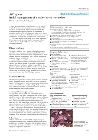
Initial management of a major burn: I—overview
Shehan Hettiaratchy, Remo Papini
A major burn is defined as a burn covering 25% or more of
total body surface area, but any injury over more than 10%
should be treated similarly. Rapid assessment is vital. The
general approach to a major burn can be extrapolated to
managing any burn. The most important points are to take an
accurate history and make a detailed examination of the patient
and the burn, to ensure that key information is not missed.
This article outlines the structure of the initial assessment.
The next article will cover the detailed assessment of burn
surface area and depth and how to calculate the fluid
resuscitation formula.
History taking
The history of a burn injury can give valuable information
about the nature and extent of the burn, the likelihood of
inhalational injury, the depth of burn, and probability of other
injuries. The exact mechanism of injury and any prehospital
treatment must be established.
A patient’s history must be obtained on admission, as this
may be the only time that a first hand history is obtainable.
Swelling may develop around the airway in the hours after
injury and require intubation, making it impossible for the
patient to give a verbal history. A brief medical history should
be taken, outlining previous medical problems, medications,
allergies, and vaccinations. Patients’ smoking habits should be
determined as these may affect blood gas analyses.
Primary survey
The initial management of a severely burnt patient is similar to
that of any trauma patient. A modified “advanced trauma life
support” primary survey is performed, with particular emphasis
on assessment of the airway and breathing. The burn injury
must not distract from this sequential assessment, otherwise
serious associated injuries may be missed.
A—Airway with cervical spine control
An assessment must be made as to whether the airway is
compromised or is at risk of compromise. The cervical spine
should be protected unless it is definitely not injured. Inhalation
of hot gases will result in a burn above the vocal cords. This
burn will become oedematous over the following hours,
especially after fluid resuscitation has begun. This means that an
airway that is patent on arrival at hospital may occlude after
admission. This can be a particular problem in small children.
Direct inspection of the oropharynx should be done by a
senior anaesthetist. If there is any concern about the patency of
the airway then intubation is the safest policy. However, an
unnecessary intubation and sedation could worsen a patient’s
condition, so the decision to intubate should be made carefully.
B—Breathing
All burn patients should receive 100% oxygen through a
humidified non-rebreathing mask on presentation. Breathing
problems are considered to be those that affect the respiratory
system below the vocal cords. There are several ways that a burn
injury can compromise respiration.
Initial assessment of a major burn
x Perform an ABCDEF primary survey
A—Airway with cervical spine control, B—Breathing,
C—Circulation, D—Neurological disability, E—Exposure with
environmental control, F—Fluid resuscitation
x Assess burn size and depth (see later article for detail)
x Establish good intravenous access and give fluids
x Give analgesia
x Catheterise patient or establish fluid balance monitoring
x Take baseline blood samples for investigation
x Dress wound
x Perform secondary survey, reassess, and exclude or treat associated
injuries
x Arrange safe transfer to specialist burns facility
Key points of a burn history
Exact mechanism
x Type of burn agent (scald,
flame, electrical, chemical)
x How did it come into contact
with patient?
x What first aid was performed?
x What treatment has been
started?
x Is there risk of concomitant
injuries (such as fall from height,
road traffic crash, explosion)?
x Is there risk of inhalational
injuries (did burn occur in an
enclosed space)?
Exact timings
x When did the injury occur?
x How long was patient exposed
to energy source?
x How long was cooling applied?
x When was fluid resuscitation
started?
Exact injury
Scalds
x What was the liquid? Was it
boiling or recently boiled?
x If tea or coffee, was milk in it?
x Was a solute in the liquid?
(Raises boiling temperature
and causes worse injury, such
as boiling rice)
Electrocution injuries
x What was the voltage (domestic
or industrial)?
x Was there a flash or arcing?
x Contact time
Chemical injuries
x What was the chemical?
Is there any suspicion of non-accidental injury?
x See previous article
Airway management
Signs of inhalational injury
x History of flame burns or burns in
an enclosed space
x Full thickness or deep dermal burns
to face, neck, or upper torso
x Singed nasal hair
x Carbonaceous sputum or carbon
particles in oropharynx
Indications for intubation
x Erythema or swelling of
oropharynx on direct
visualisation
x Change in voice, with
hoarseness or harsh cough
x Stridor, tachypnoea, or
dyspnoea
Carbonaceous particles
staining a patient’s face after
a burn in an enclosed space.
This suggests there is
inhalational injury
This is the fourth in a series of 12 articles
Clinical review
1555BMJ VOLUME 328 26 JUNE 2004 bmj.com
2. Mechanical restriction of breathing—Deep dermal or full
thickness circumferential burns of the chest can limit chest
excursion and prevent adequate ventilation. This may require
escharotomies (see next article).
Blast injury—If there has been an explosion, blast lung can
complicate ventilation. Penetrating injuries can cause tension
pneumothoraces, and the blast itself can cause lung contusions
and alveolar trauma and lead to adult respiratory distress
syndrome.
Smoke inhalation—The products of combustion, though
cooled by the time they reach the lungs, act as direct irritants to
the lungs, leading to bronchospasm, inflammation, and
bronchorrhoea. The ciliary action of pneumocytes is impaired,
exacerbating the situation. The inflammatory exudate created is
not cleared, and atelectasis or pneumonia follows. The situation
can be particularly severe in asthmatic patients. Non-invasive
management can be attempted, with nebulisers and positive
pressure ventilation with some positive end-expiratory pressure.
However, patients may need a period of ventilation, as this
allows adequate oxygenation and permits regular lung toileting.
Carboxyhaemoglobin—Carbon monoxide binds to
deoxyhaemoglobin with 40 times the affinity of oxygen. It also
binds to intracellular proteins, particularly the cytochrome
oxidase pathway. These two effects lead to intracellular and
extracellular hypoxia. Pulse oximetry cannot differentiate
between oxyhaemoglobin and carboxyhaemoglobin, and may
therefore give normal results. However, blood gas analysis will
reveal metabolic acidosis and raised carboxyhaemoglobin levels
but may not show hypoxia. Treatment is with 100% oxygen,
which displaces carbon monoxide from bound proteins six
times faster than does atmospheric oxygen. Patients with
carboxyhaemoglobin levels greater than 25-30% should be
ventilated. Hyperbaric therapy is rarely practical and has not
been proved to be advantageous. It takes longer to shift the
carbon monoxide from the cytochrome oxidase pathway than
from haemoglobin, so oxygen therapy should be continued
until the metabolic acidosis has cleared.
C—Circulation
Intravenous access should be established with two large bore
cannulas preferably placed through unburnt tissue. This is an
opportunity to take blood for checking full blood count, urea
and electrolytes, blood group, and clotting screen. Peripheral
circulation must be checked. Any deep or full thickness
circumferential extremity burn can act as a tourniquet,
especially once oedema develops after fluid resuscitation. This
may not occur until some hours after the burn. If there is any
suspicion of decreased perfusion due to circumferential burn,
the tissue must be released with escharotomies (see next article).
Profound hypovolaemia is not the normal initial response
to a burn. If a patient is hypotensive then it is may be due to
delayed presentation, cardiogenic dysfunction, or an occult
source of blood loss (chest, abdomen, or pelvis).
D—Neurological disability
All patients should be assessed for responsiveness with the
Glasgow coma scale; they may be confused because of hypoxia
or hypovolaemia.
E—Exposure with environment control
The whole of a patient should be examined (including the back)
to get an accurate estimate of the burn area (see later) and to
check for any concomitant injuries. Burn patients, especially
children, easily become hypothermic. This will lead to
hypoperfusion and deepening of burn wounds. Patients should
be covered and warmed as soon as possible.
Acute bronchoscopy being performed to assess amount of damage to the
bronchial tree. Patient has been covered in a blanket and a heat lamp placed
overhead to prevent excessive cooling
Signs of carboxyhaemoglobinaemia
COHb levels Symptoms
0-10% Minimal (normal level in heavy smokers)
10-20% Nausea, headache
20-30% Drowsiness, lethargy
30-40% Confusion, agitation
40-50% Coma, respiratory depression
> 50% Death
COHb = Carboxyhaemoglobin
Airway
Compromised or at
risk of compromise?
No
Yes
Yes
No
Yes
Yes
Circulation
Compromised perfusion
to an extremity?
Neurological disability
Impaired score on
Glasgow coma scale?
No
Breathing
Compromised?
Cause:
Mechanical
Carboxyhaemoglobin
Smoke inhalation
Blast injury
Consider:
Hypoxia (carboxyhaemoglobin level?)
Hypovolaemia
Escharotomies
Intubate and ventilate
Nebulisers
Non-invasive ventilation
Invasive ventilation
Invasive ventilation
Chest drains
Intubate
Escharotomies
Go back and re-evaluate
No
Exposure
Fully assess burn area and depth
Full examination for concomitant injuries
Keep warm
Fluids
Calculate resuscitation formula based on
surface area and time since burn
Algorithm for primary survey of a major burn injury
Clinical review
1556 BMJ VOLUME 328 26 JUNE 2004 bmj.com
3. F—Fluid resuscitation
The resuscitation regimen should be determined and begun.
This is based on the estimation of the burn area, and the
detailed calculation is covered in the next article. A urinary
catheter is mandatory in all adults with injuries covering > 20%
of total body surface area to monitor urine output. Children’s
urine output can be monitored with external catchment devices
or by weighing nappies provided the injury is < 20% of total
body area. In children the interosseous route can be used for
fluid administration if intravenous access cannot be obtained,
but should be replaced by intravenous lines as soon as possible.
Analgesia
Superficial burns can be extremely painful. All patients with
large burns should receive intravenous morphine at a dose
appropriate to body weight. This can be easily titrated against
pain and respiratory depression. The need for further doses
should be assessed within 30 minutes.
Investigations
The amount of investigations will vary with the type of burn.
Secondary survey
At the end of the primary survey and the start of emergency
management, a secondary survey should be performed. This is
a head to toe examination to look for any concomitant injuries.
Dressing the wound
Once the surface area and depth of a burn have been estimated,
the burn wound should be washed and any loose skin removed.
Blisters should be deroofed for ease of dressing, except for
palmar blisters (painful), unless these are large enough to
restrict movement. The burn should then be dressed.
For an acute burn which will be referred to a burn centre,
cling film is an ideal dressing as it protects the wound, reduces
heat and evaporative losses, and does not alter the wound
appearance. This will permit accurate evaluation by the burn
team later. Flamazine should not be used on a burn that is to be
referred immediately, since it makes assessment of depth more
difficult.
Referral to a burns unit
The National Burn Care Review has established referral
guidelines to specialist units. Burns are divided into complex
burns (those that require specialist intervention) and
non{complex burns (those that do not require immediate
admission to a specialist unit). Complex burns should be
referred automatically. If you are not sure whether a burn
should be referred, discuss the case with your local burns unit.
It is also important to discuss all burns that are not healed
within two weeks.
Shehan Hettiaratchy is specialist registrar in plastic and reconstructive
surgery, Pan-Thames Training Scheme, London; Remo Papini is
consultant and clinical lead in burns, West Midlands Regional Burn
Unit, Selly Oak University Hospital, Birmingham.
The ABC of burns is edited by Shehan Hettiaratchy; Remo Papini;
and Peter Dziewulski, consultant burns and plastic surgeon, St
Andrews Centre for Plastic Surgery and Burns, Broomfield Hospital,
Chelmsford. The series will be published as a book in the autumn.
Competing interests: RP has been reimbursed by Johnson & Johnson,
manufacturer of Integra, and Smith & Nephew, manufacturer of Acticoat
and TransCyte, for attending symposia on burn care.
BMJ 2004;328:1555–7
Investigations for major burns*
General
x Full blood count, packed cell volume, urea and electrolyte
concentration, clotting screen
x Blood group, and save or crossmatch serum
Electrical injuries
x 12 lead electrocardiography
x Cardiac enzymes (for high tension injuries)
Inhalational injuries
x Chest x ray
x Arterial blood gas analysis
Can be useful in any burn, as the base excess is predictive of the
amount of fluid resuscitation required
Helpful for determining success of fluid resuscitation and essential
with inhalational injuries or exposure to carbon monoxide
*Any concomitant trauma will have its own investigations
Indications for referral to a burns unit
All complex injuries should be referred
A burn injury is more likely to be complex if associated with:
x Extremes of age—under 5 or over 60 years
x Site of injury
Face, hands, or perineum
Feet (dermal or full thickness loss)
Any flexure, particularly the neck or axilla
Circumferential dermal or full thickness burn of limb, torso, or neck
x Inhalational injury
Any substantial injury, excluding pure carbon monoxide poisoning
x Mechanism of injury
Chemical injury > 5% of total body surface area
Exposure to ionising radiation
High pressure steam injury
High tension electrical injury
Hydrofluoric acid burn >1% of total body surface area
Suspicion of non-accidental injury
x Large size (dermal or full thickness loss)
Paediatric ( < 16 years old) > 5% of total body surface area
Adult ( ≥ 16 years) > 10% of total body surface area
x Coexisting conditions
Any serious medical conditions (cardiac dysfunction,
immunosuppression, pregnancy)
Any associated injuries (fractures, head injuries, crush injuries)
Key points
x Perform a systematic assessment as with any
trauma patient (don’t get distracted by the burn)
x Beware of airway compromise
x Provide adequate analgesia
x Exclude any concomitant injuries
x Discuss with a burns unit early
x If in doubt, reassess
Further reading
x Sheridan R. Burns. Crit Care Med 2002;30:S500-14
x British Burn Association. Emergency management of severe burns
course manual, UK version. Wythenshawe Hospital, Manchester,
1996
x Herndon D. Total burn care. 2nd ed. London: WB Saunders, 2002
x Kao CC, Garner WL. Acute burns. Plast Reconstr Surg 2000;105:
2482-93
x Burnsurgery.org. www.burnsurgery.org
Clinical review
1557BMJ VOLUME 328 26 JUNE 2004 bmj.com