Case of odontogeic fibromyxoma of maxilla case report: a rare entity.
•
1 like•385 views

Dr Bhavik Miyani, Resident Doctor in Department of Oral & Maxillofacial Surgery, Narsinhbhai Patel Dental College & Hospital, SPU, Visnagar.

Recommended
More Related Content
What's hot
What's hot (20)
Similar to Case of odontogeic fibromyxoma of maxilla case report: a rare entity.
Similar to Case of odontogeic fibromyxoma of maxilla case report: a rare entity. (20)
More from Dr Bhavik Miyani
More from Dr Bhavik Miyani (20)
Recently uploaded
Recently uploaded (20)
Case of odontogeic fibromyxoma of maxilla case report: a rare entity.
- 1. Odontogenic Fibromyxoma of Maxilla Case Report- A Rare Entity DR. BHAVIK MIYANI, DEPARTMENT OF OMFS, NPDCH
- 2. CONTENTS Introduction Case Report Conclusion References
- 3. Fibromyxomas of head and neck are rare, non capsulated, benign neoplasms which consist either partly or wholly of myxomatous tissue, depending upon the amount of collagen which is present. The term “odontogenic myxoma” is often applied when the tumor occurs in the jaws to reflect its odontogenic origin. INTRODUCTION Thoma and Goldman, 1947
- 4. Myxomas can be found in heart, skin, and subcutaneous tissue and centrally in the bone, but myxomas of the jaws are encountered rarely. It accounts for only 1% to 3% of all cysts and tumors of the jaws. We report a case of odontogenic fibromyxoma of the maxilla in a 25-years old female patient.
- 5. A 25 years old female patient reported to the Department of Oral and Maxillofacial Surgery with a chief complaint of swelling in upper right front region of jaw since 1 year. CASE REPORT
- 6. Patient was relatively asymptomatic before one year. Then patient developed swelling in right front side of upper jaw which was an symptomatic. The swelling was initially small and gradually increased to its present size of approximately 3 x 2 cm. There was no preceding history of trauma, fever, dental trouble or the nasal obstruction. H/O - Maxillary sinusitis since six months. HISTORY OF PRESENET ILLNESS
- 9. Adenomatoid Odontogenic Tumor PROVISIONAL DIAGNOSIS
- 11. DIFFERENTIAL DIAGNOSIS ADENOMATOID ODONTOGENIC TUMOR ANTRAL POLYP FIBRO- OSSEOUS LESION AMELOBLASTIC FIBROMA ODONTOGENIC FIBROMYXOMA AMELOBLASTIC FIBROMA × Etiology: Arising from the enamel organ or dental lamina. × Age: 1st and 2nd Decades of life. × Gender Predilection: Male> Female. × Site: Posterior Mandible F/B Posterior Maxilla. Painless, Slow Growing Swelling. × Associated with Impacted tooth. × Radiographic Findings: Unilocular Lesion.
- 12. DIFFERENTIAL DIAGNOSIS ADENOMATOID ODONTOGENIC TUMOR ANTRAL POLYP FIBRO- OSSEOUS LESION AMELOBLASTIC FIBROMA ODONTOGENIC FIBROMYXOMA ADENOMATOID ODONTOGENIC TUMOR × Etiology: Arising from the enamel organ or dental lamina. Age: Young People. Gender Predilection: Female: Male = 2:1. Site: Anterior Maxilla: Anterior Mandible = 2:1. × Associated with Impacted tooth (Mostly Canine). Painless Swelling, Slow Growing. × Radiographic Findings: Radiolucency extends apically beyond CEJ of unerupted tooth.
- 13. DIFFERENTIAL DIAGNOSIS ADENOMATOID ODONTOGENIC TUMOR ANTRAL POLYP FIBRO- OSSEOUS LESION AMELOBLASTIC FIBROMA ODONTOGENIC FIBROMYXOMA ANTRAL POLYP × Etiology: Recurrent Allergic Diseases. × Age: Children and Young Adult. × Gender Predilection: Female: Male = 1:2. Site: Unilaterally in Maxillary Sinus. Nasal Obstruction. CT Scan Findings: Expansion or Erosion of bony wall.
- 14. DIFFERENTIAL DIAGNOSIS ADENOMATOID ODONTOGENIC TUMOR ANTRAL POLYP FIBRO- OSSEOUS LESION AMELOBLASTIC FIBROMA ODONTOGENIC FIBROMYXOMA FIBRO- OSSEOUS LESION × Etiology: Genetic. × Age: Children are more affected. Gender Predilection: Female: Male = 1:1. × Site: Maxilla< Mandible. × Associated with Impacted tooth. Painless Swelling, Slow Growing. × Radiographic Findings: Expansion of Bony walls and Cotton wool appearance.
- 15. DIFFERENTIAL DIAGNOSIS ADENOMATOID ODONTOGENIC TUMOR ANTRAL POLYP FIBRO- OSSEOUS LESION AMELOBLASTIC FIBROMA ODONTOGENIC FIBROMYXOMA ODONTOGENIC FIBROMYXOMA Etiology: Arising from Odontogenic epithelium or ectomesenchymal origin. Age: 2nd to 3rd decades of life. Gender Predilection: Female: Male = 2:1. Site: Posterior Mandible F/B Posterior Maxilla. Painless, Slow Growing. Radiographic Findings: Honey Comb Appearance and Expansion of Bony walls. ODONTOGENIC FIBROMYXOMA
- 16. INVESTIGATIONS Routine blood investigations. Radiological investigations. 1. OPG 2. CT Scan Histopathological investigations.
- 17. Complete Obliteration of Right Maxillary Sinus with irregular, diffuse haziness OPG
- 18. CT Coronal and Axial Scan Showing Complete Obliteration of Right Maxillary Sinus And Lesion size of 5 X 4 cm. CT SCANCT SCAN
- 19. HISTOPATHOLOGY Histopathology Photograph showing Loose Myxoid Stroma with Interspersed Dense Collagen Bundles and Numerous Stellate to Plump Spindle Shaped Cells in the Stroma
- 20. Odontogenic Fibromyxoma of Maxilla FINAL DIAGNOSIS
- 21. Enucleation and Curettage of lesion including radical resection including a margin of 1.5-2 mm healthy bone using crevicular incision under general anesthesia. Long term follow up because higher recurrence rate. TREATMENT PLAN
- 22. 1. Incision Marking 2. Reflection and Exposure of Site 4. Complete Curettage of Lesion Showing Empty Cavity 6. Closure5. Resected Specimen 3. Enucleation of Lesion TREATMENT GIVEN
- 23. Healing after 1 month POST OPERATIVE
- 24. Reported cases of fibromyxoma of the maxilla in literature
- 25. Myxomas of head and neck are rare tumors and the maxilla is a rare location of a fibromyxoma. It poses a diagnostic and therapeutic challenge hence correlation of clinical, radiological and histopathological features are essential when trying to diagnose lesions which lack the characteristic appearance. Its management is surgical, and ranges from enucleation and curettage to complete resection and peripheral osteotomy according to its size. In this young married woman as the extent of lesion was confined to the maxillary sinus, we have operated for an intraoral surgical approach. CONCLUSION
- 26. 1. Kaffe I, Noor H, Buchner A. Clinical and radiological features of odontogenicmyxoma of the jaws. Dentomaxillofac Radiol 1997;26:299-303. 2. Chuchurru JA, Luberti R, Cornicelli JC, Dominguez FV. Myxoma of the mandible with unusual radiographic appearance. J Oral Maxillofac Surg 1985;43:987-90. 3. Asaumi J, Konouchi H, Hisatomi M, Kishi K. Odontogenic myxoma of maxillary sinus: CT and MR – pathologic correlation. Eur J Radiol 2001;37:1-4. 4. Peltola J, Magnusson B, Happonen RP, Borrman H. Odontogenic myxoma: A radiographic study of 21 tumours. Br J Oral Maxillofac Surg 1994;32:298-302. 5. Frezzini C, Maglione M, Rizzardi C, Melato M. Odontogenic myxoma recurring after 11 years: Case reportand observations on this unusual neoplasm. Minerva Stomatol 2003;52:247-51. 6. Chen CT, Chen YR, Lai JP, Tung TC. Maxillary myxoma treated with wide resection and immediate reconstruction: A case report. Ann Plast Surg 1997;39:87-93. 7. Kumar N, Jain S, Gupta S. Maxillary odontogenic myxoma: A diagnostic pitfall on aspiration cytology. Diagn Cytopathol 2002;27:111-4. 8. Keszler A, Dominguez FV, Giannunzio G. Myxoma in childhood: An analysis of 10 cases. J Oral Maxillofac Surg 1995;53:518-21. 9. Fenton S, Slooturg PJ, Dunnehier EA, Mouritis MP. Odontogenic myxoma in a 17 month-old 10.Sivakumar G, Kavitha B, Saraswathi TR, Sivapathasundharam B. Odontogenic REFERENCES
Editor's Notes
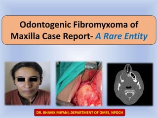
- Facial asymmetry due to swelling over the right zygoma region. Small, diffuse, bony hard, painless, immobile swelling over the right cheek. Skin over the swelling was normal without any tenderness or surface discoloration
- Intraoral examination revealed a firm to hard swelling with buccal cortical plate expansion in relation to right upper canine-premolar region. Mucosa over the swelling was normal without any draining sinuses.
- Suspecting impacted tooth,
- Suspecting impacted tooth,
- Suspecting impacted tooth,
- Suspecting impacted tooth,
- Suspecting impacted tooth,
- Suspecting impacted tooth,