This document reviews recent advances in the management of pulmonary embolism (PE). Risk-adapted treatment and follow-up has led to more favorable outcomes for PE patients. Age-adjusted D-dimer cutoff levels can decrease overuse of imaging tests by increasing the specificity of D-dimer. New oral anticoagulants are effective alternatives to standard treatments. However, areas that require more research include the implications of subsegmental PE, diagnosing PE in pregnant patients, and the efficacy of new oral anticoagulants in cancer patients. Improving guideline implementation will further optimize PE management.
Background: There is a global resolve among Clinicians towards adoption of imaging modalities in the evaluation of appendicitis because clinical algorithms have been disappointing. We sought to determine the authenticity of interobserver variability in ultrasound scan interpretation in a resourceconstrained mission hospital settings, northwestern region of Cameroon. Methods: In this study, we reviewed the standardized diagnostic approach in acute appendicitis and also performed prospective cross observational qualitative testing using sensitivity, specifi city, positive predictive value, negative predictive value, and accuracy to determine the interobserver variability of ultrasonography using the medical database of the two Mission Hospitals, northwestern region of Cameroon from January 2012 to December 2016. A sequential non-randomized convenient sampling was used and data was analyzed using the Statistical Package for the Social Sciences version 22.
Safety, risk of complications and the functional feasibility among different kinds of central venous access are still a matter of debate.Not many clinical trials have reported a comparison of complications and patency of CVCs versus Peripherally Inserted Catheters (PICC) as central venous access for indoor patients with advanced gastrointestinal disorder. The aim of the present study was to compare CVCs and PICCs regarding function, complications and convenience in a controlled clinical study on patients aimed for oncology surgery aimed for cure.
Distributions of patients were comparable. Malignant diagnoses were significantly higher among CVC-patients. CVCs and PICCs were used for treatment during equal number of days, without any signifi cant complication rates and with comparable number of days on antibiotics and other potent drugs. The overall cumulative hazard (risk) for treatment interruptions, due to either full-filled clinical indications or due to any complication among the subgroups of patients did not differ.Central Venous Catheter and Peripheral Inserted Central Venous Catheter, for central venous access, did not differ among consecutive unselected patients with serious gastro-intestinal disorders.
Letter: Is the Stupp Protocol an expensive and unsustainable standard of care...Ahmad Ozair
Glioblastoma multiforme (GBM) is the most common and aggressive primary adult brain neoplasm with an age-adjusted incidence rate of 3.22 per 100 000 individuals and a 5-yr survival rate of 6.8%.1 In 2005, Stupp and colleagues proposed maximal safe resection, concomitant temozolomide (TMZ) with radiotherapy, and adjuvant TMZ as the optimal treatment. Implementation of the Stupp protocol in high-income countries (HICs) has resulted in increased survival compared to previous regimens. With little-to-no literature on the management and outcomes of patients with GBM in low- and middle-income countries (LMICs), it is unclear whether the Stupp protocol is being adopted or whether it is, or ever can be, the optimal strategy in LMICs...
Number of Pages 4 (Double Spaced)Number of sources 8Writi.docxcherishwinsland
Number of Pages: 4 (Double Spaced)
Number of sources: 8
Writing Style: APA
Type of document: Coursework
Category: Healthcare
Order Instructions:
Comprehensive Article Review
Caverly, T.J., Fagerlin, A, & Wiener, R.S. (2018, January 22). Comparison of observed harms and expected mortality benefit for persons in the Veterans Health Affairs Lung Cancer Screening Demonstration Project. JAMA Internal Medicine.
1. What research questions are addressed in this study and what is their purpose (5 points)?
2. What type of research design was used (experimental, quasi-experimental, correlational) in this study and what led you to your decision (5 points)?
3. Are the instruments in this study valid and reliable, why or why not (10 points)?
4. Discuss the specific results of each of the ANCOVAs (analysis of covariance) done in this study. What was the purpose of"each" of the ANCOVAs? What was the covariate in each and why did they do an ANCOVA in each case (5 points)?
5. In the Tables, results are presented, Please explain the tables and summarize the results (15 points).
6. Explain, in simple language, any significant results of this study (25 points)?
7. Identify and discuss any threats to internal and/or external validity in this study (10 points).
8. If you could redesign this study correcting anything you have found wrong with the research, what would you correct and how would you do it (20 points)?
Opinion
EDITORIAL
Reducing Harms in Lung Cancer Screening
Bach to the Future
Michael ln cze, MD, MSEd: Rita F. Redberg, MD, MSc
TbeUS PreventativeServices Task Force cmrcntly recom mends si:;ree ning (grade Brecommendation)for lung canc er witha nnuallow-dose computed tomo graph}' for high-risk in dividuals ages55 to 80 years, defined as those having greate r
gLblefor LCS using the Bach risk tool,11 a vaJidatcd risk model usingsex,age, smokingduration, durationof abstinence from smoking and number of cigarettes smoked per day as inpu ts.
The asto undingly high ratesof false-pos itiveresults in the low
=Related attid e
than a 30 pack-year cumula tivesmoking historyand h av• ing quit with in the past 15 years.1 The evide nce to sup
est risk quintiles (eg, 2221false-positive resul ts per lung ca n cer death averted and a NNS of nearly 5600 in quintile1), as well as extremelylow ratesoflungcancerincidencein the low est-risk groups, confirm trends illustrated in previous stud
port thisrecommendation overwhelminglycomes rrom the Na
tional Lung CancerScreenfngTrial(NL ST). While3 other large randomized clinical trials failed to show any mortality ben efit tolung cancer screening (LCS), the NLST demonstrateda 20% reduction in lungcan ce r mortality,a lo ng with a 6.7% re duction in .ill-ca use mortality, when compared with an an nual chest radiograph, witb a number needed toscreen (NNS} of256to prevent I lung-cancerassociated death over3years.-2 5 Real-worldapplication ofLCS has been particularly .
Secondary Malignancy after Treatment of Prostate Cancer. Radical Prostatectom...asclepiuspdfs
Background: This study aims to determine whether the treatment of locally confined prostate cancer (PCa) with external radiotherapy (EBRT) increases the risk to develop secondary malignancies (SM) compared to radical prostatectomy (RPE). Materials and Methods: Data from patients who were treated curatively with RPE or EBRT from 2010 to 2018 and who did not have distant metastases, previous malignancy, or previous treatment with radiotherapy or chemotherapy at the time of diagnosis were reviewed to determine the incidence of SM over a median follow-up period of 47 months (range 12–96 months). Regression models were used to correlate the clinicopathological factors with the incidence of SM.
Presentatie Prof. dr. Deckers en Prof. dr. BotsCVON
Combining Atherosclerosis Imaging and New and Novel Markers in Asymptomatic Subjects at Intermediate CVD Risk: Implications for Pathophysiology, Prediction and Prevention.
Objective: The prognostic indictors of age-related poor outcomes in patients with acute myeloid leukemia (AML) are still controversial. The aim of this work was to provide comprehensive insights into the effect of different hemocytes and to investigate the association between age and clinical features in adult patients with AML.
Study Design: A retrospective study was performed to determine the role of age in the therapeutic outcomes of AML. A total of 166 newly diagnosed adult patients’ data from January 2015 to November 2019 in Zhongshan Hospital of Xiamen University were collected and analyzed.
Results: Older patients presented a poorer prognosis (p=0.001) with shorter overall survival, which is served as age-related outcomes. Binary logistic regression demonstrated that cytogenetic risk (OR=4.508, 95% CI 2.733–7.435), leukocyte (OR=7.410, 95% CI 1.139–5.910), and bone marrow blast cells (OR=3.261, 95% CI 1.075–5.615) were independent indictors for age-related prognosis. In addition, Kaplan-Meier curve also revealed that the above factors were associated with overall survival (all p values <0.001).
Conclusion: Cytogenetic risk, leukocyte, and bone marrow blast cells are dominant factors which account for the age-related poor outcomes and shorter overall survival in AML.
Keywords: acute myeloid leukemia, adult, cytogenetic risk, hemocyte, leukemia, overall survival
The Impact of Lymph Node Dissection on Survival in Intermediate- and High-Ris...semualkaira
Aimed to evaluate the therapeutic effect of pelvic lymph node dissection (PLND) on survival and determine the predictors of lymph node involvement (LNI) in patients with intermediate- or high-risk prostate cancer (PCa) treated with Radical Prostatectomy
The Impact of Lymph Node Dissection on Survival in Intermediate- and High-Ris...semualkaira
Aimed to evaluate the therapeutic effect of pelvic lymph node dissection (PLND) on survival and determine the
predictors of lymph node involvement (LNI) in patients with intermediate- or high-risk prostate cancer (PCa) treated with Radical
Prostatectomy
Recomendações da OMS sobre cuidados maternos e neonatais para uma experiência pós-natal positiva.
Em consonância com os ODS – Objetivos do Desenvolvimento Sustentável e a Estratégia Global para a Saúde das Mulheres, Crianças e Adolescentes, e aplicando uma abordagem baseada nos direitos humanos, os esforços de cuidados pós-natais devem expandir-se para além da cobertura e da simples sobrevivência, de modo a incluir cuidados de qualidade.
Estas diretrizes visam melhorar a qualidade dos cuidados pós-natais essenciais e de rotina prestados às mulheres e aos recém-nascidos, com o objetivo final de melhorar a saúde e o bem-estar materno e neonatal.
Uma “experiência pós-natal positiva” é um resultado importante para todas as mulheres que dão à luz e para os seus recém-nascidos, estabelecendo as bases para a melhoria da saúde e do bem-estar a curto e longo prazo. Uma experiência pós-natal positiva é definida como aquela em que as mulheres, pessoas que gestam, os recém-nascidos, os casais, os pais, os cuidadores e as famílias recebem informação consistente, garantia e apoio de profissionais de saúde motivados; e onde um sistema de saúde flexível e com recursos reconheça as necessidades das mulheres e dos bebês e respeite o seu contexto cultural.
Estas diretrizes consolidadas apresentam algumas recomendações novas e já bem fundamentadas sobre cuidados pós-natais de rotina para mulheres e neonatos que recebem cuidados no pós-parto em unidades de saúde ou na comunidade, independentemente dos recursos disponíveis.
É fornecido um conjunto abrangente de recomendações para cuidados durante o período puerperal, com ênfase nos cuidados essenciais que todas as mulheres e recém-nascidos devem receber, e com a devida atenção à qualidade dos cuidados; isto é, a entrega e a experiência do cuidado recebido. Estas diretrizes atualizam e ampliam as recomendações da OMS de 2014 sobre cuidados pós-natais da mãe e do recém-nascido e complementam as atuais diretrizes da OMS sobre a gestão de complicações pós-natais.
O estabelecimento da amamentação e o manejo das principais intercorrências é contemplada.
Recomendamos muito.
Vamos discutir essas recomendações no nosso curso de pós-graduação em Aleitamento no Instituto Ciclos.
Esta publicação só está disponível em inglês até o momento.
Prof. Marcus Renato de Carvalho
www.agostodourado.com
Flu Vaccine Alert in Bangalore Karnatakaaddon Scans
As flu season approaches, health officials in Bangalore, Karnataka, are urging residents to get their flu vaccinations. The seasonal flu, while common, can lead to severe health complications, particularly for vulnerable populations such as young children, the elderly, and those with underlying health conditions.
Dr. Vidisha Kumari, a leading epidemiologist in Bangalore, emphasizes the importance of getting vaccinated. "The flu vaccine is our best defense against the influenza virus. It not only protects individuals but also helps prevent the spread of the virus in our communities," he says.
This year, the flu season is expected to coincide with a potential increase in other respiratory illnesses. The Karnataka Health Department has launched an awareness campaign highlighting the significance of flu vaccinations. They have set up multiple vaccination centers across Bangalore, making it convenient for residents to receive their shots.
To encourage widespread vaccination, the government is also collaborating with local schools, workplaces, and community centers to facilitate vaccination drives. Special attention is being given to ensuring that the vaccine is accessible to all, including marginalized communities who may have limited access to healthcare.
Residents are reminded that the flu vaccine is safe and effective. Common side effects are mild and may include soreness at the injection site, mild fever, or muscle aches. These side effects are generally short-lived and far less severe than the flu itself.
Healthcare providers are also stressing the importance of continuing COVID-19 precautions. Wearing masks, practicing good hand hygiene, and maintaining social distancing are still crucial, especially in crowded places.
Protect yourself and your loved ones by getting vaccinated. Together, we can help keep Bangalore healthy and safe this flu season. For more information on vaccination centers and schedules, residents can visit the Karnataka Health Department’s official website or follow their social media pages.
Stay informed, stay safe, and get your flu shot today!
Ozempic: Preoperative Management of Patients on GLP-1 Receptor Agonists Saeid Safari
Preoperative Management of Patients on GLP-1 Receptor Agonists like Ozempic and Semiglutide
ASA GUIDELINE
NYSORA Guideline
2 Case Reports of Gastric Ultrasound
These simplified slides by Dr. Sidra Arshad present an overview of the non-respiratory functions of the respiratory tract.
Learning objectives:
1. Enlist the non-respiratory functions of the respiratory tract
2. Briefly explain how these functions are carried out
3. Discuss the significance of dead space
4. Differentiate between minute ventilation and alveolar ventilation
5. Describe the cough and sneeze reflexes
Study Resources:
1. Chapter 39, Guyton and Hall Textbook of Medical Physiology, 14th edition
2. Chapter 34, Ganong’s Review of Medical Physiology, 26th edition
3. Chapter 17, Human Physiology by Lauralee Sherwood, 9th edition
4. Non-respiratory functions of the lungs https://academic.oup.com/bjaed/article/13/3/98/278874
These lecture slides, by Dr Sidra Arshad, offer a quick overview of physiological basis of a normal electrocardiogram.
Learning objectives:
1. Define an electrocardiogram (ECG) and electrocardiography
2. Describe how dipoles generated by the heart produce the waveforms of the ECG
3. Describe the components of a normal electrocardiogram of a typical bipolar leads (limb II)
4. Differentiate between intervals and segments
5. Enlist some common indications for obtaining an ECG
Study Resources:
1. Chapter 11, Guyton and Hall Textbook of Medical Physiology, 14th edition
2. Chapter 9, Human Physiology - From Cells to Systems, Lauralee Sherwood, 9th edition
3. Chapter 29, Ganong’s Review of Medical Physiology, 26th edition
4. Electrocardiogram, StatPearls - https://www.ncbi.nlm.nih.gov/books/NBK549803/
5. ECG in Medical Practice by ABM Abdullah, 4th edition
6. ECG Basics, http://www.nataliescasebook.com/tag/e-c-g-basics
Prix Galien International 2024 Forum ProgramLevi Shapiro
June 20, 2024, Prix Galien International and Jerusalem Ethics Forum in ROME. Detailed agenda including panels:
- ADVANCES IN CARDIOLOGY: A NEW PARADIGM IS COMING
- WOMEN’S HEALTH: FERTILITY PRESERVATION
- WHAT’S NEW IN THE TREATMENT OF INFECTIOUS,
ONCOLOGICAL AND INFLAMMATORY SKIN DISEASES?
- ARTIFICIAL INTELLIGENCE AND ETHICS
- GENE THERAPY
- BEYOND BORDERS: GLOBAL INITIATIVES FOR DEMOCRATIZING LIFE SCIENCE TECHNOLOGIES AND PROMOTING ACCESS TO HEALTHCARE
- ETHICAL CHALLENGES IN LIFE SCIENCES
- Prix Galien International Awards Ceremony
TEST BANK for Operations Management, 14th Edition by William J. Stevenson, Ve...kevinkariuki227
TEST BANK for Operations Management, 14th Edition by William J. Stevenson, Verified Chapters 1 - 19, Complete Newest Version.pdf
TEST BANK for Operations Management, 14th Edition by William J. Stevenson, Verified Chapters 1 - 19, Complete Newest Version.pdf
Pulmonary Thromboembolism - etilogy, types, medical- Surgical and nursing man...VarunMahajani
Disruption of blood supply to lung alveoli due to blockage of one or more pulmonary blood vessels is called as Pulmonary thromboembolism. In this presentation we will discuss its causes, types and its management in depth.
Report Back from SGO 2024: What’s the Latest in Cervical Cancer?bkling
Are you curious about what’s new in cervical cancer research or unsure what the findings mean? Join Dr. Emily Ko, a gynecologic oncologist at Penn Medicine, to learn about the latest updates from the Society of Gynecologic Oncology (SGO) 2024 Annual Meeting on Women’s Cancer. Dr. Ko will discuss what the research presented at the conference means for you and answer your questions about the new developments.
ARTIFICIAL INTELLIGENCE IN HEALTHCARE.pdfAnujkumaranit
Artificial intelligence (AI) refers to the simulation of human intelligence processes by machines, especially computer systems. It encompasses tasks such as learning, reasoning, problem-solving, perception, and language understanding. AI technologies are revolutionizing various fields, from healthcare to finance, by enabling machines to perform tasks that typically require human intelligence.
2. management strategies for acute PE in past years,
while also highlighting areas that remain to be clari-
fied or resolved by ongoing and future research.
EVOLVING STRATEGIES FOR DIAGNOSIS AND
RISK ASSESSMENT
APPROPRIATE TRIAGE OF PATIENTS FOR IMAGING
TESTS. In the absence of hemodynamic instability at
presentation, the diagnostic work-up of a patient
with suspected acute PE begins with the assessment
of the clinical or pre-test probability of PE (Figure 1).
Standardized prediction rules integrating baseline
clinical parameters and the patient’s history permit
the classification of patients into distinct categories of
clinical probability of the disease. Whichever predic-
tion rule (e.g., Wells or revised Geneva) or version
(original or simplified) is used, the proportion of
patients with confirmed PE is expected to be
approximately 10% in the low-probability category,
30% in the intermediate-probability category, and
65% in the high-clinical probability category when
using the 3-level classification; if a 2-level classifica-
tion is used, PE will be confirmed in around 12% and
50% of patients in the PE-unlikely and PE-likely cat-
egories, respectively (3).
Whereas patients with high clinical probability for
PE (or “PE-likely,” if dichotomized prediction rules
are used) should directly undergo an imaging test, D-
dimer testing is recommended as the next diagnostic
step in patients with low or intermediate pre-test
probability (or PE-unlikely, if dichotomized predic-
tion rules are used) (4). Although a negative test
safely excludes PE without further testing in these
cases, the specificity of a positive D-dimer test is low
and decreases steadily with age. This might result in
too many positive tests and, consequently, in over-
use of diagnostic imaging, particularly in the elderly.
A multicenter, prospective management study eval-
uated age-adjusted (age  10 mg/l, above 50 years)
cutoff levels in a cohort of 3,346 patients with
suspected PE. Patients with a normal age-adjusted
D-dimer value did not undergo computed tomo-
graphic (CT) pulmonary angiography; these patients
were left untreated and followed over a 3-month
period. Using the age-adjusted (instead of the stan-
dard 500 mg/l) D-dimer cutoff increased the number
of patients in whom PE could be excluded from 6.4%
to 30% in patients aged 75 years or older, without a
significant increase in the rate of VTE events during
follow-up (5).
In most countries and hospitals, CT pulmonary
angiography has become the preferred imaging
method for the diagnosis of acute PE in patients with
either a high clinical (pre-test) probability or
low/intermediate probability and elevated D-
dimer levels (Figure 1). However, pitfalls and
errors resulting in misdiagnosis of PE may be
frequent in clinical practice. In a retrospec-
tive review of 937 pulmonary CT examina-
tions performed in a tertiary-care university
hospital over a 12-month period, 3 experi-
enced chest radiologists retrospectively
reinterpreted studies originally reported as
positive for PE (174, 19%). Overall, discor-
dance between the reviewing subspecialists
and the original radiologist was reported in
45 (26%) cases. Retrospective rejection of the
diagnosis occurred more often when the re-
ported PE was solitary (46% of original PE
diagnoses) or located in a segmental or sub-
segmental pulmonary artery (27% and 59%,
respectively). The most frequent explanation
for discrepancies was breathing motion arti-
fact, followed by beam-hardening artifact
from adjacent high-density structures (supe-
rior vena cava, right atrium, right ventricle)
(6). The debate surrounding the perceived
trends in (over)diagnosis of PE in the era of
CT angiography, and the clinical significance
of isolated subsegmental PE, will be dis-
cussed later.
Planar ventilation/perfusion (V/Q) lung
scans represent an alternative imaging
method to CT angiography in patients with a
high clinical probability of PE or those with a
positive D-dimer test. The main limitations of
planar V/Q scans are the high proportion of non-
conclusive results, especially in patients older than 75
years of age, often requiring additional testing. V/Q
single-photon emission computed tomography
(SPECT) is a new method of scintigraphic acquisition
that has been reported to improve diagnostic perfor-
mance and reduce the number of nonconclusive tests
(7). However, the diagnostic criteria need to be stan-
dardized (8). Moreover, head-to-head comparison of
V/Q SPECT with CT pulmonary angiography needs to
be performed, including follow-up of patients left
without anticoagulant treatment on the basis of
negative V/Q SPECT findings.
Of note, V/Q lung scans are an important early step
for ruling out chronic thromboembolic pulmonary
hypertension (CTEPH) in the diagnostic work-up of
patients with persistent dyspnea after acute PE and at
least 3 months of anticoagulation treatment (4).
RISK STRATIFICATION: IDENTIFYING HIGH- AND
LOW-RISK PE. Clinical classification of the severity
A B B R E V I A T I O N S
A N D A C R O N Y M S
ASO = antisense
oligonucleotide
CI = confidence interval
CT = computed tomography
CTEPH = chronic
thromboembolic pulmonary
hypertension
DVT = deep vein thrombosis
ESC = European Society of
Cardiology
FXI = factor XI
NOAC = non–vitamin
K-dependent oral
anticoagulant(s)
NT-proBNP = N-terminal pro–
B-type natriuretic peptide
OR = odds ratio
PE = pulmonary embolism
PESI = Pulmonary Embolism
Severity Index
RR = relative risk
rtPA = recombinant tissue-
type plasminogen activator
SPECT = single-photon
emission computed
tomography
sPESI = simplified Pulmonary
Embolism Severity Index
VKA = vitamin K antagonist
V/Q = ventilation/perfusion
VTE = venous
thromboembolism
J A C C V O L . 6 7 , N O . 8 , 2 0 1 6 Konstantinides et al.
M A R C H 1 , 2 0 1 6 : 9 7 6 – 9 0 Pulmonary Embolism Update
977
3. of acute PE is on the basis of the estimated early
death risk (Figure 1). It has been established that the
presence of right ventricular dysfunction and failure
resulting from acute pressure overload is the prin-
cipal determinant of the patient’s early clinical
course and risk of an adverse outcome (reviewed
in [4,9]). Accordingly, high-risk or massive PE refers
to the presence of shock or persistent arterial
hypotension as a result of overt right ventricular
failure. This is clearly a life-threatening situation, in
which prompt reperfusion treatment (as discussed
later) is needed, along with circulatory and res-
piratory support in order to break the spiral of
hemodynamic deterioration and to increase the
chances of survival (4,10,11).
More than 95% of patients with acute PE are (or
appear to be) hemodynamically stable at presentation
and are thus not considered to be at high risk (12).
Within this large group, the next challenging step is
to determine which patients will need hospitalization
and possibly initial monitoring, and to distinguish
them from those who are at truly low risk and may
qualify for early discharge and outpatient treatment.
To be used as risk stratification tools for this purpose,
baseline clinical parameters and prediction scores
derived from them should reliably exclude severe
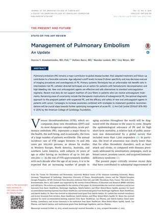
FIGURE 1 PE: Risk-Adjusted Management in the Acute Phase and Over the Long Term
PREDICTORS OF
EARLY ADVERSE OUTCOME
RISK FACTORS FOR
RECURRENT VTERISK FACTORS
FOR FIRST VTE
HORMONAL
CONTRACEPTION
TRAUMA OR FRACTURE
SURGERY
HORMONAL REPLACEMENT
TREATMENT
PREGNANCY
AND POSTPARTUM
IMMOBILIZATION
AGE
INFLAMMATORY
BOWEL DISEASE
THROMBOPHILIA†
OBESITY
CANCER
CHEMOTHERAPY
PRIOR VTE
D-DIMERS
FIRST UNPROVOKED
VTE EVENT
MALE SEX
CHRONIC HEART FAILURE
CHRONIC LUNG DISEASE
ACTIVE CANCER
VITAL SIGNS
HYPOXEMIA
RV DYSFUNCTION
(CT/ECHO)
BIOCHEMICAL MARKERS*
PRE-TEST
CLINICAL ASSESSMENT DIAGNOSIS
ACUTE RISK
STRATIFICATION TREATMENT
LONG-TERM
CLINICAL COURSE
Revised Geneva score
Wells rule
Empirical assessment
(Age-adjusted) D-dimers
CTPA
V/Q scan
Echocardiography
CUS
PESI and sPESI
Biochemical markers*
RV dysfunction
(echocardiography)
RV enlargment (CTPA)
Parenteral anticoagulants
Oral anticoagulants
Fibrinolytics
Catheter-directed
techniques
Surgical embolectomy
Vena cava filters
Assess bleeding risk
Predict VTE
recurrence
Focused screening
for CTEPH in
symptomatic patients
HIGH CLINICAL
PROBABILITY
LOW OR
INTERMEDIATE
CLINICAL
PROBABILITY
Hemodynamic
instability
Absence of
hemodynamic
instability
Age-adjusted
positive
D-dimers
ALGORITHM FOR
HIGH-RISK PE
CTPA
CTPA
ALGORITHM FOR
NON HIGH-RISK PE
V/Q scan
HIGH RISK
INTERMEDIATE RISK
LOW RISK
PRIMARY
REPERFUSION
ANTICOAGULANT
THERAPY
ANTICOAGULANT
THERAPY
ANTICOAGULANT
THERAPY
BLEEDING
RECURRENT VTE
CTEPH
Echocardiography (if
CTPA not readily
available or uncontrolled
hypotension)
CUS-based algorithms
Hemodynamic
instability
INTERMEDIATE-HIGH
INTERMEDIATE-LOW
plus
(Rescue reperfusion)
(Early discharge)
No validated prediction
models for VTE patients
Standard-duration vs.
extended (indefinite)
treatment
Individualized follow-up
programs and intervals
*Biochemical markers include markers of myocardial injury (troponins, heart-type fatty acid-binding protein) and markers of heart failure (BNP or N-terminal-proBNP).
†Only antiphospholipid syndrome and high-risk inherited thrombophilia (i.e., homozygosity for factor V Leiden, homozygosity for prothrombin G20210A mutation,
double heterozygosity, antithrombin deficiency) are considered. Nevertheless, routine thrombophilia testing is not indicated in PE patients. BNP ¼ B-type natriuretic
peptide; CT ¼ computed tomography; CTEPH ¼ chronic thromboembolic pulmonary hypertension; CTPA ¼ computed tomographic pulmonary angiogram; CUS ¼
compression ultrasound; Echo ¼ echocardiography; N-terminal-proBNP ¼ N-terminal pro–B-type natriuretic peptide; PE ¼ pulmonary embolism; PESI ¼ pulmonary
embolism severity index; RV ¼ right ventricular; sPESI ¼ simplified pulmonary embolism severity index; V/Q scan ¼ ventilation/perfusion lung scan; VTE ¼ venous
thromboembolism.
Konstantinides et al. J A C C V O L . 6 7 , N O . 8 , 2 0 1 6
Pulmonary Embolism Update M A R C H 1 , 2 0 1 6 : 9 7 6 – 9 0
978
4. acute illness and the presence of significant comor-
bidity. The Pulmonary Embolism Severity Index
(PESI) has been extensively validated and shown to
fulfill these requirements; patients in PESI risk strata I
and II were at low risk of 30-day mortality (13). The
simplified version of the Pulmonary Embolism
Severity Index (sPESI) also possessed a high negative
predictive value for ruling out an adverse early
outcome (14,15). Thus, a substantial proportion (be-
tween 25% and 46%, depending on the cohort stud-
ied) of all patients with acute PE can be classified as
being at low risk on the basis of a PESI risk class of I or
II, or a sPESI score of 0 (14–16). Although the negative
predictive value of the index may rise even further
when it is combined with the (negative) result of a
high-sensitivity cardiac troponin assay (17), it is un-
certain how often this extra reassurance is really
needed in clinical practice.
Despite the uncontested prognostic value of the
PESI, it should be kept in mind that this was primarily
designed as an epidemiological tool, and not as a direct
guide to PE management. The only prospective trial
that used this severity index to randomize patients to
outpatient versus in-hospital treatment of PE required
numerous additional eligibility criteria, including a
supportive social environment (18). Other groups,
particularly in the Netherlands, chose to develop
explicit home treatment–oriented clinical criteria,
either alone (Hestia criteria) (19) or in combination
with biomarker testing (N-terminal pro–B-type natri-
uretic peptide [NT-proBNP] plasma levels <500 pg/ml)
(20), which they tested successfully in small- to
medium-sized (150 to 300 patients) prospective cohort
trials. These criteria await validation in larger cohorts
and further countries. Importantly, and in view of first
reports that severe right ventricular dysfunction may
occasionally be present in a patient with a negative
sPESI (21), it will also need to be determined in this
context whether CT or echocardiographic imaging of
the right ventricle should be added to clinical eligi-
bility criteria for immediate or early discharge in order
to maximize patient safety.
EVOLVING DEFINITION OF INTERMEDIATE-RISK PE.
What are the next steps if a normotensive patient is
not classified into the low-risk category on the basis
of the clinical criteria described previously? With the
objective of further developing the concept of
intermediate-risk PE, the 2014 European Society of
Cardiology (ESC) guidelines critically reviewed the
combinations of imaging (echocardiographic or CT
angiographic) parameters and laboratory biomarkers
that can be used to detect right ventricular dysfunc-
tion and/or myocardial injury (4). Taking into account
that imaging and laboratory tests have consistently
been shown to have prognostic values additive to
each other and to those of clinical parameters (22,23),
and aiming to discourage uncritical time- and
resource-consuming laboratory and/or echocardio-
graphic testing in every patient with confirmed PE
without prior clinical triage, the updated ESC guide-
lines proposed a stepwise classification of early risk,
as displayed in the Central Illustration (4). Although
supported by evidence from cohort studies and vali-
dated (at least in a modified form) in a large ran-
domized therapeutic trial (24), the current risk
stratification scheme will almost certainly need
further improvement in the following years. In
particular, the definition and positive prognostic
value of the intermediate-high-risk class must be
optimized to better identify candidates for reperfu-
sion treatment among normotensive patients with
PE. Promising steps in this direction include the use
of age-adjusted cutoff values for high-sensitivity
cardiac troponin T in patients age 75 years or older
(25) and laboratory biomarkers more specific for
relevant neurohumoral activation or for myocardial
injury (26). In 2,874 normotensive PE patients
derived from 6 cohort studies, a multidimensional
prognostic model on the basis of 4 variables (systolic
blood pressure 90 to 100 mm Hg; heart rate $110
beats/min; elevated cardiac troponin; right ventricu-
lar dysfunction on imaging) was constructed, yielding
3 risk strata (27). The rate of an adverse 30-day
outcome was 29% in the high-risk stratum (>4
points) of the derivation population (27), and up to
42% in a validation cohort of 1,083 patients (28).
External validation and implementation of new pre-
diction rules in prospective management trials are
necessary steps before these can be integrated into
future risk stratification algorithms.
ADVANCES IN ANTICOAGULATION
TREATMENT
In patients with acute VTE (presenting either as PE or
proximal DVT), the duration of anticoagulation treat-
ment should cover at least 3 months (4,10,11,29).
Within this period, traditional regimens of acute-
phase treatment consist of parenteral anticoagulation
(intravenous unfractionated heparin, subcutaneous
low-molecular-weight heparin, or fondaparinux) over
the first 5 to 10 days, overlapping and followed by a
vitamin K antagonist (VKA), which is adjusted to
obtain a therapeutic (2.0 to 3.0) international normal-
ized ratio. Advances in knowledge and the remaining
open questions related to determining the optimal
duration (beyond the first 3 months) of anticoagulation
after PE are discussed separately in this review, in the
J A C C V O L . 6 7 , N O . 8 , 2 0 1 6 Konstantinides et al.
M A R C H 1 , 2 0 1 6 : 9 7 6 – 9 0 Pulmonary Embolism Update
979
5. section titled Current Controversies and Areas of
Ongoing Research.
Phase 3 trials investigating the new, non–vitamin K-
dependent oral anticoagulant agents (NOACs) apix-
aban (30), dabigatran (31,32), edoxaban (33), and
rivaroxaban (34,35) in the treatment of VTE have been
completed and published. A meta-analysis showed
that these agents are noninferior to the standard hep-
arin/VKA regimen, in terms of prevention of VTE
recurrence (relative risk [RR]: 0.90; 95% confidence
interval [CI]: 0.77 to 1.06), and that they are probably
safer in terms of major bleeding (RR: 0.61; 95% CI: 0.45
to 0.83), particularly intracranial (RR: 0.37; 95% CI:
0.21 to 0.68) and fatal (RR: 0.36; 95% CI: 0.15 to 0.84)
hemorrhage (36). As a result, NOACs are recommended
in the 2014 ESC Guidelines as an alternative to the
standard heparin/VKA treatment (4). All 4 NOACs
mentioned earlier are now licensed for treatment of
VTE in the United States and the European Union
(edoxaban still awaits approval in Canada); the
approved regimens are summarized in Table 1.
Post-marketing experience with these drugs in clinical
practice (under “real-world” conditions) appears
reassuring in the setting of stroke prevention in atrial
fibrillation, and has also begun to accumulate in VTE.
In a prospective German registry of patients treated
with rivaroxaban, rates of major bleeding for patients
with VTE were 4.1% per year (95% CI: 2.5% to 6.4% per
year), and case fatality rates were low (approximately
5% at 30 days) (37). Importantly, available data suggest
that the first reversal agent against a NOAC, the
monoclonal antibody idarucizumab, which binds the
thrombin inhibitor dabigatran, is effective in emer-
gency situations (38); this agent is expected to obtain
U.S. Food and Drug Administration approval soon. In
parallel, phase 3 clinical trials are currently being
conducted with andexanet, a modified recombinant
form of factor Xa, which is catalytically inactive (39)
and may serve as a reversal agent for rivaroxaban,
apixaban, and edoxaban.
Single oral drug regimens for PE might be expected
to improve (reduce) patients’ perceived burden of
CENTRAL ILLUSTRATION Acute PE: Current Risk Stratification
Risk Parameters and Scores
Early Mortality Risk
+
+
+
+ (+)(+)
Either 1 (or none) positive
Assessment optional:
If assessed, both negative-
Both positive
Shock or
Hypotension
PESI Class III-V
or sPESI ≥1
Signs of RV
Dysfunction on
an Imaging Test
Cardiac
Laboratory
Biomarkers*
-
-
-Low
Intermediate-
high
Intermediate-
low
High
Intermediate
Konstantinides, S.V. et al. J Am Coll Cardiol. 2016; 67(8):976–90.
*Markers of myocardial injury (e.g., elevated cardiac troponin or heart type-fatty acid-binding [H-FABP] plasma concentrations), or of right ventricular dysfunction
(elevated natriuretic peptide plasma concentrations). Adapted with permission from the 2014 European Society of Cardiology Guidelines on the Diagnosis and Man-
agement of Pulmonary Embolism (4). PE ¼ pulmonary embolism; PESI ¼ Pulmonary Embolism Severity Index; RV ¼ right ventricular; sPESI ¼ simplified Pulmonary
Embolism Severity Index.
Konstantinides et al. J A C C V O L . 6 7 , N O . 8 , 2 0 1 6
Pulmonary Embolism Update M A R C H 1 , 2 0 1 6 : 9 7 6 – 9 0
980
6. anticoagulation therapy and, possibly, the costs
related to prolonged hospitalization and bleeding
complications. As part of the open-label EINSTEIN-PE
(Oral Direct Factor Xa Inhibitor Rivaroxaban in
Patients With Acute Symptomatic Pulmonary Embo-
lism) rivaroxaban phase 3 trial, 2,397 patients in
7 countries completed a validated measure of treat-
ment satisfaction, the Anti-Clot Treatment Scale
(ACTS); rivaroxaban treatment was reported to result
in improved treatment satisfaction compared with
enoxaparin/VKA, particularly by reducing the patient-
reported anticoagulation burden (40). In an analysis of
data from the EINSTEIN trials, rivaroxaban was asso-
ciated with greater discounted quality-adjusted life-
expectancy, as well as per-patient cost savings for each
treatment duration modeled (3, 6, and 12 months); the
benefits were greatest with shorter durations (41).
Further specific aspects of NOAC treatment that
were beyond the scope of the phase 3 trials are
currently under investigation. For example, the
safety and efficacy of NOACs in patients with
intermediate-risk PE have not been systematically
addressed thus far (42). One of the phase 3 clinical
trials investigating the use of NOACs in patients with
VTE reported efficacy results for the subgroup of
patients with acute PE and right ventricular
dysfunction; the latter was defined either as NT-
proBNP levels >500 pg/ml, or as a right-to-left
ventricular dimension ratio >0.9 on CT pulmonary
angiography (33). Among patients with elevated
NT-proBNP levels, recurrent VTE occurred in 15 of 454
patients in the edoxaban arm and in 30 of 484 patients
in the warfarin arm, with a hazard ratio of 0.52
(95% CI: 0.28 to 0.98) (33). These findings are to be
regarded as hypothesis-generating at the present
stage; a prospective, multicenter management trial
will focus on the safety, efficacy, and cost-
effectiveness of dabigatran in the treatment of pa-
tients with acute intermediate-risk PE, defined by
imaging (echocardiographic or CT) and laboratory
TABLE 1 Non–Vitamin K-Dependent Oral Anticoagulant Agents in the Treatment and Secondary Prevention of VTE
Dosage and Interval
Not Recommended or Contraindicated*Initial Phase Long-Term Phase Extended Phase
Rivaroxaban† 15 mg twice daily with
food for 21 days
20 mg once daily with food CrCl 30 ml/min
Moderate or severe hepatic impairment
(Child-Pugh B and C), or hepatic disease
associated with coagulopathy
Concomitant use of combined P-gp and
strong CYP3A4 inhibitors or inducers
Dabigatran
etexilate‡
Initial therapy with
parenteral
anticoagulation for
5–10 days should
precede
administration of
dabigatran etexilate
150 mg twice daily CrCl 30 ml/min
Concomitant treatment with P-gp inhibitors
in patients with CrCl 50 ml/min
Concomitant treatment with P-gp inducers
(i.e., rifampin)
Apixaban 10 mg twice daily
for 7 days
5 mg twice daily 2.5 mg twice daily after at
least 6 months of treatment
CrCl 15 ml/min
Severe hepatic impairment (Child-Pugh C),
or hepatic disease associated with
coagulopathy
Strong dual inhibitors or inducers of
CYP3A4 and P-gp
Edoxaban§ Initial therapy with
parenteral
anticoagulation for
5–10 days should
precede
administration
of edoxaban
60 mg once daily
30 mg once daily can be considered in patients
with $1 of the following factors: CrCl
15–50 ml/min; body weight #60 kg;
concomitant use of P-gp inhibitors,
cyclosporin, dronedarone, erythromycin,
or ketoconazole
CrCl 15 ml/min
Moderate or severe hepatic impairment
(Child-Pugh B and C), or hepatic disease
associated with coagulopathy
Concomitant treatment with rifampin
The table displays drugs and regimens on the basis of U.S. FDA approval for the treatment of acute VTE. *In addition to the specific conditions listed here, all mentioned
anticoagulant agents should be avoided in patients: 1) with hemodynamically unstable acute pulmonary embolism for whom thrombolysis or pulmonary embolectomy may be
required; 2) requiring dialysis; 3) at significant risk of bleeding or with active pathological bleeding; 4) treated with a concomitant anticoagulant agent; 5) with known hy-
persensitivity to the agent; and 6) in pregnant women or during breast feeding. Moreover, all mentioned anticoagulant agents should be administered with caution in patients
with an increased bleeding risk, including those receiving concomitant treatment with NSAIDs, acetylsalicylic acid, and platelet aggregation inhibitors. †According to the EMA
product information, rivaroxaban 15 mg should be considered for the long-term phase if the patient’s assessed risk for bleeding outweighs the risk for recurrent venous
thromboembolism. In the European Union, rivaroxaban is contraindicated in patients with CrCl 15 ml/min and should be used with caution in patients with CrCl 15-30 ml/min.
‡According to the EMA product information, dabigatran etexilate 110 mg twice daily can be considered in patients $80 years of age; for those under concomitant treatment
with moderate P-gp inhibitors (i.e., amiodarone, quinidine, verapamil); at higher risk of bleeding, including elderly patients 75 years of age with 1 risk factor for bleeding; and
with CrCl 30-50 ml/min. In the European Union, dabigatran etexilate is not recommended in patients with elevated liver enzymes 2Â upper limit of normal or with liver
disease expected to have any impact on survival. §Although a separate extension trial was not conducted for edoxaban, 40% of patients included in the HOKUSAI-VTE study
received an extended anticoagulant treatment with edoxaban for up to 12 months.
CrCl ¼ creatinine clearance; CYP3A4 ¼ cytochrome P450-3A4; EMA ¼ European Medicines Agency; FDA ¼ Food and Drug Administration; NSAID ¼ nonsteroidal anti-
inflammatory drug(s); P-gp ¼ P-glycoprotein; VTE ¼ venous thromboembolism.
J A C C V O L . 6 7 , N O . 8 , 2 0 1 6 Konstantinides et al.
M A R C H 1 , 2 0 1 6 : 9 7 6 – 9 0 Pulmonary Embolism Update
981
7. (circulating levels of cardiac troponins and natriuretic
peptides) parameters, and their combinations, as
proposed by the ESC guidelines (4). The study plan-
ned to enroll its first patient in the fourth quarter of
2015 (EudraCT 2015-001830-12).
The available data from cohort studies suggest, as a
whole, that a shift towards ambulatory treatment
might affect a substantial proportion (up to 50%) of
patients with PE (reviewed in [43]). A prospective
multicenter management trial has set out to deter-
mine whether early discharge and out-of-hospital
treatment with rivaroxaban of patients with “low-
risk” PE (on the basis of the Hestia criteria (19),
combined with the exclusion of right ventricular
dysfunction and intracardiac thrombi) is feasible and
safe; the trial will also obtain health economic vari-
ables as the basis for description of resource utiliza-
tion (EudraCT 2013-001657-28).
The advances and outlook of anticoagulation in
patients with PE and cancer are discussed separately
in the section Current Controversies and Areas of
Ongoing Research.
REPERFUSION STRATEGIES
SYSTEMIC FIBRINOLYTIC TREATMENT. Fibrinolytic
agents have been tested in randomized trials over
almost one-half a century, and those currently
approved for clinical use were recently reviewed (4).
A meta-analysis of 15 trials involving a total of
2,057 patients showed that fibrinolysis reduced overall
mortality (odds ratio [OR]: 0.59; 95% CI: 0.36 to 0.96)
and achieved a significant reduction in the combined
endpoint of death or treatment escalation (OR: 0.34;
95% CI: 0.22 to 0.53), PE-related mortality (OR: 0.29;
95% CI: 0.14 to 0.60), and PE recurrence (OR: 0.50;
95% CI: 0.27 to 0.94). At the same time, however, major
hemorrhage (OR: 2.91; 95% CI: 1.95 to 4.36) and fatal or
intracranial bleeding (OR: 3.18; 95% CI: 1.25 to 8.11)
were significantly more frequent among patients
receiving thrombolysis (44). Of note, interpretation of
meta-analyses in this field should be extremely
cautious, considering the marked heterogeneity
of: 1) trial size and patient selection (PE severity)
criteria; 2) the fibrinolytic agents, doses, and regimens
tested; and 3) drug application modalities and dura-
tion of treatment. These differences become even
more pronounced and critical if trials on full-dose or
reduced-dose (see later discussion) fibrinolysis, and
on systemically or locally infused fibrinolytic agents,
are analyzed together (45).
With all of the previously mentioned limitations in
mind, there is consensus that immediate reperfusion
treatment using systemic fibrinolysis is indicated in
patients who present with high-risk or massive PE,
that is, those with persistent arterial hypotension or
shock (4,10,11). This is in contrast to the controversy
that, until recently, surrounded the possible net
clinical benefit of fibrinolysis in apparently stable
patients with intermediate-risk or submassive PE.
The international PEITHO (Pulmonary Embolism
Thrombolysis) trial (24) compared a single intrave-
nous bolus of tenecteplase plus heparin with placebo
plus heparin in 1,006 patients with confirmed PE,
right ventricular dysfunction detected by echocardi-
ography or CT, and a positive troponin I or T test
(partly corresponding to the ESC category of
intermediate-high-risk PE [4]). In the fibrinolysis
group, the primary outcome of all-cause death
or hemodynamic decompensation/collapse within
7 days occurred less frequently than in the group
receiving heparin alone (2.6% vs. 5.6%; OR: 0.44;
95% CI: 0.23 to 0.88). In parallel, a higher incidence of
hemorrhagic stroke (2.0%) and major nonintracranial
bleeding (6.3%) was observed in patients allocated to
tenecteplase than in the placebo group (0.2% and
1.5%, respectively) (24). In view of these latter data, it
becomes clear that full-dose systemic thrombolysis
cannot be recommended as routine primary treat-
ment for patients with intermediate-risk or sub-
massive PE, even if signs of both right ventricular
dysfunction and myocardial injury are initially pre-
sent. Patients belonging to this risk group should
receive parenteral heparin anticoagulation and be
monitored closely over at least 48 to 72 h, and rescue
fibrinolysis should be considered if clinical signs of
hemodynamic decompensation appear (4).
WEAK EVIDENCE FOR REDUCED-DOSE FIBRINOLYSIS.
The life-threatening bleeding complications that
have consistently been associated with full-dose
systemic fibrinolysis (reviewed in [46]) raise the
question of whether reduced doses might improve
safety without loss of efficacy. In a randomized pilot
trial of 118 patients with either hemodynamic insta-
bility or “massive” pulmonary artery obstruction
(without a standardized definition of clinical
severity), half-dose recombinant tissue-type plas-
minogen activator (rtPA) was noninferior to the full
dose in terms of improving pulmonary vascular
obstruction, and it appeared to cause less bleeding
(47). In another small study of 121 patients with
“moderate” PE (also without a standardized defini-
tion of clinical severity), reduced-dose rtPA appeared
to be safe in the acute phase and for reducing the
persistence of echocardiographically assessed pul-
monary hypertension at 28 Æ 5 months of follow-up
(48). The safety and efficacy of reduced-dose intra-
venous fibrinolytic regimens in patients 75 years of
Konstantinides et al. J A C C V O L . 6 7 , N O . 8 , 2 0 1 6
Pulmonary Embolism Update M A R C H 1 , 2 0 1 6 : 9 7 6 – 9 0
982
8. age or older are also indirectly supported by the
findings of a randomized controlled trial on acute
ST-segment elevation myocardial infarction (49).
Although “half-dose” systemic fibrinolytic treat-
ment appears appealing to many physicians, the ev-
idence in its favor should be considered preliminary
at best, and such off-label regimens cannot be rec-
ommended at the present stage. As an alternative
option for PE patients who need reperfusion treat-
ment due to initial or evolving hemodynamic
decompensation, but present with absolute or rela-
tive contraindications to systemic fibrinolysis,
catheter-based techniques may be considered, as will
be explained later.
CATHETER-DIRECTED REPERFUSION TECHNIQUES, WITH
OR WITHOUT FIBRINOLYSIS. Catheter-directed reper-
fusion techniques for removal of obstructing thrombi
from the main pulmonary arteries may be an alter-
native to surgical embolectomy for patients with
absolute or relative contraindications to thrombolysis
(50). The phase 2 ULTIMA (Ultrasound Accelerated
Thrombolysis of Pulmonary Embolism) trial random-
ized 59 patients with acute main- or lower-lobe
PE and an echocardiographic right-to-left ventricular
dimension ratio $1.0 to receive unfractionated
heparin plus a catheter-directed, ultrasound-assisted
thrombolytic regimen of 10 to 20 mg rtPA over 15 h, as
opposed to heparin alone (51). Reduced-dose local
thrombolysis significantly reduced the subannular
right-to-left ventricular dimension ratio between
baseline and 24-h follow-up, without an increase in
bleeding complications (51). The efficacy and safety
of pharmacomechanical thrombolysis is further sup-
ported by the results of a prospective, single-arm,
multicenter trial from the United States that
enrolled 150 patients with submassive or massive
PE (52). A recent multicenter registry on
catheter-directed mechanical or pharmacomechanical
thrombectomy reported clinical success (defined as
all of the following: stabilization of hemodynamics;
improvement in pulmonary hypertension and/or
right heart strain; and survival to hospital discharge)
in 86% of 28 included patients with massive PE and
97% of 73 patients with submassive PE. No hemor-
rhagic strokes were observed (53).
The obvious need for local expertise and a high
institutional volume to ensure satisfactory outcomes
of catheter-directed treatment (54), along with the
high costs of the equipment for ultrasound-assisted
pharmacomechanical fibrinolysis and the lack of
reimbursement by the health systems of several
countries, still limit the widespread use of this tech-
nique outside of selected specialized centers. More-
over, it remains to be confirmed that ultrasound is
indeed necessary to enhance the efficacy of the locally
delivered low-dose fibrinolytic agent. In a recently
published controlled clinical trial, 48 patients with
acute iliofemoral DVT (but not PE) were randomized to
receive ultrasound-assisted catheter-directed fibri-
nolysis versus catheter-directed fibrinolysis alone
(55). The thrombolysis regimen (20 mg alteplase over
15 h) was identical in all patients. The percentage of
thrombus load reduction was 55 Æ 27% in the
ultrasound-assisted versus 54 Æ 27% in the conven-
tional catheter-directed thrombolysis group (p ¼ 0.91).
At the 3-month follow-up, primary venous patency
was 100% in the ultrasound-assisted and 96% in the
conventional catheter-directed thrombolysis group
(p ¼ 0.33), and there was no difference in the severity
of the post-thrombotic syndrome (55).
CONTINUING DISCUSSION ON THE UTILITY
OF INFERIOR VENA CAVA FILTERS
Venous filters are usually placed in the infrarenal
portion of the inferior vena cava. A reasonable general
recommendation is to use them in patients with acute
PE who have absolute contraindications to anticoag-
ulant drugs, in those experiencing major bleeding
events during the acute phase, and in patients with
objectively confirmed recurrent PE, despite adequate
anticoagulation treatment (4). However, in some
countries, particularly in the United States, a growing
liberalization of indications for both permanent and
retrievable filters is observed; this trend is highlighted
by a 3-fold increase in their use between 2001 and
2006 according to data from the National Hospital
Discharge Survey (56). Epidemiological data from the
U.S. Nationwide Inpatient Sample, analyzing almost
298,000 filter implantations, suggest that cava filters
may be associated with an improved outcome (57);
registry data from Europe (albeit with fewer patients)
have been less convincing (58). The PREPIC (Préven-
tion du Risque d’Embolie Pulmonaire par Interruption
Cave) 2 trial, a randomized, open-label, blinded
endpoint trial with a 6-month follow-up, was more
recently published (59). Hospitalized patients with
acute, symptomatic PE associated with lower-limb
vein thrombosis and at least 1 criterion for severity
were assigned to retrievable inferior vena cava filter
implantation plus anticoagulation (n ¼ 200) or anti-
coagulation alone with no filter implantation (n ¼ 199).
Anticoagulant treatment was not interrupted during
filter placement, and access site hematomas were
observed in only 2.6% of the patients. By 3 months,
recurrent PE had occurred in 6 patients (3.0%; all
events fatal) in the filter group and in 3 patients
(1.5%; 2 fatal) in the control group (RR with filter: 2.0;
J A C C V O L . 6 7 , N O . 8 , 2 0 1 6 Konstantinides et al.
M A R C H 1 , 2 0 1 6 : 9 7 6 – 9 0 Pulmonary Embolism Update
983
9. 95% CI: 0.51 to 7.89); results were similar at 6 months
(59). In this context, it should also be borne in
mind that cava filter placement is not free of compli-
cations, which may include penetration of the caval
wall or embolization to the right heart cavities
and occasionally require emergency treatment (60).
Moreover, and importantly, the high success rates of
filter retrieval (153 of 164 patients in whom it was
attempted) reported in the PREPIC 2 trial (59) will
be very difficult to reproduce in the real world,
probably increasing the rate of long-term complica-
tions. In conclusion, the evidence derived from
trial data does not support the liberalization of
cava filter use beyond the strict indications listed
previously.
IMPACT OF EVOLVING MANAGEMENT
STRATEGIES: TRENDS IN MORTALITY
AND THE ECONOMIC BURDEN OF
PULMONARY EMBOLISM
Evidence published in the past decade and continuing
to accumulate consistently indicates a progressive
reduction of case fatality rates among patients with
acute PE (Figure 2). Data obtained from the U.S.
Nationwide Inpatient Sample during the 8-year period
between 1998 and 2005 were used to investigate the
outcomes of patients with a primary or secondary PE
diagnosis who had been discharged from acute care
hospitals. The number of patients increased from
126,546 to 229,637 annually during that period; at the
same time, in-hospital case fatality rates for these pa-
tients decreased from 12.3% to 8.2%, and the length of
hospital stay decreased from 9.4 to 8.6 days (65).
Another study, using both the U.S. Nationwide Inpa-
tient Sample cohort and the Multiple Cause-of-Death
database, reported that the incidence of diagnosed
PE increased by as much as 81% (from 62.1 to 112.3 per
100,000) following the introduction of CT angiog-
raphy, in comparison to the earlier reference period
(1998 to 2006 vs. 1993 to 1998); in parallel, case fatality
rates decreased before (from 13.2% to 12.1%) and,
particularly, in the era of CT angiography (from 12.1%
to 7.8%). Over the entire observation period, mortality
related to PE dropped from 13.4 to 11.9 per 100,000
(66). Similar trends were reported from Germany (67),
and also on the basis of the National Hospital Discharge
Database, covering the entire Spanish population (68).
In the latter study, in-hospital case fatality rates of PE
decreased from 12.9% in 2002 to 8.3% in 2011 in parallel
with a decrease in mean length of hospital stay from
12.7 to 10 days.
FIGURE 2 Global Trends in PE Incidence and Case Fatality Rates
120
110
100
90
80
70
60
50
40
30
20
10
1997 1999 2001 2003 2005 2007 2009 2011 2013
Year
NumberofPulmonaryEmbolismDiagnoses/100,000Inhabitants
1997 1999 2001 2003 2005 2007 2009 2011 2013
Year
26
24
22
20
18
16
14
12
10
8
6
4
2
NumberofIn-hospitalDeaths/100PulmonaryEmbolismDiagnoses(%)
Incidence Rate Case Fatality Rate
U.S. (66)†
U.S. (66)*
Italy (62)*
Australia (61)*
Spain (68)*
China (64)*
Italy (62)*
Spain (68)*
U.S. (66)†
U.S. (70)†
U.S. (66)*
U.S. (70)*
U.S. (63)*
(Left) Pulmonary embolism (PE) incidence. (Right) Case fatality rates. Data shown here were retrieved from studies of trends in pulmonary embolism (61–64,66,68,70).
In case of duplicate or overlapping data, only the most recent publication was included. *Pulmonary embolism was listed as principal diagnosis. †Any listed code for
pulmonary embolism was considered.
Konstantinides et al. J A C C V O L . 6 7 , N O . 8 , 2 0 1 6
Pulmonary Embolism Update M A R C H 1 , 2 0 1 6 : 9 7 6 – 9 0
984
10. An obvious conclusion from dropping case-fatality
rates could be that diagnosis and treatment of PE
have both improved substantially, likely due to a
combination of a higher level of suspicion, stan-
dardized clinical prediction rules and use of D-dimer
testing, high accuracy of multidetector CT angiog-
raphy, and the efficacy of low-molecular-weight
heparins (population data reflecting the impact of
NOACs on VTE-related mortality are not yet avail-
able). Evolution and broad implementation of multi-
disciplinary programs, such as the Pulmonary
Embolism Response Team (PERT), which bring
together a team of specialists to rapidly evaluate
intermediate- and high-risk patients with PE, formu-
late a treatment plan, and mobilize the necessary
resources, may contribute to even better patient
outcomes in the future (69). It can also be argued that
the parallel increase in the annual incidence of VTE
(68), and the rather subtle (66) or absent (70) changes
in PE-related annual mortality over time, reflect the
increasing comorbidity in aging populations. How-
ever, overdiagnosis of PE with the liberal use of
multidetector CT is an alarming alternative explana-
tion (66,71), and it needs to be addressed by
concerted efforts to increase the implementation of
evidence-based guidelines.
Costs related to the management of acute PE (and
VTE in general) have long been assumed to be sub-
stantial, but it was only recently that they were sys-
tematically calculated in the United States (72). In a
cost model built on adult incidence-based events and
potential complications, total annual VTE costs
ranged from $13.5 to $69.3 billion, with preventable
costs of $4.5 to $39.3 billion (72). These data highlight
the potential for cost savings in the future by:
1) improving VTE preventive measures in hospital-
ized patients; 2) implementing evidence-based, risk-
adjusted management algorithms, as recommended
by current guidelines; 3) identifying candidates for
early discharge and ambulatory treatment; 4) using
anticoagulant agents with an improved safety
profile; and 5) increasing VTE awareness in the pop-
ulation (2).
CURRENT CONTROVERSIES AND AREAS
OF ONGOING RESEARCH
MAGNETIC RESONANCE IMAGING. Five years after
the disappointingly low rate of technically adequate
images reported in the PIOPED (Prospective Investi-
gation of Pulmonary Embolism Diagnosis) III study,
an ongoing prospective management study, is inves-
tigating the performance of magnetic resonance im-
aging in combination with lower-limb compression
ultrasound in the diagnostic workup of suspected PE
(NCT02059551). Beyond angiographic techniques and
the detection of filling defects, a study using an
experimental mouse model of VTE, as well as
ex vivo-generated human clots, investigated a novel
technique for the sensitive and specific identification
of developing thrombi (73). The study used
background-free 19
F magnetic resonance imaging,
together with alpha2-antiplasmin peptide-targeted
perfluorocarbon nanoemulsions (PFCs) as a contrast
agent cross-linked to fibrin by active factor XIII.
Developing thrombi with a diameter 0.8 mm could
be visualized in vivo in the murine inferior vena cava
as hot spots by simultaneous acquisition of anatomic
matching (73). If further developed and tested in
humans, this method may offer the potential to
visualize fresh, developing thrombi that are still
susceptible to pharmacological intervention.
SIGNIFICANCE OF SUBSEGMENTAL PE. The clinical
significance and therapeutic implications (i.e., need
for anticoagulation treatment) of isolated sub-
segmental PE on CT pulmonary angiography remain
subjects of debate. A meta-analysis reported that the
rate of subsegmental PE diagnosis has doubled in
parallel with advances in CT technology, rising from
4.7% (95% CI: 2.5% to 7.6%) of patients undergoing
single-detector CT angiography to 9.4% (5.5% to
14.2%) of those submitted to multidetector CT (71).
The 3-month VTE recurrence risk in patients who
were left untreated on the basis of a negative CT
angiography remained unaffected by the use of
multidetector CT (71), and it is possible that at least
some of the tests were false positive because the
positive predictive value is low and the interobserver
agreement poor at this distal level (6,74). Compres-
sion ultrasound of the leg veins can be helpful in this
situation, because the confirmation of proximal DVT
in a patient with isolated subsegmental PE sets the
indication for anticoagulant treatment, whereas the
exclusion of DVT could support a decision against
treatment; these latter cases should be managed on
an individual basis, taking the clinical probability and
the bleeding risk into account (4). An ongoing pro-
spective cohort study is currently investigating the
safety of withholding anticoagulation in patients with
subsegmental PE and no cancer, who have negative
serial bilateral lower extremity ultrasound tests and
are carefully followed over 3 months (NCT01455818).
SUSPECTED VTE IN PREGNANCY. PE is the leading
cause of pregnancy-related maternal death in devel-
oped countries; at the same time, pregnancy is 1 of
the main risk factors for inappropriate management
of suspected PE (75). These facts emphasize the
J A C C V O L . 6 7 , N O . 8 , 2 0 1 6 Konstantinides et al.
M A R C H 1 , 2 0 1 6 : 9 7 6 – 9 0 Pulmonary Embolism Update
985
11. importance of the recommendation that suspicion of
PE in pregnancy warrants formal diagnostic assess-
ment with validated methods (4). This may be easy to
say, but it is much more difficult to implement in
practice, because individual symptoms and clinical
signs are even less specific than in nonpregnant pa-
tients, and clinical prediction rules are lacking. As
D-dimer levels increase during pregnancy, adapted
cutoff values might help increase specificity, but
these have not been tested in outcome trials. Finally,
controversy persists on the appropriate imaging
test(s) for PE, and VTE in general, due to the ques-
tionable performance of leg vein compression ultra-
sound in this setting and the (partly justified) fears of
fetal and maternal irradiation (overview of absorbed
doses provided in [4]). An ongoing multicenter
outcome study in pregnant patients is currently
investigating a PE diagnostic strategy on the basis of 4
sequential steps: assessment of clinical probability;
D-dimer measurement; compression ultrasonogra-
phy; and CT pulmonary angiography (NCT00771303).
OPTIMAL MANAGEMENT OF PATIENTS WITH
CANCER. The epidemiological and clinical relevance
of the association between VTE and cancer is well
documented. Overall, the risk of VTE in cancer pa-
tients is 4Â as high as in the general population (76).
Conversely, the proportion of a first VTE event that
could be attributed to cancer was consistently around
20% in population-based studies and registries
(reviewed in [77]). Importantly, unprovoked VTE may
be the earliest sign of cancer, and up to 10% of pa-
tients with VTE may be diagnosed with cancer within
1 year following the index event. Nevertheless, a
multicenter randomized open-label trial of 854 pa-
tients with first unprovoked VTE recently showed
that extended occult cancer screening, including
comprehensive CT of the abdomen and pelvis, does
not reduce the number of missed occult cancers, the
mean time to cancer diagnosis, or cancer-related
mortality by the end of the 1-year follow-up,
compared with a limited screening strategy (78).
The consensus that weight-adjusted subcutaneous
low-molecular-weight heparin should be considered
for the first 3 to 6 months, instead of oral anticoagu-
lant agents, for patients with PE and cancer has
remained unchanged in the past years (4,10). The
results of the recently published CATCH (Comparison
of Acute Treatments in Cancer Hemostasis) trial on
900 patients randomized to tinzaparin (175 IU/kg
once daily) versus international normalized ratio–
adjusted warfarin treatment over 6 months generally
support the efficacy and safety of this approach,
although the study did not, by itself, show superior
efficacy of tinzaparin over warfarin in preventing VTE
recurrence or overall mortality (79). Post hoc analysis
of the patients with active cancer or history of cancer
included in the EINSTEIN-PE and -DVT phase 3
rivaroxaban trials (80), as well as a meta-analysis of
the cancer patients included in all phase 3 NOAC trials
on the treatment of VTE (81), suggest a good efficacy
and safety profile for target-specific oral anticoagu-
lant agents as compared with VKA. However, the
existing evidence must be considered preliminary,
and further data, including a comparison between
NOACs and low-molecular-weight heparins, are
needed to determine the optimal anticoagulation
strategy in this patient population. The ongoing
HOKUSAI-cancer (Cancer Venous Thromboembolism
[VTE]) randomized controlled trial is aiming to eval-
uate whether the oral factor Xa inhibitor edoxaban is
noninferior to the low-molecular-weight heparin
dalteparin for treating acute VTE in cancer patients
over a 12-month period (NCT02073682) (82).
ANTICOAGULATION IN THE POST-NOAC ERA. The
ideal anticoagulant agent could be expected to effec-
tively prevent or treat thrombosis without (signifi-
cantly) increasing the risk of bleeding. Experimental
data suggest that reduction of factor XI (FXI) levels
might be a promising target in this regard (83,84). An
open-label, parallel-group study randomly assigned
300 patients, who were undergoing elective primary
unilateral total knee arthroplasty, to receive 1 of 2
doses of FXI antisense oligonucleotide (ASO) (200 mg
or 300 mg), or 40 mg of enoxaparin once daily (85).
The primary efficacy outcome, incidence of VTE
(assessed by bilateral venography or report of symp-
tomatic events), occurred in 36 of 134 patients (27%)
who received 200 mg and 3 of 71 (4%) who received
300 mg of FXI-ASO, compared with 21 of 69 patients
(30%) who received enoxaparin. The 200-mg regimen
was thus noninferior, and the 300-mg regimen was
superior, to enoxaparin (p 0.001). Bleeding occurred
in 3%, 3%, and 8% of the patients in the 3 study
groups, respectively (85). These results await confir-
mation in larger trials and in further prophylactic or
therapeutic indications.
FOLLOW-UP AFTER PE: DURATION OF ANTICOAGULATION
AND SEARCH FOR CHRONIC THROMBOEMBOLIC
PULMONARY VASCULAR DISEASE. The optimal
duration of anticoagulation following the first
episode of acute unprovoked VTE (i.e., VTE occurring
in the absence of reversible risk factors such
as surgery, trauma, immobilization, or contra-
ception/hormonal replacement therapy in the 3
weeks to 6 months preceding diagnosis [29]) remains
controversial. Beyond the “compulsory” 3-month
anticoagulation treatment, decisions on extending
VTE secondary prophylaxis for an indefinite period
Konstantinides et al. J A C C V O L . 6 7 , N O . 8 , 2 0 1 6
Pulmonary Embolism Update M A R C H 1 , 2 0 1 6 : 9 7 6 – 9 0
986
12. should be made on an individual basis, after taking
into consideration the patient’s risk profile for
recurrence without anticoagulant treatment and
weighing it against the bleeding risk under anti-
coagulation (4,29) (Figure 1). Although this approach
sounds quite reasonable, it is often difficult to
translate into clinical practice, because the proposed
recurrence scores (86–88) still lack external valida-
tion, and preliminary data suggest that the bleeding
scores derived from atrial fibrillation may not perform
adequately in patients with VTE (89,90). In this re-
gard, it has been proposed that D-dimer testing may
identify patients in whom anticoagulation can be
safely discontinued, particularly with serial mea-
surements combined with evidence of residual
thrombosis (91) or when integrated into recurrence
scores (86–88). However, a recently published pro-
spective management study questioned the safety of
this strategy (92). In 410 adults aged 75 years or
younger with a first unprovoked proximal DVT or PE,
who had completed 3 to 7 months of anticoagulant
therapy, warfarin was stopped if D-dimer test results
were negative and was not restarted if results were
still negative after 1 month. The study showed that
the risk for recurrence was, at least in men, not low
enough to justify the discontinuation of anticoagu-
lant therapy; there was imprecision and uncertainty
in the female population (92). An ongoing prospective
cohort study is investigating the value of a rule
combining clinical data and D-dimer levels for pre-
diction of a low recurrence risk after unprovoked VTE
(NCT00261014).
In summary, considering: 1) the inconsistent re-
ports on the influence of specific baseline or follow-
up parameters on the risk of VTE recurrence
(reviewed in [29]); 2) the lack of externally validated
recurrence or bleeding scores for VTE; 3) the rise
in recurrence risk as soon as anticoagulation is dis-
continued, regardless of its previous duration
(93–95); and 4) the good safety profile of extended
NOAC treatment (35,94,96), but also of contemporary
VKA-based anticoagulation (93), a progressive shift of
anticoagulation strategies towards indefinite treat-
ment periods after a first unprovoked PE can be
anticipated, provided that it can be confirmed with
real-world data that the favorable risk-to-benefit ratio
of anticoagulation is maintained over the long term.
An ongoing randomized double-blind study is inves-
tigating the efficacy and safety of reduced-dose
(10 mg once daily) versus standard-dose (20 mg
once daily) rivaroxaban versus aspirin in the long-
term prevention of recurrent symptomatic VTE in
patients who have completed 6 or 12 months of
anticoagulation after DVT or PE (NCT02064439).
Long-term follow-up studies indicate that as many
as 50% of patients who have survived an acute PE
episode report persistent functional limitation and/or
reduced quality of life for long periods (up to several
years) after the index event. We are still far from
understanding how many of these patients experi-
ence a “post-PE syndrome,” and what the definition
and pathophysiology of such a syndrome might be
(97). So far, we have made progress in understanding
and managing the far end of its severity spectrum,
namely CTEPH. This debilitating and potentially life-
threatening disease is caused by chronic obstruction
of major pulmonary arteries and has a reported cu-
mulative incidence of 0.1% to 9.1% within the first
2 years after a symptomatic PE event (98). This large
margin of error is probably due to referral bias,
absence of early symptoms, and the occasional diffi-
culty in differentiating acute PE from an episode
superimposed on pre-existing CTEPH (99). The 2014
ESC guidelines specify that CTEPH should be ruled
out in patients with persistent dyspnea after acute PE
and at least 3 months of anticoagulation treatment;
however, routine screening for CTEPH is not recom-
mended in asymptomatic survivors of PE on the basis
of currently available data (4). Adequately powered
multicenter prospective cohort studies with system-
atic, multimodality follow-up programs are needed
to identify the determinants of progressive clinical,
functional, and hemodynamic impairment after PE,
and the population in whom early signs of developing
CTEPH should be sought.
CONCLUSIONS
Recently published randomized trials and major
cohort studies were able to clarify several important
aspects related to the management of acute PE. In
particular, age-adjusted D-dimer cutoff levels were
helpful in optimizing the use of imaging procedures
in patients with low pre-test clinical probability
for the disease; the risks of full-dose systemic fibri-
nolysis, administered as primary treatment, were
shown to outweigh its benefits in patients with
intermediate-risk PE; catheter-directed pharmaco-
mechanical techniques emerged as a promising op-
tion for patients with indications to reperfusion
treatment and a high bleeding risk; 4 new oral anti-
coagulant agents were approved for treatment and
secondary prevention of VTE, showing noninferior
efficacy and probably superior safety compared with
traditional heparin/VKA regimens; retrievable vena
cava filters were not found to improve 3-month or 6-
month prognoses when inserted on top of anti-
coagulation treatment; and further evidence was
J A C C V O L . 6 7 , N O . 8 , 2 0 1 6 Konstantinides et al.
M A R C H 1 , 2 0 1 6 : 9 7 6 – 9 0 Pulmonary Embolism Update
987
13. provided in favor of extended or even indefinite
anticoagulation for secondary prophylaxis after
unprovoked PE. However, important issues that
remain to be resolved include the therapeutic impli-
cations of subsegmental PE on CT pulmonary angi-
ography; diagnostic algorithms adapted to pregnant
patients; the efficacy and safety of new oral antico-
agulant agents in patients with cancer; and the elab-
oration of follow-up strategies, in order to determine
the optimal duration of anticoagulation and identify
patients at risk of developing CTEPH. Overdiagnosis
of PE is a growing challenge, as is the need to
demonstrate the cost-effectiveness of new drugs and
interventions. Updated guidelines recommending
risk-adjusted diagnostic and therapeutic algorithms
need to be implemented in clinical practice and
accompanied by campaigns to increase the awareness
of the disease; these measures will provide the basis
for a sustainable decrease of both incidence and case
fatality rates in the years to come.
REPRINT REQUESTS AND CORRESPONDENCE: Prof.
Stavros V. Konstantinides, Center for Thrombosis
and Hemostasis, University Medical Center Mainz,
Langenbeckstrasse 1, Building 403, 55131 Mainz, Germany.
E-mail: stavros.konstantinides@unimedizin-mainz.de.
R E F E R E N C E S
1. Raskob GE, Angchaisuksiri P, Blanco AN, et al.,
for the ISTH Steering Committee for World
Thrombosis Day. Thrombosis: a major contributor
to global disease burden. Arterioscler Thromb
Vasc Biol 2014;34:2363–71.
2. Wendelboe AM, McCumber M, Hylek EM,
et al., for the ISTH Steering Committee for World
Thrombosis Day. Global public awareness of
venous thromboembolism. J Thromb Haemost
2015;13:1365–71.
3. Ceriani E, Combescure C, Le Gal G, et al. Clinical
prediction rules for pulmonary embolism: a sys-
tematic review and meta-analysis. J Thromb
Haemost 2010;8:957–70.
4. Konstantinides SV, Torbicki A, Agnelli G, et al.
2014 ESC guidelines on the diagnosis and man-
agement of acute pulmonary embolism. Eur Heart
J 2014;35:3033–69, 3069a–3069k.
5. Righini M, Van Es J, den Exter PL, et al. Age-
adjusted D-dimer cutoff levels to rule out pul-
monary embolism: the ADJUST-PE study. JAMA
2014;311:1117–24.
6. Hutchinson BD, Navin P, Marom EM, et al.
Overdiagnosis of pulmonary embolism by pulmo-
nary CT angiography. AJR Am J Roentgenol 2015;
205:271–7.
7. Phillips JJ, Straiton J, Staff RT. Planar and
SPECT ventilation/perfusion imaging and com-
puted tomography for the diagnosis of pulmonary
embolism: a systematic review and meta-analysis
of the literature, and cost and dose comparison.
Eur J Radiol 2015;84:1392–400.
8. Le Duc-Pennec A, Le Roux PY, Cornily JC, et al.
Diagnostic accuracy of single-photon emission
tomography ventilation/perfusion lung scan in the
diagnosis of pulmonary embolism. Chest 2012;141:
381–7.
9. Konstantinides S, Goldhaber SZ. Pulmonary
embolism: risk assessment and management. Eur
Heart J 2012;33:3014–22.
10. Kearon C, Akl EA, Comerota AJ, et al. Antith-
rombotic Therapy for VTE Disease: Antithrombotic
Therapy and Prevention of Thrombosis, 9th ed:
American College of Chest Physicians Evidence-
Based Clinical Practice Guidelines. Chest 2012;
141:e419S–e94S.
11. Jaff MR, McMurtry MS, Archer SL, et al., for the
American Heart Association Council on Cardiopul-
monary, Critical Care, Perioperative and Resusci-
tation, Council on Peripheral Vascular Disease, and
Council on Arteriosclerosis, Thrombosis and
Vascular Biology. Management of massive and
submassive pulmonary embolism, iliofemoral deep
vein thrombosis, and chronic thromboembolic
pulmonary hypertension: a scientific statement
from the American Heart Association. Circulation
2011;123:1788–830.
12. Laporte S, Mismetti P, Décousus H, et al., for
the RIETE Investigators. Clinical predictors for fatal
pulmonary embolism in 15,520 patients with
venous thromboembolism: findings from the
Registro Informatizado de la Enfermedad Trombo-
Embolica venosa (RIETE) Registry. Circulation
2008;117:1711–6.
13. Aujesky D, Obrosky DS, Stone RA, et al. Deri-
vation and validation of a prognostic model for
pulmonary embolism. Am J Respir Crit Care Med
2005;172:1041–6.
14. Jiménez D, Aujesky D, Moores L, et al.
Simplification of the pulmonary embolism severity
index for prognostication in patients with acute
symptomatic pulmonary embolism. Arch Intern
Med 2010;170:1383–9.
15. Righini M, Roy PM, Meyer G, et al. The
Simplified Pulmonary Embolism Severity Index
(PESI): validation of a clinical prognostic model for
pulmonary embolism. J Thromb Haemost 2011;9:
2115–7.
16. Aujesky D, Roy PM, Le Manach CP, et al.
Validation of a model to predict adverse outcomes
in patients with pulmonary embolism. Eur Heart J
2006;27:476–81.
17. Lankeit M, Jiménez D, Kostrubiec M, et al.
Predictive value of the high-sensitivity troponin T
assay and the simplified Pulmonary Embolism
Severity Index in hemodynamically stable patients
with acute pulmonary embolism: a prospective
validation study. Circulation 2011;124:2716–24.
18. Aujesky D, Roy PM, Verschuren F, et al.
Outpatient versus inpatient treatment for patients
with acute pulmonary embolism: an international,
open-label, randomised, non-inferiority trial.
Lancet 2011;378:41–8.
19. Zondag W, Mos IC, Creemers-Schild D, et al.,
for the Hestia Study Investigators. Outpatient
treatment in patients with acute pulmonary em-
bolism: the Hestia Study. J Thromb Haemost 2011;
9:1500–7.
20. Agterof MJ, Schutgens RE, Snijder RJ, et al.
Out of hospital treatment of acute pulmonary
embolism in patients with a low NT-proBNP level.
J Thromb Haemost 2010;8:1235–41.
21. Hellenkamp K, Kaeberich A, Schwung J, et al.
Risk stratification of normotensive pulmonary
embolism based on the sPESI: does it work for all
patients? Int J Cardiol 2015;197:162–3.
22. Lankeit M, Jiménez D, Kostrubiec M, et al.
Validation of N-terminal pro-brain natriuretic pep-
tide cut-off values for risk stratification of pulmo-
nary embolism. Eur Respir J 2014;43:1669–77.
23. Jiménez D, Kopecna D, Tapson V, et al., for the
PROTECT Investigators. Derivation and validation of
multimarker prognostication for normotensive pa-
tients with acute symptomatic pulmonary embo-
lism. Am J Respir Crit Care Med 2014;189:718–26.
24. Meyer G, Vicaut E, Danays T, et al., for the
PEITHO Investigators. Fibrinolysis for patients
with intermediate-risk pulmonary embolism.
N Engl J Med 2014;370:1402–11.
25. Kaeberich A, Seeber V, Jiménez D, et al. Age-
adjusted high-sensitivity troponin T cut-off value
for risk stratification of pulmonary embolism. Eur
Respir J 2015;45:1323–31.
26. Dellas C, Tschepe M, Seeber V, et al. A novel
H-FABP assay and a fast prognostic score for risk
assessment of normotensive pulmonary embolism.
Thromb Haemost 2014;111:996–1003.
27. Bova C, Sanchez O, Prandoni P, et al. Identifi-
cation of intermediate-risk patients with acute
symptomatic pulmonary embolism. Eur Respir J
2014;44:694–703.
28. Fernández C, Bova C, Sanchez O, et al. Vali-
dation of a model for identification of patients at
intermediate to high risk for complications asso-
ciated with acute symptomatic pulmonary embo-
lism. Chest 2015;148:211–8.
29. Kearon C, Akl EA. Duration of anticoagulant
therapy for deep vein thrombosis and pulmonary
embolism. Blood 2014;123:1794–801.
Konstantinides et al. J A C C V O L . 6 7 , N O . 8 , 2 0 1 6
Pulmonary Embolism Update M A R C H 1 , 2 0 1 6 : 9 7 6 – 9 0
988
14. 30. Agnelli G, Buller HR, Cohen A, et al., for the
AMPLIFY Investigators. Oral apixaban for the
treatment of acute venous thromboembolism.
N Engl J Med 2013;369:799–808.
31. Schulman S, Kakkar AK, Goldhaber SZ, et al.,
for the RE-COVER II Trial Investigators. Treatment
of acute venous thromboembolism with dabiga-
tran or warfarin and pooled analysis. Circulation
2014;129:764–72.
32. Schulman S, Kearon C, Kakkar AK, et al., for
the RE-COVER Study Group. Dabigatran versus
warfarin in the treatment of acute venous throm-
boembolism. N Engl J Med 2009;361:2342–52.
33. Hokusai-VTE Investigators. Edoxaban versus
warfarin for the treatment of symptomatic venous
thromboembolism. N Engl J Med 2013;369:
1406–15.
34. EINSTEIN–PE Investigators. Oral rivaroxaban
for the treatment of symptomatic pulmonary
embolism. N Engl J Med 2012;366:1287–97.
35. EINSTEIN Investigators. Oral rivaroxaban for
symptomatic venous thromboembolism. N Engl J
Med 2010;363:2499–510.
36. van Es N, Coppens M, Schulman S, et al. Direct
oral anticoagulants compared with vitamin K an-
tagonists for acute venous thromboembolism:
evidence from phase 3 trials. Blood 2014;124:
1968–75.
37. Beyer-Westendorf J, Förster K, Pannach S,
et al. Rates, management, and outcome of rivar-
oxaban bleeding in daily care: results from the
Dresden NOAC registry. Blood 2014;124:955–62.
38. Pollack CV Jr., Reilly PA, Eikelboom J, et al.
Idarucizumab for dabigatran reversal. N Engl J
Med 2015;373:511–20.
39. Lu G, DeGuzman FR, Hollenbach SJ, et al.
A specific antidote for reversal of anticoagulation
by direct and indirect inhibitors of coagulation
factor Xa. Nat Med 2013;19:446–51.
40. Prins MH, Bamber L, Cano SJ, et al. Patient-
reported treatment satisfaction with oral rivarox-
aban versus standard therapy in the treatment of
pulmonary embolism; results from the EINSTEIN
PE trial. Thromb Res 2015;135:281–8.
41. Bamber L, Muston D, McLeod E, et al. Cost-
effectiveness analysis of treatment of venous
thromboembolism with rivaroxaban compared
with combined low molecular weight heparin/
vitamin K antagonist. Thromb J 2015;13:20.
42. Klok FA, Meyer G, Konstantinides S. Manage-
ment of intermediate-risk pulmonary embolism:
uncertainties and challenges. Eur J Haematol
2015;95:489–97.
43. Meyer G, Planquette B, Sanchez O. Pulmonary
embolism: whom to discharge and whom to
thrombolyze? J Thromb Haemost 2015;13 Suppl 1:
S252–8.
44. Marti C, John G, Konstantinides S, et al. Sys-
temic thrombolytic therapy for acute pulmonary
embolism: a systematic review and meta-analysis.
Eur Heart J 2015;36:605–14.
45. Chatterjee S, Chakraborty A, Weinberg I, et al.
Thrombolysis for pulmonary embolism and risk of
all-cause mortality, major bleeding, and intracra-
nial hemorrhage: a meta-analysis. JAMA 2014;311:
2414–21.
46. Konstantinides S. Clinical practice. Acute pul-
monary embolism. N Engl J Med 2008;359:
2804–13.
47. Wang C, Zhai Z, Yang Y, et al., for the China
Venous Thromboembolism (VTE) Study Group.
Efficacy and safety of low dose recombinant
tissue-type plasminogen activator for the treat-
ment of acute pulmonary thromboembolism: a
randomized, multicenter, controlled trial. Chest
2010;137:254–62.
48. Sharifi M, Bay C, Skrocki L, et al., for the
“MOPETT” Investigators. Moderate pulmonary
embolism treated with thrombolysis (from the
“MOPETT” Trial). Am J Cardiol 2013;111:273–7.
49. Armstrong PW, Gershlick AH, Goldstein P,
et al., for the STREAM Investigative Team. Fibri-
nolysis or primary PCI in ST-segment elevation
myocardial infarction. N Engl J Med 2013;368:
1379–87.
50. Engelberger RP, Kucher N. Ultrasound-
assisted thrombolysis for acute pulmonary em-
bolism: a systematic review. Eur Heart J 2014;
35:758–64.
51. Kucher N, Boekstegers P, Müuller OJ, et al.
Randomized, controlled trial of ultrasound-
assisted catheter-directed thrombolysis for acute
intermediate-risk pulmonary embolism. Circula-
tion 2014;129:479–86.
52. Piazza G, Hohlfelder B, Jaff MR, et al., for the
SEATTLE II Investigators. A Prospective, Single-
Arm, Multicenter Trial of Ultrasound-Facilitated,
Catheter-Directed, Low-Dose Fibrinolysis for
Acute Massive and Submassive Pulmonary Embo-
lism: the SEATTLE II study. J Am Coll Cardiol Intv
2015;8:1382–92.
53. Kuo WT, Banerjee A, Kim PS, et al. Pulmonary
Embolism Response to Fragmentation, Embolec-
tomy, and Catheter Thrombolysis (PERFECT):
initial results from a prospective multicenter reg-
istry. Chest 2015;148:667–73.
54. Jarrett H, Zack CJ, Aggarwal V, et al. Impact of
institutional volume on outcomes of catheter
directed thrombolysis in the treatment of acute
proximal deep vein thrombosis: a 6-year United
States experience (2005-2010). Circulation 2015;
132:1127–35.
55. Engelberger RP, Spirk D, Willenberg T, et al.
Ultrasound-assisted versus conventional catheter-
directed thrombolysis for acute iliofemoral deep
vein thrombosis. Circ Cardiovasc Interv 2015;8:
e002027.
56. Stein PD, Matta F, Hull RD. Increasing use of
vena cava filters for prevention of pulmonary
embolism. Am J Med 2011;124:655–61.
57. Stein PD, Matta F, Keyes DC, et al. Impact of
vena cava filters on in-hospital case fatality rate
from pulmonary embolism. Am J Med 2012;125:
478–84.
58. Muriel A, Jiménez D, Aujesky D, et al. Survival
effects of inferior vena cava filter in patients with
acute symptomatic venous thromboembolism and
a significant bleeding risk. J Am Coll Cardiol 2014;
63:1675–83.
59. Mismetti P, Laporte S, Pellerin O, et al., for the
PREPIC2 Study Group. Effect of a retrievable
inferior vena cava filter plus anticoagulation vs
anticoagulation alone on risk of recurrent pulmo-
nary embolism: a randomized clinical trial. JAMA
2015;313:1627–35.
60. Jia Z, Wu A, Tam M, et al. Caval penetration by
inferior vena cava filters: a systematic literature
review of clinical significance and management.
Circulation 2015;132:944–52.
61. Shiraev TP, Omari A, Rushworth RL. Trends in
pulmonary embolism morbidity and mortality in
Australia. Thromb Res 2013;132:19–25.
62. Dentali F, Ageno W, Pomero F, et al. Time
trends and case fatality rate of in-hospital treated
pulmonary embolism during 11 years of observa-
tion in Northwestern Italy. Thromb Haemost 2016;
115:399–405.
63. Agarwal S, Clark D III, Sud K, et al. Gender
disparities in outcomes and resource utilization for
acute pulmonary embolism hospitalizations in the
United States. Am J Cardiol 2015;116:1270–6.
64. Yang Y, Liang L, Zhai Z, et al., for the In-
vestigators for National Cooperative Project for
Prevention and Treatment of PTE-DVT. Pulmonary
embolism incidence and fatality trends in Chinese
hospitals from 1997 to 2008: a multicenter
registration study. PLoS One 2011;6:e26861.
65. Park B, Messina L, Dargon P, et al. Recent
trends in clinical outcomes and resource utilization
for pulmonary embolism in the United States:
findings from the nationwide inpatient sample.
Chest 2009;136:983–90.
66. Wiener RS, Schwartz LM, Woloshin S. Time
trends in pulmonary embolism in the United
States: evidence of overdiagnosis. Arch Intern Med
2011;171:831–7.
67. Pütter C, von Beckerath O, Sobik HM, et al.
Prescription of enoxaparin is associated with
decreasing pulmonary embolism mortality rate in
Germany. J Thromb Thrombolysis 2015;40:468–73.
68. de Miguel-Diez J, Jiménez-Garcia R,
Jiménez D, et al. Trends in hospital admissions for
pulmonary embolism in Spain from 2002 to 2011.
Eur Respir J 2014;44:942–50.
69. Provias T, Dudzinski DM, Jaff MR, et al. The
Massachusetts General Hospital Pulmonary Em-
bolism Response Team (MGH PERT): creation of a
multidisciplinary program to improve care of pa-
tients with massive and submassive pulmonary
embolism. Hosp Pract (1995) 2014;42:31–7.
70. Tsai J, Grosse SD, Grant AM, et al. Trends in in-
hospital deaths among hospitalizations with pul-
monary embolism. Arch Intern Med 2012;172:
960–1.
71. Carrier M, Righini M, Wells PS, et al. Sub-
segmental pulmonary embolism diagnosed by
computed tomography: incidence and clinical im-
plications. A systematic review and meta-analysis
of the management outcome studies. J Thromb
Haemost 2010;8:1716–22.
72. Mahan CE, Borrego ME, Woersching AL, et al.
Venous thromboembolism: annualised United
States models for total, hospital-acquired and
preventable costs utilising long-term attack rates.
Thromb Haemost 2012;108:291–302.
73. Temme S, Grapentin C, Quast C, et al. Nonin-
vasive imaging of early venous thrombosis by 19
F
magnetic resonance imaging with targeted
J A C C V O L . 6 7 , N O . 8 , 2 0 1 6 Konstantinides et al.
M A R C H 1 , 2 0 1 6 : 9 7 6 – 9 0 Pulmonary Embolism Update
989
15. perfluorocarbon nanoemulsions. Circulation 2015;
131:1405–14.
74. Stein PD, Goodman LR, Hull RD, et al. Diagnosis
and management of isolated subsegmental pul-
monary embolism: review and assessment of the
options. Clin Appl Thromb Hemost 2012;18:20–6.
75. Le Gal G, Righini M. Controversies in the
diagnosis of venous thromboembolism. J Thromb
Haemost 2015;13 Suppl 1:S259–65.
76. Heit JA, Silverstein MD, Mohr DN, et al. Risk
factors for deep vein thrombosis and pulmonary
embolism: a population-based case-control study.
Arch Intern Med 2000;160:809–15.
77. Timp JF, Braekkan SK, Versteeg HH, et al.
Epidemiology of cancer-associated venous
thrombosis. Blood 2013;122:1712–23.
78. Carrier M, Lazo-Langner A, Shivakumar S,
et al., for the SOME Investigators. Screening for
occult cancer in unprovoked venous thromboem-
bolism. N Engl J Med 2015;373:697–704.
79. Lee AY, Kamphuisen PW, Meyer G, et al., for
the CATCH Investigators. Tinzaparin vs warfarin
for treatment of acute venous thromboembolism
in patients with active cancer: a randomized clin-
ical trial. JAMA 2015;314:677–86.
80. Prins MH, Lensing AWA, Brighton TA, et al.
Oral rivaroxaban versus enoxaparin with
vitamin K antagonist for the treatment of symp-
tomatic venous thromboembolism in patients
with cancer (EINSTEIN-DVT and EINSTEIN-PE): a
pooled subgroup analysis of two randomised
controlled trials. Lancet Haematol 2014;1:
e37–46.
81. Vedovati MC, Germini F, Agnelli G, et al. Direct
oral anticoagulants in patients with VTE and can-
cer: a systematic review and meta-analysis. Chest
2015;147:475–83.
82. van Es N, Di Nisio M, Bleker SM, et al. Edox-
aban for treatment of venous thromboembolism in
patients with cancer. Rationale and design of the
Hokusai VTE-cancer study. Thromb Haemost 2015;
114:1268–76.
83. Crosby JR, Marzec U, Revenko AS, et al.
Antithrombotic effect of antisense factor XI
oligonucleotide treatment in primates. Arte-
rioscler Thromb Vasc Biol 2013;33:1670–8.
84. Zhang H, Löwenberg EC, Crosby JR, et al. In-
hibition of the intrinsic coagulation pathway factor
XI by antisense oligonucleotides: a novel antith-
rombotic strategy with lowered bleeding risk.
Blood 2010;116:4684–92.
85. Büller HR, Bethune C, Bhanot S, et al., for the
FXI-ASO TKA Investigators. Factor XI antisense
oligonucleotide for prevention of venous throm-
bosis. N Engl J Med 2015;372:232–40.
86. Tosetto A, Iorio A, Marcucci M, et al. Predict-
ing disease recurrence in patients with previous
unprovoked venous thromboembolism: a pro-
posed prediction score (DASH). J Thromb Haemost
2012;10:1019–25.
87. Eichinger S, Heinze G, Jandeck LM, et al. Risk
assessment of recurrence in patients with unpro-
voked deep vein thrombosis or pulmonary embo-
lism: the Vienna prediction model. Circulation
2010;121:1630–6.
88. Rodger MA, Kahn SR, Wells PS, et al. Identi-
fying unprovoked thromboembolism patients at
low risk for recurrence who can discontinue anti-
coagulant therapy. CMAJ 2008;179:417–26.
89. Klok FA, Niemann C, Dellas C, et al. Performance
of five different bleeding-prediction scores in pa-
tients with acute pulmonary embolism. J Thromb
Thrombolysis 2015 Jun 20 [E-pub ahead of print].
90. Riva N, Bellesini M, Di Minno MN, et al. Poor
predictive value of contemporary bleeding risk
scores during long-term treatment of venous
thromboembolism. A multicentre retrospective
cohort study. Thromb Haemost 2014;112:511–21.
91. Palareti G, Cosmi B, Legnani C, et al., for the
DULCIS (D-dimer and ULtrasonography in Combi-
nation Italian Study) Investigators. D-dimer to
guide the duration of anticoagulation in patients
with venous thromboembolism: a management
study. Blood 2014;124:196–203.
92. Kearon C, Spencer FA, O’Keeffe D, et al., for
the D-Dimer Optimal Duration Study Investigators.
D-dimer testing to select patients with a first
unprovoked venous thromboembolism who can
stop anticoagulant therapy: a cohort study. Ann
Intern Med 2015;162:27–34.
93. Couturaud F, Sanchez O, Pernod G, et al., for
the PADIS-PE Investigators. Six months vs
extended oral anticoagulation after a first episode
of pulmonary embolism: the PADIS-PE random-
ized clinical trial. JAMA 2015;314:31–40.
94. Schulman S, Kearon C, Kakkar AK, et al., for
the RE-MEDY Trial Investigators, RE-SONATE Trial
Investigators. Extended use of dabigatran,
warfarin, or placebo in venous thromboembolism.
N Engl J Med 2013;368:709–18.
95. Agnelli G, Prandoni P, Becattini C, et al.
Extended oral anticoagulant therapy after a first
episode of pulmonary embolism. Ann Intern Med
2003;139:19–25.
96. Agnelli G, Buller HR, Cohen A, et al., for
the AMPLIFY-EXT Investigators. Apixaban for
extended treatment of venous thromboembolism.
N Engl J Med 2013;368:699–708.
97. Klok FA, van der Hulle T, den Exter PL, et al.
The post-PE syndrome: a new concept for chronic
complications of pulmonary embolism. Blood Rev
2014;28:221–6.
98. Lang IM, Pesavento R, Bonderman D, et al.
Risk factors and basic mechanisms of chronic
thromboembolic pulmonary hypertension: a
current understanding. Eur Respir J 2013;41:
462–8.
99. Guérin L, Couturaud F, Parent F, et al. Preva-
lence of chronic thromboembolic pulmonary hy-
pertension after acute pulmonary embolism.
Thromb Haemost 2014;112:598–605.
KEY WORDS anticoagulation, cava filters,
prognosis, reperfusion, risk assessment,
venous thromboembolism
Konstantinides et al. J A C C V O L . 6 7 , N O . 8 , 2 0 1 6
Pulmonary Embolism Update M A R C H 1 , 2 0 1 6 : 9 7 6 – 9 0
990