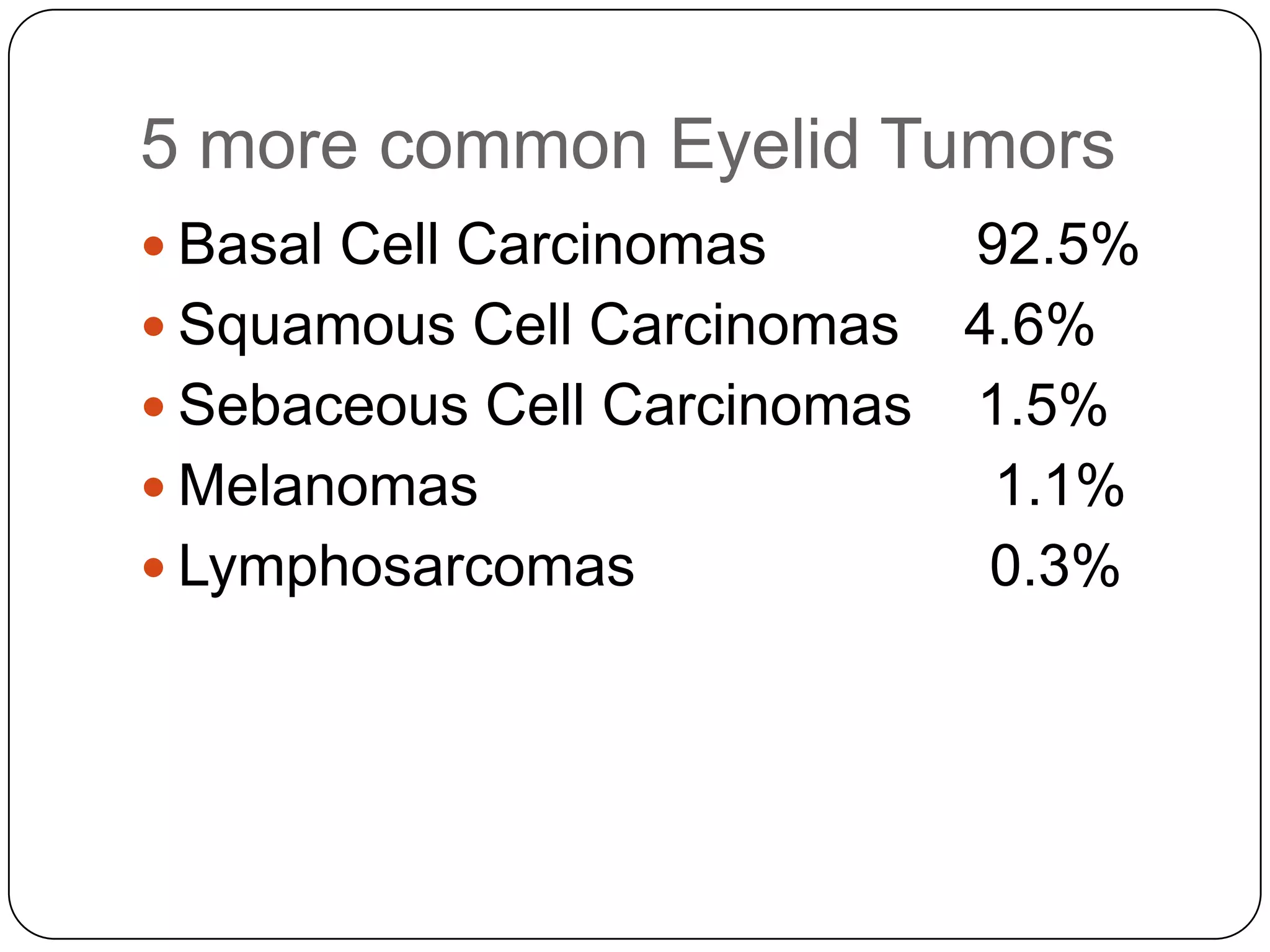
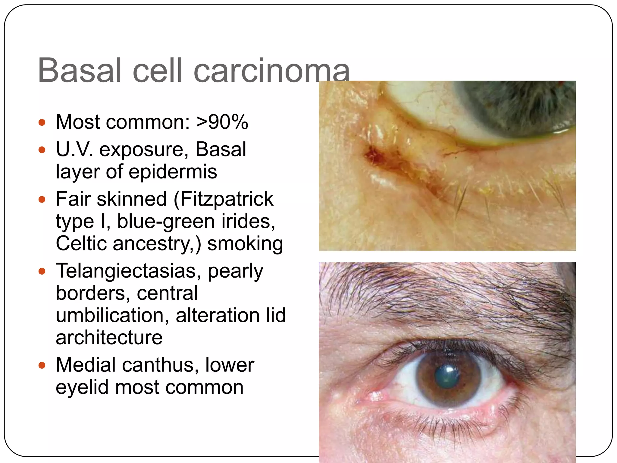
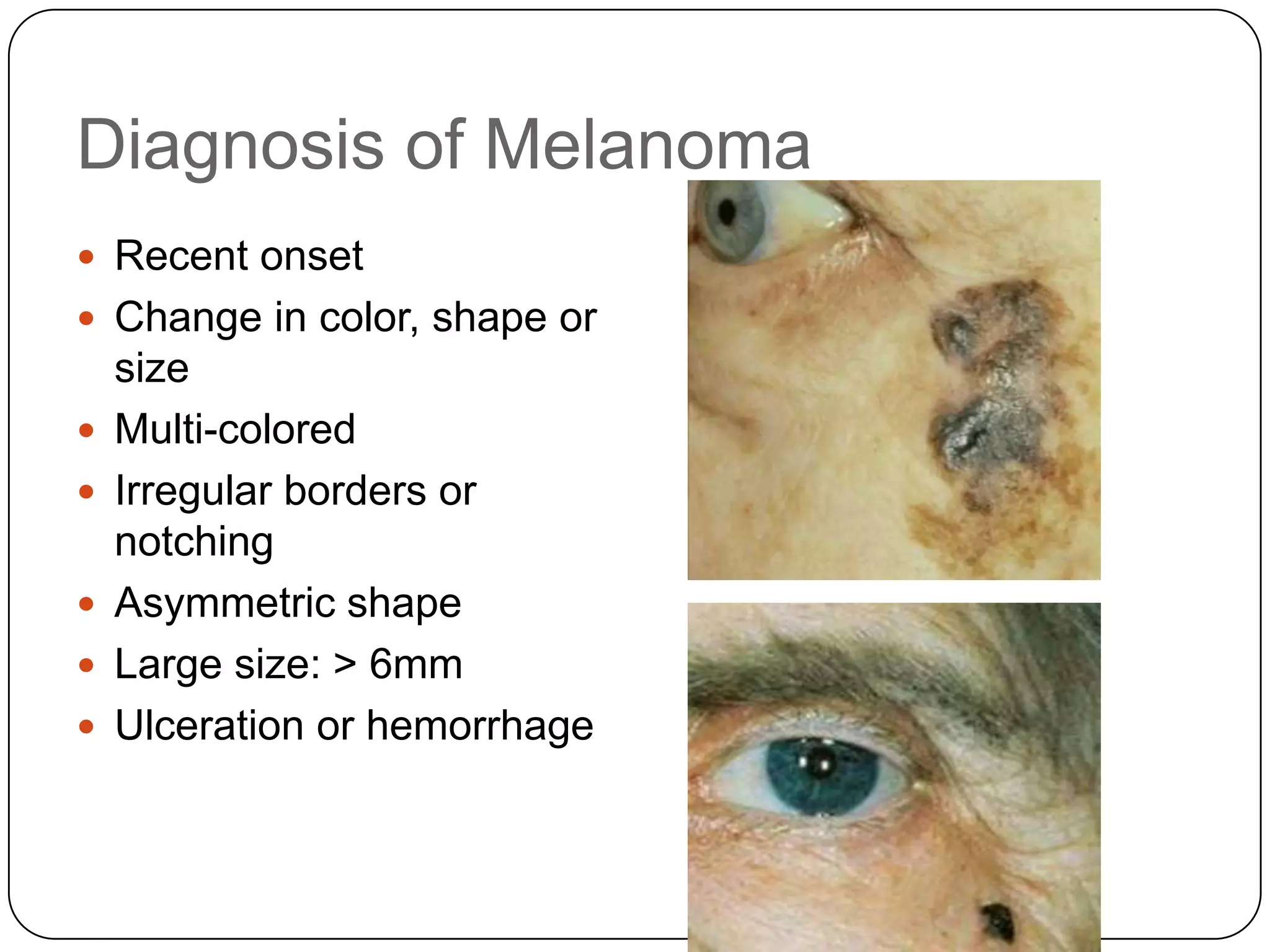
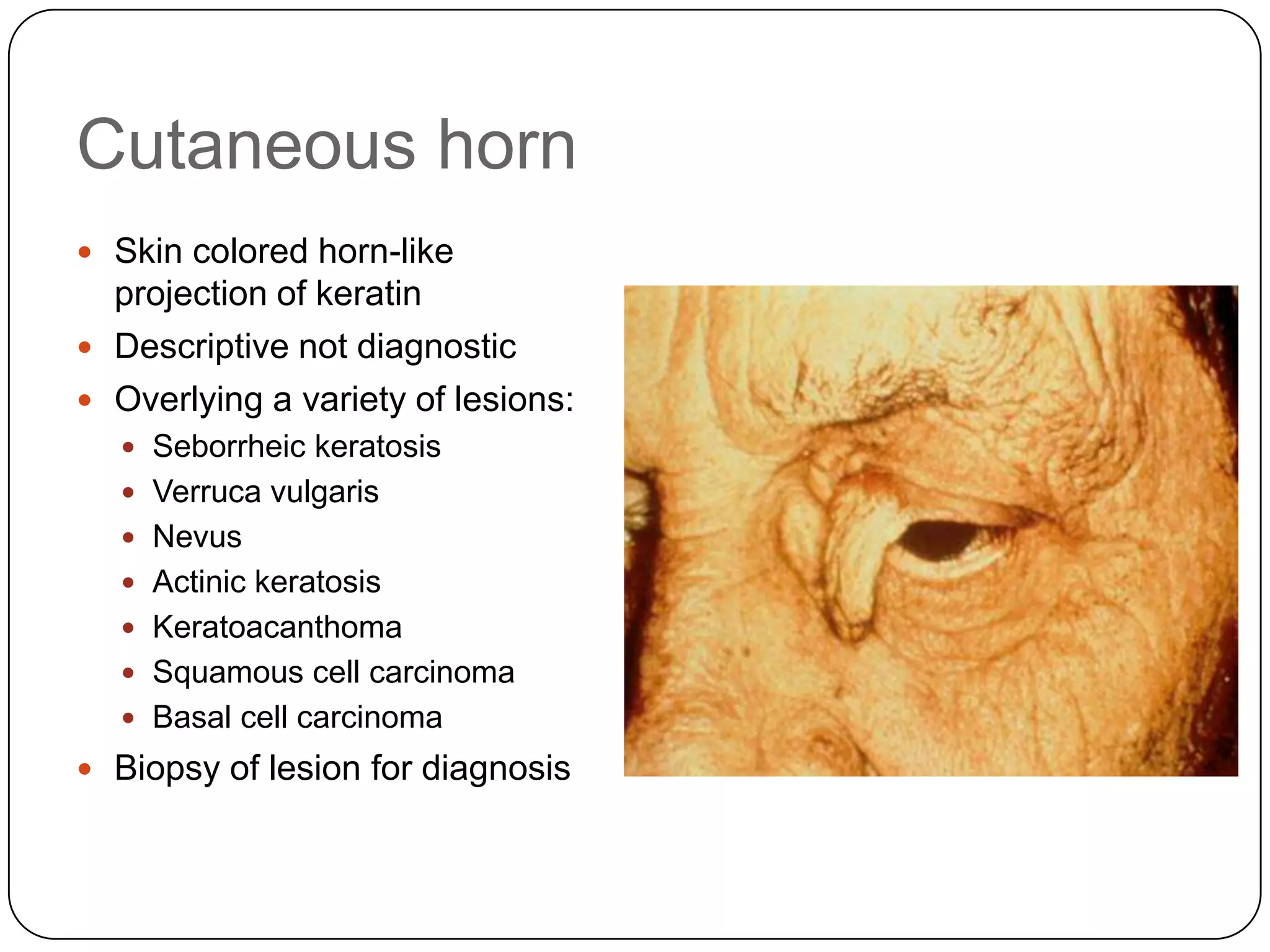
Fritz Allen MD

Visionary Ophthalmology
     March 3rd 2013
Review of ocular malignancies

       focused on - The Eyelids
                - The Cornea and the
             Conjunctiva
                      - The Iris
Malignancies of the Eyelids
5 more common Eyelid Tumors
 Basal Cell Carcinomas     92.5%
 Squamous Cell Carcinomas 4.6%
 Sebaceous Cell Carcinomas 1.5%
 Melanomas                  1.1%
 Lymphosarcomas            0.3%
4 Types of Basal cell Carcinomas
 Nodular
 Diffuse
 Superficial
 Barzexsydrome (autosomal dominant) multiple
 Basal cell carcinomas
Basal cell carcinoma
 Most common: >90%
 U.V. exposure, Basal
  layer of epidermis
 Fair skinned (Fitzpatrick
  type I, blue-green irides,
  Celtic ancestry,) smoking
 Telangiectasias, pearly
  borders, central
  umbilication, alteration lid
  architecture
 Medial canthus, lower
  eyelid most common
Cystic BCC
Unusual BCC
Basal cell carcinoma
Sebaceous cell carcinoma

Irregularity

Induration

Ulceration

Telangiectasia

Alteration normal
      architecture
Sebaceous   secretions
      inflammation
Sebaceous cell carcinoma
 Appearance not typical of
  other periocular neoplasms
 Growth characteristics
   Pagetoid spread
   Late ulceration
   Multifocal origin
 Upper eyelid
 Masquerade syndrome
   Mimics chalazia, unilateral
    blepharitis or conjunctivitis
   Often associated with lash loss
SCCS


       MimicingChalazion
Pigmented lesions
 Benign features
   Uniform color
   Symmetric shape
   Regular border


 Malignant features
   A- asymmetry
   B- borders (irregular)
   C- color (non-uniform)
   D- diameter > 6mm
   E- evolving
Diagnosis of Melanoma
 Recent onset
 Change in color, shape or
    size
   Multi-colored
   Irregular borders or
    notching
   Asymmetric shape
   Large size: > 6mm
   Ulceration or hemorrhage
Differential Diagnosis
 Chalazia
 Benign lesions
Chalazion

 Lipogranulomatous
    inflammation of sebaceous
    gland
   Anterior (Zeis) or posterior
    (meibomian)
   Associated MGD and rosacea
   Pyogenic granuloma if erupts
    posteriorly
   Multiple lesions, recurrences
   Treatment: heat, Doxycycline,
    omega-3 fatty acids, I&D,
    steroid injection
   Limited role for topical gtts/ung
Hordeola
           Internal: meibomian gland
Apocrine hydrocystoma
– Solitary smooth cyst near
  lid margin
– Common middle age and
  older
– Translucent, may be bluish
– Adenoma of gland of Moll
– Often called a
  „sudoriferous cyst‟
Benign eyelid lesions


 Benign epithelial
  proliferations
 “papillomas”
Papilloma
 Pedunculated flesh-
  colored tumor
 Descriptive, not diagnostic
  term
 Used to describe
     Acrochordon
     “skin tag”
     Intradermal nevus
     Seborrheic keratosis
     Verruca vulgaris (wart)
     Actinic keratosis
Seborrheic keratosis

 Greasy, keratotic plaque with
    “stuck on” appearance
   Early – small 1-3 mm well
    outlined, oval flat lesion
   Later – larger, thicker more
    verrucous lesions
   Varigated color, light to dark
    brown
   Multiple keratin plugs on surface
   Usually multiple lesions
   Extremely common
   Benign proliferation of normal
    epithelial cells
   Shave biopsy or excision
Acrochordon “skin tag”
 “Fibroepithelioma”
 Pedunculated flesh-
  colored tumor
 Common on eyelid, neck,
  axillae, groin
 Small, 2-3 mm, often
  multiple
 Snip off at base
Cutaneous horn
 Skin colored horn-like
  projection of keratin
 Descriptive not diagnostic
 Overlying a variety of lesions:
   Seborrheic keratosis
   Verruca vulgaris
   Nevus
   Actinic keratosis
   Keratoacanthoma
   Squamous cell carcinoma
   Basal cell carcinoma
 Biopsy of lesion for diagnosis
Epidermal inclusion cyst
 Whitish dermal – or
    subcutaneous, round cyst
   Contains cheesy,
    desquamated keratin
   Not a “sebaceous cyst”
   May become infected or
    rupture and cause
    inflammation
   Must excise, destroy, or
    marsupialize lining or will
    recur
Epidermal inclusion cyst
Tumors of the Cornea and
Conjunctiva
Conjunctival Intraepithelial Neoplasia
(CIN)
 Bowen‟s disease
 Conjunctival dysplasia
 Intraepithelial epithelioma
 Dyskeratosis
Etiology of CIN

 Uncertain, usually unilateral in fair-skinned men in
  mid 60‟s
 Smoking
 Human Papillo Virus (HPV)
Management and Treatment
 Local excision and Cryotherapy
  with double or triple freeze-thaw
 Interferon
Invasive Squamous Cell Carcinoma
 Replacement of normal epithelium by bizarre
  pleomorphic cells
 10-40% recurrence
Less Common Neoplasms of the
Conjunctiva
 Mucoepidermoid Carcinoma
  (anywhere from the conjunctiva)
 Spindle Cell Carcinoma
 Sebaceous Cell Carcinoma (50-
  60% Upper lids, 20% Lower lids)
  Asians
 Melanocytic Tumors
Miscellaneous Conditions
 Kaposi‟s Sarcoma (in
  immunosupressed , AIDS)
 Lymphoid Tumors
 Granulomas
Tumors of the Iris
 Nevi and Melanomas (most common primary
  tumors of the iris 49-72% of all iris tumors
 Melanocytomas (Magnocellular nevus)
 Melanocytosis(Nevus of Ota)
Pathology
 13% Melanomas (Spindle cells or epithelial cells)
 87% Benign Nevi
 Medium age 40-50 yo
 More common in light Irides, rare in black and
 Asian
Examination and Evaluation
 Slit Lamp, Gonioscopy
 OCT/UBM
 Fluoresceine angiogram
Management
 Conservative approach
 (observation and growth documentation)
 Iridectomy and
 Iridocyclectomy(no glaucoma surgery
 in proven malignant iris tumors)
Differential Diagnosis
 Benign Nevi (less than 3mm in diameter-1mm
    thickness)
   Iris Cyst (OCT)
   Iris nodules (Sarcoidosis, Tuberculosis)
   Hamartomas (Lisch nodules)
   Metastatic lesions
Congenital Disorders


 NEUROFIBROMATOSIS NF1


          Café-au-lait spots   Iris Lisch nodules
Neoplastic Disorders


                       Iris metastasis from lung carcinoma
Malignancies 2013

Malignancies 2013

  • 1.
    Fritz Allen MD VisionaryOphthalmology March 3rd 2013
  • 2.
    Review of ocularmalignancies focused on - The Eyelids - The Cornea and the Conjunctiva - The Iris
  • 3.
  • 4.
    5 more commonEyelid Tumors  Basal Cell Carcinomas 92.5%  Squamous Cell Carcinomas 4.6%  Sebaceous Cell Carcinomas 1.5%  Melanomas 1.1%  Lymphosarcomas 0.3%
  • 5.
    4 Types ofBasal cell Carcinomas  Nodular  Diffuse  Superficial  Barzexsydrome (autosomal dominant) multiple Basal cell carcinomas
  • 6.
    Basal cell carcinoma Most common: >90%  U.V. exposure, Basal layer of epidermis  Fair skinned (Fitzpatrick type I, blue-green irides, Celtic ancestry,) smoking  Telangiectasias, pearly borders, central umbilication, alteration lid architecture  Medial canthus, lower eyelid most common
  • 7.
  • 8.
  • 9.
  • 10.
  • 11.
    Sebaceous cell carcinoma Appearance not typical of other periocular neoplasms  Growth characteristics  Pagetoid spread  Late ulceration  Multifocal origin  Upper eyelid  Masquerade syndrome  Mimics chalazia, unilateral blepharitis or conjunctivitis  Often associated with lash loss
  • 12.
    SCCS MimicingChalazion
  • 13.
    Pigmented lesions  Benignfeatures  Uniform color  Symmetric shape  Regular border  Malignant features  A- asymmetry  B- borders (irregular)  C- color (non-uniform)  D- diameter > 6mm  E- evolving
  • 14.
    Diagnosis of Melanoma Recent onset  Change in color, shape or size  Multi-colored  Irregular borders or notching  Asymmetric shape  Large size: > 6mm  Ulceration or hemorrhage
  • 15.
  • 16.
    Chalazion  Lipogranulomatous inflammation of sebaceous gland  Anterior (Zeis) or posterior (meibomian)  Associated MGD and rosacea  Pyogenic granuloma if erupts posteriorly  Multiple lesions, recurrences  Treatment: heat, Doxycycline, omega-3 fatty acids, I&D, steroid injection  Limited role for topical gtts/ung
  • 17.
    Hordeola Internal: meibomian gland
  • 18.
    Apocrine hydrocystoma – Solitarysmooth cyst near lid margin – Common middle age and older – Translucent, may be bluish – Adenoma of gland of Moll – Often called a „sudoriferous cyst‟
  • 19.
    Benign eyelid lesions Benign epithelial proliferations  “papillomas”
  • 20.
    Papilloma  Pedunculated flesh- colored tumor  Descriptive, not diagnostic term  Used to describe  Acrochordon  “skin tag”  Intradermal nevus  Seborrheic keratosis  Verruca vulgaris (wart)  Actinic keratosis
  • 21.
    Seborrheic keratosis  Greasy,keratotic plaque with “stuck on” appearance  Early – small 1-3 mm well outlined, oval flat lesion  Later – larger, thicker more verrucous lesions  Varigated color, light to dark brown  Multiple keratin plugs on surface  Usually multiple lesions  Extremely common  Benign proliferation of normal epithelial cells  Shave biopsy or excision
  • 22.
    Acrochordon “skin tag” “Fibroepithelioma”  Pedunculated flesh- colored tumor  Common on eyelid, neck, axillae, groin  Small, 2-3 mm, often multiple  Snip off at base
  • 23.
    Cutaneous horn  Skincolored horn-like projection of keratin  Descriptive not diagnostic  Overlying a variety of lesions:  Seborrheic keratosis  Verruca vulgaris  Nevus  Actinic keratosis  Keratoacanthoma  Squamous cell carcinoma  Basal cell carcinoma  Biopsy of lesion for diagnosis
  • 24.
    Epidermal inclusion cyst Whitish dermal – or subcutaneous, round cyst  Contains cheesy, desquamated keratin  Not a “sebaceous cyst”  May become infected or rupture and cause inflammation  Must excise, destroy, or marsupialize lining or will recur
  • 25.
  • 28.
    Tumors of theCornea and Conjunctiva
  • 29.
    Conjunctival Intraepithelial Neoplasia (CIN) Bowen‟s disease  Conjunctival dysplasia  Intraepithelial epithelioma  Dyskeratosis
  • 33.
    Etiology of CIN Uncertain, usually unilateral in fair-skinned men in mid 60‟s  Smoking  Human Papillo Virus (HPV)
  • 34.
    Management and Treatment Local excision and Cryotherapy with double or triple freeze-thaw  Interferon
  • 35.
    Invasive Squamous CellCarcinoma  Replacement of normal epithelium by bizarre pleomorphic cells  10-40% recurrence
  • 36.
    Less Common Neoplasmsof the Conjunctiva  Mucoepidermoid Carcinoma (anywhere from the conjunctiva)  Spindle Cell Carcinoma  Sebaceous Cell Carcinoma (50- 60% Upper lids, 20% Lower lids) Asians  Melanocytic Tumors
  • 39.
    Miscellaneous Conditions  Kaposi‟sSarcoma (in immunosupressed , AIDS)  Lymphoid Tumors  Granulomas
  • 46.
    Tumors of theIris  Nevi and Melanomas (most common primary tumors of the iris 49-72% of all iris tumors  Melanocytomas (Magnocellular nevus)  Melanocytosis(Nevus of Ota)
  • 47.
    Pathology  13% Melanomas(Spindle cells or epithelial cells)  87% Benign Nevi  Medium age 40-50 yo  More common in light Irides, rare in black and Asian
  • 51.
    Examination and Evaluation Slit Lamp, Gonioscopy  OCT/UBM  Fluoresceine angiogram
  • 52.
    Management  Conservative approach (observation and growth documentation)  Iridectomy and Iridocyclectomy(no glaucoma surgery in proven malignant iris tumors)
  • 56.
    Differential Diagnosis  BenignNevi (less than 3mm in diameter-1mm thickness)  Iris Cyst (OCT)  Iris nodules (Sarcoidosis, Tuberculosis)  Hamartomas (Lisch nodules)  Metastatic lesions
  • 58.
    Congenital Disorders NEUROFIBROMATOSISNF1 Café-au-lait spots Iris Lisch nodules
  • 59.
    Neoplastic Disorders Iris metastasis from lung carcinoma

Editor's Notes