STRATEGY OF ADAPTIVE
IMMUNERESPONSE
First response to particular antigen called
primary response
May take a week or more to develop
Immune system remembers pathogen on
subsequent exposure
Termed secondary response
Adaptive immunity divided into
Humoral immunity
Eliminates extracellular pathogens
Cellular immunity
Eliminates intracellular pathogens
3.
HUMORAL IMMUNITY
B LYMPHOCYTE
Overview of humoral immunity
Mediated by B lymphocytes
a.k.a B cells
Develops in bone marrow
B cells may be triggered to proliferate into plasma
cells
Plasma cells produce antibodies
Antibodies produce when antigen bonds B cell receptor
Some B cells produce memory cells
4.
Nature of Antigens
Coined from compounds that elicit antibody
production
Antibody generator
Includes an enormous variety of materials
Today, term used to describe any compound
that elicits an immune response
Antigen that causes immune response termed
immunogen
Proteins and polysaccharides induce string
response
Lipids and nucleic acids often do not
Recognition of antigen directed at antigenic
determinant or epitope
5.
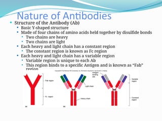
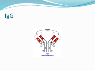
Nature of Antibodies
Structure of the Antibody (Ab)
Basic Y-shaped structure
Made of four chains of amino acids held together by disulfide bonds
Two chains are heavy
Two chains are light
Each heavy and light chain has a constant region
The constant region is known as Fc region
Each heavy and light chain has a variable region
Variable region is unique to each Ab
This region binds to a specific Antigen and is known as “Fab”
region
6.
Nature of Antibodies
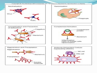
Protective outcomes of antibody-antigen
binding
› Neutralization
Prevents toxin from interacting with cell
› Immobilization and prevention of adherence
Antibody bonding to cellular structures to interfere with
function
› Agglutination and precipitation
Clumping of bacterial cells by specific antibody
Bacteria more easily phagocytized
7.
Nature of Antibodies
Protective outcomes of antibody-antigen
binding
› Opsinization
Coating of bacteria with antibody to enhance phagocytosis
› Complement activation
Antibody bonding triggers classical pathway
› Antibody-dependent cellular cytotoxicity
Multiple antibodies bind a cell which becomes target for
certain cells
9.
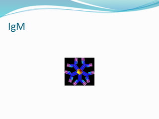
Nature of Antibodies
IgM
First Ab to respond to infection
5 – 13% of Ab in circulation
Only Ab that can be formed by the fetus
IgM expressed as membrane bound anitbodies on B-cells
Pentamer
5 units held together by disulfide bonds
J (Joining) chain functions in the polymerization of monomers
First immunoglobulin class produced in a primary response to an antigen
Has 10 anitgen binding sites
More effective at stimulating complement
Large-size - does not diffuse well
The FC receptors on phagocytes bind IgM (opsinization)
Nature of Antibodies
Five classes of Ab
IgG
Dominant Ab in circulation
• 80 – 85% Ab in circulation
Structure = monomer
The antibody of memory!!!!!
Cross placenta and play important role in protecting
fetus
Provides passive immunity to unborn fetus.
Placental cells bind the Fc portion of IgG and transfer Ab
across the placental membrane.
Activate complement system
Opsonin—phagocytosis
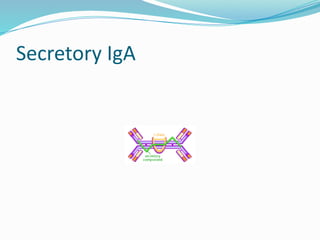
IgA
Found insecretions
10 - 13 % of Ab in circulation
Found Predominantly in external secretions i.e. Breast Milk,
Saliva, tears, mucus.
Serum form is a monomer
Plasma cells that release IgA Abs are concentrated along the
Mucus Membrane surface.
Provides passive immunity to infants through mothers breast
milk
Nature of Antibodies
Five classes of Ab
IgD
<1% of total Ab in circulation
Structure = monomer
Maturation of antibody response
Despite studies extending for more than 4 decades, a
specific role for serum IgD has not been defined
while for IgD bound to the membrane of many B
lymphocytes, several functions have been proposed.
Does NOT cross the placenta.
Does NOT fix complement.
16.
Ig E
Barelydetectable in circulation
Active in allergic reaction
Mediate the immediate hypersensitivity reactions
(hayfever, asthma, hives, anaphylactic shock)
Mast cells and basophils bind fc portion of IgE
Cross-linkage of receptor bound IgE molecules by antigen,
induces degranulaltion of the Mast and basophil cells
Parasitic response
Eosinophils express receptors for IgE
CELL MEDIATED
T LYMPHOCYTES
Overview of cellular immunity
Mediated by T lymphocytes
a.k.a T cells
Matures in thymus
Divided into 2 subsets
Cytotoxic T cells
Helper T cells
T cell receptors help with antigen recognition
19.
Two majorfunction T cell populations
Cytotoxic T cells
Proliferate and differentiate to destroy infected or
cancerous “self” cells
Have CD8 marker
Recognize MHC class I
Helper T cells
Multiply and develop into cells that activate B cells and
macrophages
Stimulate other T cells; orchestrate immune response
Have CD4 marker
Recognize antigen display by MHC class II
20.
During antigenpresentation, antigen cradled in
grove of major histocompatability complex
molecule (MHC molecule)
Two types MHC
MHC class I
Bind endogenous antigen
MHC class II
Bind exogenous antigen
21.
NATURAL KILLER CELLS
Natural killer cells descend from lymphoid stem
cells
› They lack antigen specificity
No antigen receptors
Recognize antigens by means of Fc portion of IgG antibodies
Allow NK cells to attach to antibody-coated cells
Actions augment adaptive immune response
› Important in process of antibody dependent cellular
toxicity
Enable killing of host cells with foreign protein in membrane
Natural killer cells recognize destroyed host cells
with no MHC class I surface molecules
› Important in viral infection
22.
Role ofT cells different from B cells
T cells never produce antibodies
T cells armed with effectors that interact directly
with antigen
T cell receptor does not react with free antigen
23.
Harmful effects ofImmunity
1. AUTOIMMUNITY
It may be defined as the failure of normal process
of an individual to distinguish between self and
non-self i.e when the individual fails to recognize
its own parts as self and develops an immune
response against its own cells and tissues.
Diseases that occur because of autoimmunity are
called as autoimmune diseases.
24.
They resultin structural and functional damages
to the host they include
1. Autoimmune hemolytic anemia
2. Thyrotoxicosis( graves disease)
3. Myasthenia gravis
4. Rheumatoid arthritis
25.
2.HYPERSENSITIVITY
Hypersensitivity diseasesor ailments caused by
impaired immune responses are called
hypersensitivity disorders.
The Causes of hypersensitivity diseases are
Autoimmunity
Reactions against microbes
Reactions against environmental antigens
26.
Reactions against microbes
Reactions against persistent microbial agent may
occur in the form of T-cell response.
Tuberculosis, inflammatory bowel disease and
viral hepatitis are some of the related conditions.
27.
Reactions against environmentalantigens
It does not occur in majority of the population
but very less percentage of the individuals may
show reaction against some harmless
environmental products. As a result of allergy
such patients generate immunoglobulin E (IgE)
antibodies that cause allergic reactions or
disease.
28.
Mechanism and classificationof
hypersensitivity reactions
Based on immune response and some
miscellaneous factors, hypersensitivity reactions
are classified as follows
1. Type I hypersensitivity or immediate
hypersensitivity
It is characterized by the stimulation of helper T
cells that are associated with production of IgE
antibodies and inflammation. Type I is the most
common hypersensitive reaction. Atopy or
allergic reaction is the best example of type I
reactions.
29.
2.Type II hypersensitivedisorders
This occurs due to activation of complement
system by IgG and IgM antibodies. Some of these
antibodies are specific for some antigens and the
disease caused by such antibodies are called type
II hypersensitive disorders such as Graves’
disease.
30.
3.Type III hypersensitivedisorders
Various other antibodies make immune
complexes in blood circulation and cause tissue
damage. Such immune complex diseases are
called type III hypersensitive disorders. Arthus
reaction is a type III hypersensitive disorder.
31.
4.Type IV hypersensitivedisorder/Delayed
hypersensitivity
It involves activation of phagocytes, T-
lymphocytes, and natural killer cells. Multiple
sclerosis is one of such kind.
In brief, majority of hypersensitive reactions are
caused by stimulation of subset of T helper cells.
They generally induce inflammation and tissue
damage by recruiting neutrophils and
macrophages
32.
Activated Tcells do one of two things:
• release cytokines that activate macrophages, or
• kill cells directly
This process is normally useful against intracellular organisms
(viruses, fungi, parasites)
Here, it causes bad stuff: inflammation, cell destruction,
granuloma formation