Definition
• Lung abscess:Localized, pus-filled cavity
within lung parenchyma.
• Forms due to necrosis of lung tissue from
severe infection.
• Characterized by cavity formation with air–
fluid level.
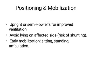
Positioning & Mobilization
•Upright or semi-Fowler’s for improved
ventilation.
• Avoid lying on affected side (risk of shunting).
• Early mobilization: sitting, standing,
ambulation.
Contraindications & Precautions
•Active hemoptysis.
• Severe uncontrolled pleuritic pain.
• Very high fever.
• Hemodynamic instability.
15.
Summary
• Lung abscess:localized necrotic cavity with
pus.
• Commonly due to aspiration or pneumonia.
• Physiotherapy focuses on airway clearance,
breathing exercises, and mobilization.
• Early intervention prevents complications
and promotes recovery.


![PERI-PROSTHETIC FRACTURE NAIL-PLATE CONSTRUCT [NPC].pptx](https://cdn.slidesharecdn.com/ss_thumbnails/drarunkumardrmohamedashrafperiprostheticfrasturenail-plateconstructnpc-260209164459-7e9d15a1-thumbnail.jpg?width=640&height=640&fit=bounds)
