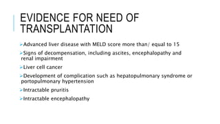
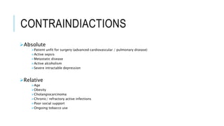
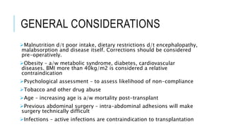
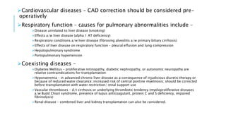
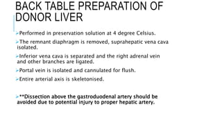
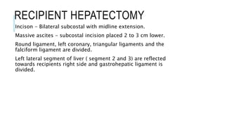
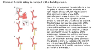
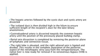
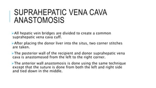
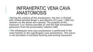
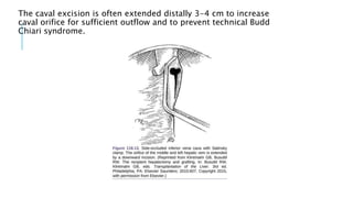
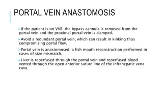
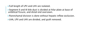
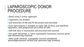
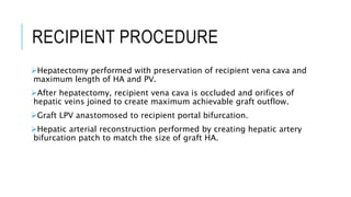
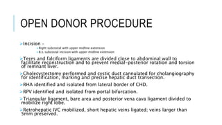
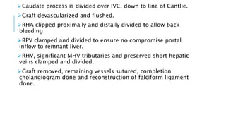
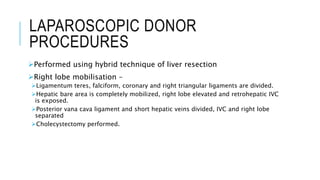
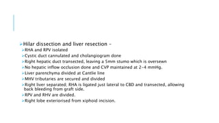
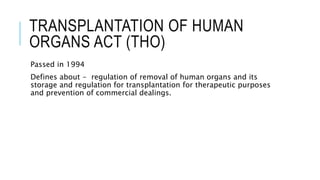
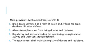
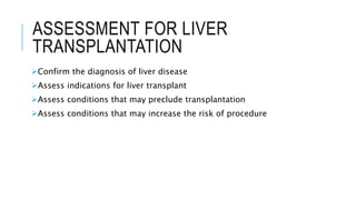
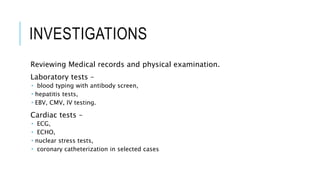
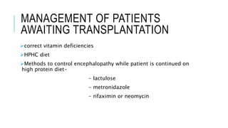
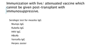
LIVER TRANSPLANTATION
 Moderator – Dr. Mandeep Singh, Dr. Prabhjot kaur
 Presenter – Dr. Venu Goyal
 Date – 02/11/2020
CONTENTS
History of transplantation
Types of donors and grafts
Organ viability
Ethical and constitutional issues
History of liver transplantation
Indications and contraindidcations
General and specific liver disease considerations
Pediatric transplantation
Pre anaesthetic and system oriented assessment
Donor work up
Anatomic variations
Small-for-size syndrome
DDLT
LDLT
Rejection and immunosuppression
Outcomes
ORGAN TRANSPLANTATION
HISTORY OF TRANSPLANTATION
1869 – first skin sutograft transplantation by Carl Bunger.
Bunger did rhinoplasty for syphilis with graft from inner
thigh.
1905 – first successful cornea transplant.
1908 – first skin allograft transplantation
1950 – first successful kidney transplant
1955 – first heart valve allograft into descending aorta
1963 – first successful lung transplant
1998 – first successful hand transplant
1999 – first successful tissue engineered bladder transplanted
TYPES OF DONORS
Living donor
Cadaveric or brain dead donors
Natural death donors
TYPES OF GRAFTS
Autograft –
Transplant of tissue to the same person.
Eg.– skin grafts and vein grafts.
Allograft –
Transplant b/w two genetically non-identical members of same
species.
Most human tissue and organ transplants.
Isograft –
Tissue is transplanted from donor to a genetically identical
recipient.
Eg: identical twins.
Do not trigger an immune response.
Xenograft –
Transplant of organs from one species to another.
Eg. Porcine heart valve.
Domino transplants –
as in CF patients, lung and heart from a deceased donor to CF
patient and heart from CF patient to heart ds patient.
ABO incompatible transplants –
as infants (under 12 months of age)do not have well-developed
immune system
TYPES OF TRANSPLANTS -
Organs – heart, kidney, liver, lung, pancreas, stomach and intestine
Tissues – cornea, bone, tendon, skin, pancreas islets, heart valves,
nerves and veins
Cells – bone marrow and stem cells
Limbs – hands, arms and feet
Multi-organ transplants – include lungs and heart or pancreas and
kidney
ORGANS AND TISSUES
TRANSPLANTED
From deceased donor only – heart, pancreas, stomach, penis, hand,
large intestine and cornea
From deceased and living donor – bone, heart valve, blood and
blood products, kidney, lung, small intestine and testis
Deceased or autograft – blood vessels
Living or autograft – bone marrow/ adult stem cell
ORGAN VIABILITY
Kidney – 30 hrs or less storage time
Pancreas and liver – less than 12 hrs
Heart and lungs – less than 6 hrs
- Preservation is done in chilled solution???????
TRANSPLANTATION OF HUMAN
ORGANS ACT (THO)
Passed in 1994
Defines about - regulation of removal of human organs and its
storage and regulation for transplantation for therapeutic purposes
and prevention of commercial dealings.
Main provisions (with amendments of 2014)
1. Brain death identified as a form of death and criteria for brain
death certification defined.
2. Allows transplantation from living donors and cadavers.
3. Regulatory and advisory bodies for monitoring transplantation
activity and their constitution defined.
4. The government shall maintain registry of donors and recipients.
REASONS FOR DONATION AND
ETHICAL ISSUES
Living related donors –
 when donors have emotional investment.
 Includes “paired exchange” technique.
Good Samaritan or ‘altruistic” donation –
 Recipient has no prior affiliation with the donor.
 Current allocation system does not assess a donor’s motive.
Financial compensation –
 legalized in certain parts of world.
 In Chennai, fishermen sold their kidneys after their livelihood was destroyed
by Indian Ocean tsunami in 2004.
Forced donation –
 lack of public organ donation program in China is used as justification.
Mostly from people deemed undesirable, such as prison populations.
ALLOCATION OF ORGANS
National Organ Transplant Act (NOTA) created Organ procurement
and Transplantation Network (OPTN)
Principles of primary importance –
1. Utility – maximization of net benefit to the community
2. Justice – fair pattern of distribution of nebefits??????
3. Respect for persons - including respect for autonomy
Organ allocation scores based on medical urgency –
1. Liver – MELD score and Child-Turcotte-Pugh score
2. Lungs – Lung Allocation Score (LAS)
3. Kidney – Estimated Post Transplant Survival (EPTS) score
ORGAN TRANSPLANTATION IN
INDIA
India’s 1st transplant – kidney transplant in 1970s
Approx 5000 kidneys, 1000livers and 50 hearts transplanted
annually in India.
India has a poor organ donation rate – 0.26 per million
Required – 1 per million-donation rate
First deceased donor liver transplantation – in 1995
First successful DDLT – 1998
First successful LDLT - 1998
LIVER TRANSPLANTATION
BROAD INDICATIONS OF
TRANSPLANTATION
For improved quality of life
For improved survival benefit
DR. THOMAS STARZL
HISTORY OF LIVER
TRANSPLANTAION
Liver transplant techniques were developed in 50’s and 60’s
with pivotal work of DR Thomas starzl.
First attempted human liver transplant - 1963
First successful liver transplant - 1967
Liver transplantation remained experimental till the
introduction of cyclosporine by Sir Roy Clane in 1981.
The first altruistic living liver donation - in December 2012 in St
James University Hospital Leeds.
EVIDENCE FOR NEED OF
TRANSPLANTATION
Advanced liver disease with MELD score more than/ equal to 15
Signs of decompensation, including ascites, encephalopathy and
renal impairment
Liver cell cancer
Development of complication such as hepatopulmonary syndrome or
portopulmonary hypertension
Intractable pruritis
Intractable encephalopathy
CONTRAINDIACTIONS
Absolute
Patient unfit for surgery (advanced cardiovascular / pulmonary disease)
Active sepsis
Metastatic disease
Active alcoholism
Severe intractable depression
Relative
Age
Obesity
Cholangiocarcinoma
Chronic/ refractory active infections
Poor social support
Ongoing tobacco use
GENERAL CONSIDERATIONS
Malnutrition d/t poor intake, dietary restrictions d/t encephalopathy,
malabsorption and disease itself. Corrections should be considered
pre-operatively.
Obesity – a/w metabolic syndrome, diabetes, cardiovascular
diseases. BMI more than 40kg/m2 is considered a relative
contraindication
Psychological assessment – to assess likelihood of non-compliance
Tobacco and other drug abuse
Age – increasing age is a/w mortality post-transplant
Previous abdominal surgery – intra-abdominal adhesions will make
surgery technically difficult
Infections – active infections are contraindication to transplantation
Cardiovascular diseases – CAD correction should be considered pre-
operatively
Respiratory function – causes for pulmonary abnormalities include –
Disease unrelated to liver disease (smoking)
Effects a/w liver disease (alpha 1 AT deficiency)
Respiratory conditions a/w liver disease (fibrosing alveolitis a/w primary biliary cirrhosis)
Effects of liver disease on respiratory function – pleural effusion and lung compression
Hepatopulmonary syndrome
Portopulmonary hypertension
Coexisting diseases –
Diabetes Mellitus – proliferative retinopathy, diabetic nephropathy, or autonomic neuropathy are
relative contraindications for transplantation
Hyponatremia – in advanced chronic liver disease as a consequence of injudicious diuretic therapy or
because of reduced water clearance; increased risk of central pontine myelinosis; should be corrected
before transplantation with water restriction/ renal support use
Vascular thromboses – d/t cirrhosis or underlying thrombotic tendency (myeloproliferative diseases
a/w Budd Chiari syndrome, presence of lupus anticoagulant, protein C and S deficiency, impaired
fibrinolysis)
Renal disease – combined liver and kidney transplantation can also be considered.
VARIANTS OF ACUTE LIVER
FAILURE
Fulminant hepatic failure
Subacute liver failure
Fulminant autoimmune hepatitis
FULMINANT HEPATIC FAILURE
Characterized by the onset of encephalopathy less than 7 days after the
development of jaundice.
Causes – acetaminophen overdose, viral infections (hep A, B, C, and E),
Wilson’s disease and liver disease of pregnancy.
Causes for death in FHF – cerebral edema, multiorgan failure, sepsis
(bacterial/ fungal), cardiac arrhythmia and respiratory failure.
Contraindications to transplantation in FHF include the onset of irreversible
complications i.e. irreversible cerebral edema, prolonged elevation of ICP or
presence of fixed, dilated pupils for more than 6 hrs.
Best guide to disease progression is estimation of PT or factor V.
Due to extrahepatic manifestations of FHF secondary to extensive necrotic
tissue, a two stage procedure may be considered and pt remains anhepatic
until suitable graft is available.
METABOLIC DISEASES
Wilson’s disease,
Haemophilia,
Hyperoxaluria requiring liver and kidney replacement,
Erythropoietic protoporphyria - likely to recur post transplant but
with decreased morbidity
Budd Chiari syndrome – transplantation considered only when portal
decompression has failed (or is not feasible) or there is established
cirrhosis.
CHRONIC LIVER DISEASE
Appropriate when estimated length of life in the absence of
transplantation is limited to 1 yr.
Child-Pugh and MELD scores are used for timing of transplantation.
PRIMARY BILIARY CIRRHOSIS
Therapies to be considered before transplantation –
 cholestyramine
 Plasmapheresis
 Antidepressants for lethargy
 Questran light (formulation without sorbitol) for cholestyramine induced nausea or diarrhea
 Extrahepatic biliary diversion
 Plasmapheresis and MARS (extracorporeal albumin perfusion)
- it does recur in allograft; at a slower rate.
PRIMARY SCLEROSING
CHOLANGITIS
- Premalignant condition for cholangiocarcinoma
- Cholangiocarcinoma is usually difficult to detect, therefore early
transplantation is considered.
ALCOHOLIC LIVER DISEASE
Candidates with good prognosis post transplant, provided,
continued abstinence from alcohol.
Abstinence period of atleast 6 months required before
transplantation
Alcoholic hepatitis is severe hepatitis characterized by high S.
bilirubin and prolonged clotting.
Patients with advanced alcoholic cardiomyopathy and pancreatitis
are unc=suitable for transplantation.
HEPATITIS VIRAL INFECTIONS
Hep A infection – few patients have recurrent infection in graft
Hep B infection – effective antiviral therapy has significantly reduced
need for transplantation; Hep B Ig and oral antiviral agents are
continued after transplantation.
Hep C infection – pegylated interferon and ribavirin are given pre-
operatively; seen in hemophilia patients secondary to transfusion of
infected blood.
HEPATOCELLULAR CARCINOMA
Accoutns for upto 30%transplants.
Other treatment approaches include – chemotherapy, resection embolization,
transarterial embolization, injection with ethanol and radiofrequency ablation.
Milan criteria (Mazzaferro et al, 1996)
- single tumor less than/equal to 5 cm, or
- 2-3 tumors none exceeding 3 cm, and
- no vascular invasion and/or extrahepatic spread
UCSF Criteria (Yao et al, 2001)
- single tumor less than / equal to 6.5 cm, or
- 2-3 lesions, none exceeding 4.5 cm, with total tumor diameter less
than/equal to 8 cm
- no vascular invasion and/or extrahepatic spread
Factors predicting poor outcome –
Tumor size more than 5 cm
Vascular invasion
Positive nodes
Histologic grade with poor differentiation
Serum marker – alpha fetoprotein
Transplantation in non-cirrhotic HCC livers is done for unresectable
tumors
Cholangiocarcinoma is a contraindication to transplantation because
of high recurrence rate.
SECONDARY LIVER CANCERS
Transplantation is not indicated for metastatic malignancy except in
carcinoid and other NETs.
Better prognostic factors – age less than 50 yrs
- primaries in lung or bowel
- pretransplant somatostatin therapy
PEDIATRIC TRANSPLANTATION
Indications other than those for adults – growth retardation and development of
metabolic bone disease.
Most common indication – disorders of biliary system
- biliary atresia (most common)
- Alagille syndrome
- non syndromic intrahepatic biliary hypoplasia
2nd MC indication – disorders of metabolism
- tyrosinemia
- glycogen storage diseases
- Wilson disease
- galactosuria
- gaucher syndrome
ASSESSMENT FOR LIVER
TRANSPLANTATION
Confirm the diagnosis of liver disease
Assess indications for liver transplant
Assess conditions that may preclude transplantation
Assess conditions that may increase the risk of procedure
INVESTIGATIONS
Reviewing Medical records and physical examination.
Laboratory tests –
 blood typing with antibody screen,
 hepatitis tests,
 EBV, CMV, IV testing.
Cardiac tests –
 ECG,
 ECHO,
 nuclear stress tests,
 coronary catheterization in selected cases
MANAGEMENT OF PATIENTS
AWAITING TRANSPLANTATION
correct vitamin deficiencies
HPHC diet
Methods to control encephalopathy while patient is continued on
high protein diet-
- lactulose
- metronidazole
- rifaximin or neomycin
Immunization with live/ attenuated vaccine which
cannot be given post-transplant with
immunosuppressive.
Serologic test for measles IgG
Mumps IgG
Rubella IgG
HAV IgG
HBsAb
Varicella IgG
Herpes zoster
Ascites
catabolic state a/w risk of SBP.
Prophylactic antibiotics such as ciprofloxacin or amoxyclav.
For severe ascites resistant to diet and diuretics, consider TIPS.
Varices
pharmacoprophylaxis with propranolol or carvedilol.
TIPS
PSC - review patients for cholangiocarcinoma/ bowel cancer
HCC patient reviewed for tumor burden.
NSAIDs avoided to reduce risk of –
- renal failure
- fluid retention
- gastritis induced haemorrhage
PREOPERATIVE ANESTHETIC
ASSESSMENT
CARDIOVASCULAR SYSTEM
Hyperdynamic circulation
In ESLD patients, with increased cardiac output secondary to decreased systemic vascular resistance and
abnormal distribution of central, splanchnic and peripheral circulation.
Cardiomyopathy
Cirrhotic, non ischemic cardiomyopathy
Treatment – sodium restriction
Also a complication of primary disease such as ethanol abuse, amyloidosis, hemochromatosis and
Wilson’s disease.
CAD
Perioperative beta-blocker has a protective effect
PULMONARY SYSTEM
Portopulmonary hypertension
With increased pulmonary vascular resistance and portal hypertension
Mean pulmonary artery pressure more than 25mmHg
PVR more than 240dynes/sec/cm5
Medical optimization with -
 prostaglandins (intravenous epoprostenol or inhaled iloprost)
Phosphodiestrase inhibitors (sildenafil)
ET-1 antagonists (bosentan)
Hepatopulmonary syndrome
Hypoxemia secondary to pulmonary capillary vasodilation, causing VP mismatch
Dyspnea with orthodeoxia
Liver transplantation is the only treatment
Hepatic hydrothorax
Pleural effusion more than 500ml with normal cardiac and pulmonary functions
HEMOSTASIS IN ESLD
Main defect – thrombocytopenia ( secondary to splenic sequestration
and decreased thrombopoetin production)
Main compensatory mechanism – increased endothelium synthesized
vWF
Reduced levels of liver synthesized factors –
Found in both acute and chronic liver failure
 plasminogen, plasmin inhibitor, thrombin activated fibrinolysis inhibitor and factor VIII
PT and aPTT– used to assess severity of synthetic dysfuntion
RENAL SYSTEM
Hepatorenal syndrome –
 marked by renal vasoconstriction and a severe reduction in GFR.
HRS-1 – rapid decline in renal function with multi-organ failure.
HRS-2 – benign course with milder elevation in S. creatinine.
Treatment – vasoconstrictors, renal replacement therapy.
Liver transplantation is the treatment of choice for patients with
cirrhosis and HRS.
DONOR WORK UP
Medical work up
BMI more than 30kg/m2 has a possibility of fatty liver.
Extensive psychological evaluation
Estimation of formula derived graft recipient volume ratio
Dtermination of future remnant liver volume
Liver biopsy may be considered in patients with steatosis
MRI with MRA, MRV and MRC may be done to define graft remnant
liver volume and vascular and biliary anatomy.
THE LIVER IS DIVIDED INTO EIGHT (8) SEGMENTS REFLECTING THE
EIGHT (8) MAJOR DIVISIONS OF THE PORTAL VEIN AND THE BILE DUCT
LIVER ANATOMY
TYPES OF LIVER GRAFTS
ANATOMIC VARIATIONS
HEPATIC ARTERY ANOMALIES
PORTAL VEIN
Normal – bifurcation into left and right common portal trunks.
Variation – trifurcation i.e right anterior and posterior sectional
branches arise separately
HEPATIC VEINS
GRAFT SIZE AND SMALL FOR SIZE
SYNDROME
Measure graft recipient weight ratio
SFSS – post-transplantation hepatic insufficiency presenting as
prolonged cholestasis, coagulopathy and ascites in the absence of
hepatic vascular insufficiency.
Other factors contributing to SFSS –
advanced cirrhosis
Portal hypertension
Associated hyperdynamic circulation
Optimizifn liver graft outflow reduces hyperdynamic liver injury.
Dual liver grafts – avoids SFSS
CADAVER DONOR
HEPATECTOMY
Incisions –
from suprasternal notch (two cavity access) to pubic symphysis
xiphoid process (one-cavity access) to pubic symphysis
Ligamentum teres, falciform ligament and gastrohepatic ligaments are
divided.
Retroperitoneal dissection done to identify left renal vein (a/w risk to injure
SMA, left renal artery and pancreas).
IMV isolated at ligament of Treitz, distally ligated.
Distal abdominal aorta exposed and encircled at bifurcation level and at level
of origin of IMA.
Porta hepatis exposed, CBD identified and encircled distally at duodenal
border and half way incised.
GB fundus is incised and flushed with saline until clear effluent comes from CBD.
Supraceliac aorta identified and encircled, cardiac catheter inserted at distal end
and aortic cross claping done.
Suprahepatic vena cava transected and dual perfusion with ice cold preservation
solution started.
The pericardium and left and right diaphragm around the liver are divided.
Gastroduodenal, Splenic vein, SMV and splenic artery are divided.
Transection of CBD Is completed and hilar dissectionncompleted.
The IMV is divided proximal to the renal vein and liver is excised and packed in
UW solution for preservation.
BACK TABLE PREPARATION OF
DONOR LIVER
Performed in preservation solution at 4 degree Celsius.
The remnant diaphragm is removed, suprahepatic vena cava
isolated.
Inferior vena cava is separated and the right adrenal vein
and other branches are ligated.
Portal vein is isolated and cannulated for flush.
Entire arterial axis is skeletonised.
**Dissection above the gastroduodenal artery should be
avoided due to potential injury to proper hepatic artery.
RECIPIENT HEPATECTOMY
Incison - Bilateral subcostal with midline extension.
Massive ascites - subcostal incision placed 2 to 3 cm lower.
Round ligament, left coronary, triangular ligaments and the
falciform ligament are divided.
Left lateral segment of liver ( segment 2 and 3) are reflected
towards recipients right side and gastrohepatic ligament is
divided.
Common hepatic artery is clamped with a bulldog clamp.
Dissection techniques of the arterial axis in the
recipient. A. Normal hepatic anatomy. RHA,
right hepatic artery; LHA, left hepatic artery;
CHA, common hepatic artery; GDA,
gastroduodenal artery. A dissection technique
that, as a first step, directly ligates (B) and
divides (C) the RHA and LHA should be avoided.
This technique can lead to a dissection of the
intima of the respective arteries due to the
preserved blood flow and subsequently result in
an occlusion of the vessels (inlay in C), which
can significantly impair the patency of the
anastomosis between the recipient and donor
arteries. (D) In contrast to this technique, at
UCLA, an atraumatic bulldog clamp (yellow) is
first placed at the CHA. Subsequently, the RHA
and LHA are ligated (E) and divided (F). In the
latter technique (D, E, and F), the risk of intima
dissection is minimized. (
The hepatic arteries followed by the cystic duct and cystic artery are
dissected
The isolated duct is then divided high in the hilum to ensure
sufficient length of the recipient’s duct for the later biliary
anastomosis.
Gastroduodenal artery is dissected towards the common hepatic
artery until the position of the previously placed bulldog clamp.
Portal vein dissection is completed by removing the posterior plane
of lymphatic and connective tissue.
The right lobe is elevated, and the right adrenal vein is ligated and
divided. This results in the complete separation of the posterior
aspect of the retrohepatic vena cava from the retroperitoneum. The
native liver is now ready for removal if no venovenous bypass (VVB) is
applied.
Vascular clamps are placed on the distal portal vein, infrahepatic and
suprahepatic vena cava.
 All clamps are oriented horizontally with the liver in the normal
anatomic position to avoid rotation of the vessels.
Large Satinsky vascular clamp for the suprahepatic vena cava, is
placed on the very edge of the diaphragmatic reflection to avoid
injuries to the phrenic nerve.
Lower cava is divided as proximal as possible and finally the portal
vein. The native liver is now removed. Hemostasis of the bare area is
achieved by retroperitonealization. For recipients with HCC, no
residual liver tissue of the native liver should remain at the
suprahepatic vena cava
VENOVENOUS BYPASS
The VVB drains the infrahepatic caval and splanchnic blood into the superior
vena cava.
Advantage :
 Maintenance of venous return and splanchnic venous drainage.
 Results in improved hemodynamic stability during the anhepatic phase .
 Reduction in mesenteric edema.
Indications:
 Patients with hemodynamic instability after clamping,
 with fulminate hepatic failure to reduce volume overload,
 with nondialysis-dependent hepatorenal syndrome.
IMPLANTATION OF CADAVER
WHOLE ALLOGRAFT
SUPRAHEPATIC VENA CAVA
ANASTOMOSIS
All hepatic vein bridges are divided to create a common
suprahepatic vena cava cuff.
After placing the donor liver into the situs, two corner stitches
are taken.
The posterior wall of the recipient and donor suprahepatic vena
cava is anastomosed from the left to the right corner.
The anterior wall anastomosis is done using the same technique
except that the suture is done from both the left and right side
and tied down in the middle.
INFRAHEPATIC VENA CAVA
ANASTOMOSIS
During the creation of the anastomosis, the liver is flushed
with chilled lactated Ringer’s and albumin (25 g per 1,000 mL)
solution via the portal vein cannula. The purpose of this
maneuver is to remove possible air and the high extracellular
potassium from the preservation solution (UW).
The infrahepatic vena cava anastomosis is performed in the
same fashion as the suprahepatic cava anastomosis. The suture
is not tied down to facilitate flushing and venting maneuvers
The caval excision is often extended distally 3-4 cm to increase
caval orifice for sufficient outflow and to prevent technical Budd
Chiari syndrome.
PORTAL VEIN ANASTOMOSIS
If the patient is on VVB, the bypass cannula is removed from the
portal vein and the proximal portal vein is clamped.
Avoid a redundant portal vein, which can result in kinking thus
compromising portal flow.
Portal vein is anastomosed, a fish mouth reconstruction performed in
cases of size mismatch.
Liver is reperfused through the portal vein and reperfused blood
vented through the open anterior suture line of the infrahepatic vena
cava.
ARTERIAL
ANASTOMOSIS
An imperfect anastomosis can result in early hepatic artery
thrombosis with graft loss.
In normal cases with type 1 anatomy of the donor and
recipient artery, the anastomosis can usually be created as end-
to-end anastomosis between a Carrel patch of the donor aorta
or artery and the common hepatic artery or branch patch of the
bifurcation of the proper hepatic and gastroduodenal artery.
Branch patch technique for
anastomosis of the donor celiac axis
with a Carrel patch to the longitudinally
opened recipient right and left hepatic
arteries
The recipient gastroduodenal artery is always ligated and divided
when the arterial anastomosis is performed with the recipient
common hepatic artery.
A replaced right hepatic donor artery stemming from the SMA (type 3
anatomy) is the most common arterial variant that requires
reconstruction.In these cases, a Carrel patch of the donor celiac artery
can be anastomosed with a Carrel patch of the donor SMA.
BILIARY
ANASTOMOSIS
Starts with cholecystectomy which includes the resection of the entire
donor cystic duct.
 Retrograde resection of the gallbladder from fundus downward done.
Both a duct-to-duct and a Roux-en-Y biliodigestive anastomosis can be
used for biliary reconstruction.
For choledocho-choledochostomy, donor and recipient bile duct are
shortened to an appropriate length.
 The choledocho-choledochostomy is performed as end-to-end
anastomosis. It is important to avoid redundancy and tension of the bile
duct to prevent biliary obstruction and duct necrosis, respectively.
If a T-tube is indicated, it is brought out of the recipient duct
approximately 1 to 1.5 cm downward the anastomotic suture
line. The T-tube exit side is narrowed with a figure-of-eight
suture .
Techniques of biliary duct-to-duct anastomosis with size
mismatch of donor and recipient bile duct (sketches A and C). For
equal but small-size bile ducts, the anastomotic diameter can be
increased by a site cut in each duct.
PIGGYBACK TECHNIQUE
The piggyback technique is used alternative to the classic
approach and preserves the native retrohepatic vena cava.
The recipient hepatectomy has a mobilization of right and left
lobes and the dissection of hepatoduodenal ligament with
division of vascular and biliary structures.
VVB is not required since venous return via IVC is preserved.
The right lobe is separated from retrohepatic vena cava and
the portal vein is clamped; the native liver is excised.
PRINCIPLES OF CLASSIC TECHNIQUE
(A) AND PIGGYBACK TECHNIQUE OF
ORTHOTOPIC LIVER TRANSPLANTATION
(B)
LIVING DONOR LIVER
TRANSPLANTATION
For LDLT, donor operation is done before recipient to ensure that
there are no obvious physical/ anatomic contraindications to safely
proceeding with donor.
In hepatic malignancy, recipient is explored first to exclude
preoperatively undetected disease that may preclude transplant.
Minimally invasive approaches in living donor hepatectomy reduce
morbidity related to abdominal wall hernia, bowel obstruction and
chronic abdominal discomfort.
Can be laproscopic/ hand assisted or robotic.
LEFT LATERAL SECTION
TRANSPLANT
OPEN DONOR PROCEDURE
Incision -
Upper midline
Left subcostal with midline extension
Left subcostal without midline extension
Bilateral subcostal with an upper midline extension
Left lateral section mobilized by dividing teres, falciform, coronary and
triangular ligaments.
Cystic duct cholangiogram done to localize left hepatic duct.
Gastrohepatic ligament (lesser omentum) divided while preserving replaced
LHA arising from left gastric artery.
Parenchymal bridge overlying umbilical fissure between segments III and IV
is divided, exposing left portal structures.
Vascular dissection is approached from LHA origin distally. Artery to
segment IV may be sacrificed as resultant ischemia is well tolerated in donor.
 Artery to segment IV is preserved when it arises separately from right hepatic artery
Full length of LPV and LHV are isolated.
Segment II and III bile duct is divided at hilar plate at base of
umbilical fissure, and distal end oversewn.
Parenchymal division is done without hepatic inflow occlusion.
LHA, LPV and LHV are divided, and graft removed.
LAPAROSCOPIC DONOR
PROCEDURE
Done using 5 trocar approach.
Ligaments are divided.
Left branches of HA and PV dissected and encircled.
Liver parenchyma is divided on right side of teres and falciform
ligaments, and progressed up to level of LHV.
Left bile duct is transected and proximal end sutured.
For graft extraction, a 10cm suprapubic cutaneous incision given
and retrieval beg inserted.
After graft retrieval, suprapubic incision completed,
pneumoperitoneum vented and specimen bag removed.
RECIPIENT PROCEDURE
Hepatectomy performed with preservation of recipient vena cava and
maximum length of HA and PV.
After hepatectomy, recipient vena cava is occluded and orifices of
hepatic veins joined to create maximum achievable graft outflow.
Graft LPV anastomosed to recipient portal bifurcation.
Hepatic arterial reconstruction performed by creating hepatic artery
bifurcation patch to match the size of graft HA.
LEFT LIVER TRANSPLANTATION
OPEN DONOR PROCEDURE
Segment IV is not devascularized.
After exposure and mobilization of left hemiliver, cholecystectomy is
performed and LHA and LPV are isolated at origin.
Left hepatic duct is divided to the left of possible anomalous entry point of
posterior right hepatic duct.
Caudate lobe is dissected off vena cava to the base of Cantlie line and
caudate vein encircled.
LHV and MHV isolated using Arantius technique.
With temporary inflow occlusion, parenchyma transected along delineation,
facilitated by hanging maneuver.
Intrahepatic MHV is divided when it enters segment IVa.
LHA and LPV ligated and divided, hepatic veins clamped and divided, and
graft removed.
LAPAROSCOPIC DONOR
PROCEDURE
Video accesss by abdominal wall lifting method or CO2
pneumoperitoneum.
Caudate lobe is dissected through foramen of Winslow.
Hepatic transection done under intermittent flow occlusion of right
liver with demarcation seen during occlusion.
RECIPIENT PROCEDURE
Caval preservation done
LHV-MHV confluence of donor and recipient are anastomosed.
Similar to all forms of LDLT
RIGHT LIVER TRANSPLANTATION
OPEN DONOR PROCEDURE
Incision –
Right subcostal with upper midline extension
B/L subcostal incision with upper midline extension
Teres and falciform ligaments are divided close to abdominal wall to
facilitate reconstruction and to prevent medial-posterior rotation and torsion
of remnant liver.
Cholecystectomy performed and cystic duct cannulated for cholangiography
for identification, marking and precise hepatic duct transection.
RHA identified and isolated from lateral border of CHD.
RPV identified and isolated from portal bifurcation.
Triangular ligament, bare area and posterior vena cava ligament divided to
mobilize right lobe.
Retrohepatic IVC mobilized, short hepatic veins ligated; veins larger than
5mm preserved.
Caudate process is divided over IVC, down to line of Cantlie.
Graft devascularized and flushed.
RHA clipped proximally and distally divided to allow back
bleeding
RPV clamped and divided to ensure no compromise portal
inflow to remnant liver.
RHV, significant MHV tributaries and preserved short hepatic
veins clamped and divided.
Graft removed, remaining vessels sutured, completion
cholangiogram done and reconstruction of falciform ligament
done.
LAPAROSCOPIC DONOR
PROCEDURES
Performed using hybrid technique of liver resection
Right lobe mobilisation –
Ligamentum teres, falciform, coronary and right triangular ligaments are divided.
Hepatic bare area is completely mobilized, right lobe elevated and retrohepatic IVC
is exposed.
Posterior vana cava ligament and short hepatic veins divided, IVC and right lobe
separated
Cholecystectomy performed.
Hilar dissection and liver resection –
RHA and RPV isolated
Cystic duct cannulated and cholangiogram done
Right hepatic duct transected, leaving a 5mm stumo which is oversewn
No hepatic inflow occlusion done and CVP maintained at 2-4 mmHg.
Liver parenchyma divided at Cantlie line
MHV tributaries are secured and divided
Right liver separated; RHA is ligated just lateral to CBD and transected, allowing
back bleeding from graft side.
RPV and RHV are divided.
Right lobe exteriorised from xiphoid incision.
IN SITU AND EX SITU
TECHNIQUE
In situ splitting minimizes cold ischemia time.
Biliary structures are better identifiable during in situ
transection.
Insitu splitting into two full hemiliver offer advantage to
apply a pringle maneuver in order to identify anatomic line
of transection.
RECIPIENT COMPLICATIONS
Primary non-function or poor function of the newly transplanted liver
occurs in approximately 1-5% of new transplants. If the function of the
liver does not improve sufficiently or quickly enough, the patient may
urgently require a second transplant to survive.
Hepatic artery thrombosis
Portal vein thrombosis
Biliary complications - Biliary leak
- Biliary stricture
Bleeding
Infection
INDUCTION AND
MAINTENANCE
IMMUNOSUPPRESSION
Triple therapy protocol – calcineurine inhibitor (tacrolimus
and cyclosporine)
- steroids
(methylprednisone)
- DNA
antimetabolite (mycophenolate mofetil).
Quadruple therapy – triple therapy plus daclizumab
(monoclonal IL 2 receptor anatagonist). For patients with
renal compromise.
Sirolimus os used in patients with intolerance to calcineurine
inhibitors or ongoing rejection with calcineurine inhibitors.
REJECTION
Rejection usually causes no symptoms. The first sign is usually
abnormally elevated liver laboratory test results.
Signs and symptoms, if present, include –
 Fever
 Headache
 Fatiguq
 Nausea
 Loss of appetite
 pruritis
 Dark-colored urine
 Jaundice
 Abdominal tenderness
OPPORTUNISTIC INFECTIONS
AND CANCER
Immunosuppression weakens a transplant recipient's
defenses against infections.
They can be divided into three periods : (1,1-6 and >6
months)
 The first month: bacteria and fungi are most common.
 first six months :Viral infections such as cytomegalovirus and other
unusual infections such as tuberculosis and pneumocystis carinii are
seen .
POST-TRANSPLANT
LYMPHOPROLIPHERATIVE
DISORDER (PTLD)
It is almost always associated with Epstein-Barr virus (EBV).
A common approach is therefore to give this drug,
rituximab, in conjunction with less drastic cuts of the
immunosuppression drugs.
The majority of PTLD cases can be successfully treated with
preservation of the transplanted organ.
OUTCOMES
Currently, the overall patient survival one year after liver
transplant is 88%. Patient survival five years after liver
transplant is 73%.
These results vary significantly based on the indication for
liver transplantation. For example, patients who underwent
transplantation for hepatocellular carcinoma had a one-year
survival of only 86% whereas patients who underwent
transplantation for biliary atresia liver disease had a one-
year survival of 94%.
THANK YOU

Liver transplant

  • 1.
    LIVER TRANSPLANTATION  Moderator– Dr. Mandeep Singh, Dr. Prabhjot kaur  Presenter – Dr. Venu Goyal  Date – 02/11/2020
  • 2.
    CONTENTS History of transplantation Typesof donors and grafts Organ viability Ethical and constitutional issues History of liver transplantation Indications and contraindidcations General and specific liver disease considerations Pediatric transplantation Pre anaesthetic and system oriented assessment Donor work up Anatomic variations Small-for-size syndrome DDLT LDLT Rejection and immunosuppression Outcomes
  • 3.
  • 4.
    HISTORY OF TRANSPLANTATION 1869– first skin sutograft transplantation by Carl Bunger. Bunger did rhinoplasty for syphilis with graft from inner thigh. 1905 – first successful cornea transplant. 1908 – first skin allograft transplantation 1950 – first successful kidney transplant 1955 – first heart valve allograft into descending aorta 1963 – first successful lung transplant 1998 – first successful hand transplant 1999 – first successful tissue engineered bladder transplanted
  • 5.
    TYPES OF DONORS Livingdonor Cadaveric or brain dead donors Natural death donors
  • 6.
    TYPES OF GRAFTS Autograft– Transplant of tissue to the same person. Eg.– skin grafts and vein grafts. Allograft – Transplant b/w two genetically non-identical members of same species. Most human tissue and organ transplants. Isograft – Tissue is transplanted from donor to a genetically identical recipient. Eg: identical twins. Do not trigger an immune response. Xenograft – Transplant of organs from one species to another. Eg. Porcine heart valve.
  • 7.
    Domino transplants – asin CF patients, lung and heart from a deceased donor to CF patient and heart from CF patient to heart ds patient. ABO incompatible transplants – as infants (under 12 months of age)do not have well-developed immune system
  • 8.
    TYPES OF TRANSPLANTS- Organs – heart, kidney, liver, lung, pancreas, stomach and intestine Tissues – cornea, bone, tendon, skin, pancreas islets, heart valves, nerves and veins Cells – bone marrow and stem cells Limbs – hands, arms and feet Multi-organ transplants – include lungs and heart or pancreas and kidney
  • 9.
    ORGANS AND TISSUES TRANSPLANTED Fromdeceased donor only – heart, pancreas, stomach, penis, hand, large intestine and cornea From deceased and living donor – bone, heart valve, blood and blood products, kidney, lung, small intestine and testis Deceased or autograft – blood vessels Living or autograft – bone marrow/ adult stem cell
  • 10.
    ORGAN VIABILITY Kidney –30 hrs or less storage time Pancreas and liver – less than 12 hrs Heart and lungs – less than 6 hrs - Preservation is done in chilled solution???????
  • 11.
    TRANSPLANTATION OF HUMAN ORGANSACT (THO) Passed in 1994 Defines about - regulation of removal of human organs and its storage and regulation for transplantation for therapeutic purposes and prevention of commercial dealings.
  • 12.
    Main provisions (withamendments of 2014) 1. Brain death identified as a form of death and criteria for brain death certification defined. 2. Allows transplantation from living donors and cadavers. 3. Regulatory and advisory bodies for monitoring transplantation activity and their constitution defined. 4. The government shall maintain registry of donors and recipients.
  • 13.
    REASONS FOR DONATIONAND ETHICAL ISSUES Living related donors –  when donors have emotional investment.  Includes “paired exchange” technique. Good Samaritan or ‘altruistic” donation –  Recipient has no prior affiliation with the donor.  Current allocation system does not assess a donor’s motive. Financial compensation –  legalized in certain parts of world.  In Chennai, fishermen sold their kidneys after their livelihood was destroyed by Indian Ocean tsunami in 2004. Forced donation –  lack of public organ donation program in China is used as justification. Mostly from people deemed undesirable, such as prison populations.
  • 15.
    ALLOCATION OF ORGANS NationalOrgan Transplant Act (NOTA) created Organ procurement and Transplantation Network (OPTN) Principles of primary importance – 1. Utility – maximization of net benefit to the community 2. Justice – fair pattern of distribution of nebefits?????? 3. Respect for persons - including respect for autonomy
  • 16.
    Organ allocation scoresbased on medical urgency – 1. Liver – MELD score and Child-Turcotte-Pugh score 2. Lungs – Lung Allocation Score (LAS) 3. Kidney – Estimated Post Transplant Survival (EPTS) score
  • 17.
    ORGAN TRANSPLANTATION IN INDIA India’s1st transplant – kidney transplant in 1970s Approx 5000 kidneys, 1000livers and 50 hearts transplanted annually in India. India has a poor organ donation rate – 0.26 per million Required – 1 per million-donation rate First deceased donor liver transplantation – in 1995 First successful DDLT – 1998 First successful LDLT - 1998
  • 18.
  • 19.
    BROAD INDICATIONS OF TRANSPLANTATION Forimproved quality of life For improved survival benefit
  • 20.
  • 21.
    HISTORY OF LIVER TRANSPLANTAION Livertransplant techniques were developed in 50’s and 60’s with pivotal work of DR Thomas starzl. First attempted human liver transplant - 1963 First successful liver transplant - 1967 Liver transplantation remained experimental till the introduction of cyclosporine by Sir Roy Clane in 1981. The first altruistic living liver donation - in December 2012 in St James University Hospital Leeds.
  • 22.
    EVIDENCE FOR NEEDOF TRANSPLANTATION Advanced liver disease with MELD score more than/ equal to 15 Signs of decompensation, including ascites, encephalopathy and renal impairment Liver cell cancer Development of complication such as hepatopulmonary syndrome or portopulmonary hypertension Intractable pruritis Intractable encephalopathy
  • 24.
    CONTRAINDIACTIONS Absolute Patient unfit forsurgery (advanced cardiovascular / pulmonary disease) Active sepsis Metastatic disease Active alcoholism Severe intractable depression Relative Age Obesity Cholangiocarcinoma Chronic/ refractory active infections Poor social support Ongoing tobacco use
  • 25.
    GENERAL CONSIDERATIONS Malnutrition d/tpoor intake, dietary restrictions d/t encephalopathy, malabsorption and disease itself. Corrections should be considered pre-operatively. Obesity – a/w metabolic syndrome, diabetes, cardiovascular diseases. BMI more than 40kg/m2 is considered a relative contraindication Psychological assessment – to assess likelihood of non-compliance Tobacco and other drug abuse Age – increasing age is a/w mortality post-transplant Previous abdominal surgery – intra-abdominal adhesions will make surgery technically difficult Infections – active infections are contraindication to transplantation
  • 26.
    Cardiovascular diseases –CAD correction should be considered pre- operatively Respiratory function – causes for pulmonary abnormalities include – Disease unrelated to liver disease (smoking) Effects a/w liver disease (alpha 1 AT deficiency) Respiratory conditions a/w liver disease (fibrosing alveolitis a/w primary biliary cirrhosis) Effects of liver disease on respiratory function – pleural effusion and lung compression Hepatopulmonary syndrome Portopulmonary hypertension Coexisting diseases – Diabetes Mellitus – proliferative retinopathy, diabetic nephropathy, or autonomic neuropathy are relative contraindications for transplantation Hyponatremia – in advanced chronic liver disease as a consequence of injudicious diuretic therapy or because of reduced water clearance; increased risk of central pontine myelinosis; should be corrected before transplantation with water restriction/ renal support use Vascular thromboses – d/t cirrhosis or underlying thrombotic tendency (myeloproliferative diseases a/w Budd Chiari syndrome, presence of lupus anticoagulant, protein C and S deficiency, impaired fibrinolysis) Renal disease – combined liver and kidney transplantation can also be considered.
  • 27.
    VARIANTS OF ACUTELIVER FAILURE Fulminant hepatic failure Subacute liver failure Fulminant autoimmune hepatitis
  • 28.
    FULMINANT HEPATIC FAILURE Characterizedby the onset of encephalopathy less than 7 days after the development of jaundice. Causes – acetaminophen overdose, viral infections (hep A, B, C, and E), Wilson’s disease and liver disease of pregnancy. Causes for death in FHF – cerebral edema, multiorgan failure, sepsis (bacterial/ fungal), cardiac arrhythmia and respiratory failure. Contraindications to transplantation in FHF include the onset of irreversible complications i.e. irreversible cerebral edema, prolonged elevation of ICP or presence of fixed, dilated pupils for more than 6 hrs. Best guide to disease progression is estimation of PT or factor V. Due to extrahepatic manifestations of FHF secondary to extensive necrotic tissue, a two stage procedure may be considered and pt remains anhepatic until suitable graft is available.
  • 29.
    METABOLIC DISEASES Wilson’s disease, Haemophilia, Hyperoxaluriarequiring liver and kidney replacement, Erythropoietic protoporphyria - likely to recur post transplant but with decreased morbidity Budd Chiari syndrome – transplantation considered only when portal decompression has failed (or is not feasible) or there is established cirrhosis.
  • 30.
    CHRONIC LIVER DISEASE Appropriatewhen estimated length of life in the absence of transplantation is limited to 1 yr. Child-Pugh and MELD scores are used for timing of transplantation.
  • 32.
    PRIMARY BILIARY CIRRHOSIS Therapiesto be considered before transplantation –  cholestyramine  Plasmapheresis  Antidepressants for lethargy  Questran light (formulation without sorbitol) for cholestyramine induced nausea or diarrhea  Extrahepatic biliary diversion  Plasmapheresis and MARS (extracorporeal albumin perfusion) - it does recur in allograft; at a slower rate.
  • 34.
    PRIMARY SCLEROSING CHOLANGITIS - Premalignantcondition for cholangiocarcinoma - Cholangiocarcinoma is usually difficult to detect, therefore early transplantation is considered.
  • 35.
    ALCOHOLIC LIVER DISEASE Candidateswith good prognosis post transplant, provided, continued abstinence from alcohol. Abstinence period of atleast 6 months required before transplantation Alcoholic hepatitis is severe hepatitis characterized by high S. bilirubin and prolonged clotting. Patients with advanced alcoholic cardiomyopathy and pancreatitis are unc=suitable for transplantation.
  • 36.
    HEPATITIS VIRAL INFECTIONS HepA infection – few patients have recurrent infection in graft Hep B infection – effective antiviral therapy has significantly reduced need for transplantation; Hep B Ig and oral antiviral agents are continued after transplantation. Hep C infection – pegylated interferon and ribavirin are given pre- operatively; seen in hemophilia patients secondary to transfusion of infected blood.
  • 37.
    HEPATOCELLULAR CARCINOMA Accoutns forupto 30%transplants. Other treatment approaches include – chemotherapy, resection embolization, transarterial embolization, injection with ethanol and radiofrequency ablation. Milan criteria (Mazzaferro et al, 1996) - single tumor less than/equal to 5 cm, or - 2-3 tumors none exceeding 3 cm, and - no vascular invasion and/or extrahepatic spread UCSF Criteria (Yao et al, 2001) - single tumor less than / equal to 6.5 cm, or - 2-3 lesions, none exceeding 4.5 cm, with total tumor diameter less than/equal to 8 cm - no vascular invasion and/or extrahepatic spread
  • 38.
    Factors predicting pooroutcome – Tumor size more than 5 cm Vascular invasion Positive nodes Histologic grade with poor differentiation Serum marker – alpha fetoprotein Transplantation in non-cirrhotic HCC livers is done for unresectable tumors Cholangiocarcinoma is a contraindication to transplantation because of high recurrence rate.
  • 39.
    SECONDARY LIVER CANCERS Transplantationis not indicated for metastatic malignancy except in carcinoid and other NETs. Better prognostic factors – age less than 50 yrs - primaries in lung or bowel - pretransplant somatostatin therapy
  • 40.
    PEDIATRIC TRANSPLANTATION Indications otherthan those for adults – growth retardation and development of metabolic bone disease. Most common indication – disorders of biliary system - biliary atresia (most common) - Alagille syndrome - non syndromic intrahepatic biliary hypoplasia 2nd MC indication – disorders of metabolism - tyrosinemia - glycogen storage diseases - Wilson disease - galactosuria - gaucher syndrome
  • 41.
    ASSESSMENT FOR LIVER TRANSPLANTATION Confirmthe diagnosis of liver disease Assess indications for liver transplant Assess conditions that may preclude transplantation Assess conditions that may increase the risk of procedure
  • 42.
    INVESTIGATIONS Reviewing Medical recordsand physical examination. Laboratory tests –  blood typing with antibody screen,  hepatitis tests,  EBV, CMV, IV testing. Cardiac tests –  ECG,  ECHO,  nuclear stress tests,  coronary catheterization in selected cases
  • 43.
    MANAGEMENT OF PATIENTS AWAITINGTRANSPLANTATION correct vitamin deficiencies HPHC diet Methods to control encephalopathy while patient is continued on high protein diet- - lactulose - metronidazole - rifaximin or neomycin
  • 44.
    Immunization with live/attenuated vaccine which cannot be given post-transplant with immunosuppressive. Serologic test for measles IgG Mumps IgG Rubella IgG HAV IgG HBsAb Varicella IgG Herpes zoster
  • 45.
    Ascites catabolic state a/wrisk of SBP. Prophylactic antibiotics such as ciprofloxacin or amoxyclav. For severe ascites resistant to diet and diuretics, consider TIPS. Varices pharmacoprophylaxis with propranolol or carvedilol. TIPS PSC - review patients for cholangiocarcinoma/ bowel cancer HCC patient reviewed for tumor burden. NSAIDs avoided to reduce risk of – - renal failure - fluid retention - gastritis induced haemorrhage
  • 46.
  • 47.
    CARDIOVASCULAR SYSTEM Hyperdynamic circulation InESLD patients, with increased cardiac output secondary to decreased systemic vascular resistance and abnormal distribution of central, splanchnic and peripheral circulation. Cardiomyopathy Cirrhotic, non ischemic cardiomyopathy Treatment – sodium restriction Also a complication of primary disease such as ethanol abuse, amyloidosis, hemochromatosis and Wilson’s disease. CAD Perioperative beta-blocker has a protective effect
  • 48.
    PULMONARY SYSTEM Portopulmonary hypertension Withincreased pulmonary vascular resistance and portal hypertension Mean pulmonary artery pressure more than 25mmHg PVR more than 240dynes/sec/cm5 Medical optimization with -  prostaglandins (intravenous epoprostenol or inhaled iloprost) Phosphodiestrase inhibitors (sildenafil) ET-1 antagonists (bosentan) Hepatopulmonary syndrome Hypoxemia secondary to pulmonary capillary vasodilation, causing VP mismatch Dyspnea with orthodeoxia Liver transplantation is the only treatment Hepatic hydrothorax Pleural effusion more than 500ml with normal cardiac and pulmonary functions
  • 49.
    HEMOSTASIS IN ESLD Maindefect – thrombocytopenia ( secondary to splenic sequestration and decreased thrombopoetin production) Main compensatory mechanism – increased endothelium synthesized vWF Reduced levels of liver synthesized factors – Found in both acute and chronic liver failure  plasminogen, plasmin inhibitor, thrombin activated fibrinolysis inhibitor and factor VIII PT and aPTT– used to assess severity of synthetic dysfuntion
  • 50.
    RENAL SYSTEM Hepatorenal syndrome–  marked by renal vasoconstriction and a severe reduction in GFR. HRS-1 – rapid decline in renal function with multi-organ failure. HRS-2 – benign course with milder elevation in S. creatinine. Treatment – vasoconstrictors, renal replacement therapy. Liver transplantation is the treatment of choice for patients with cirrhosis and HRS.
  • 51.
    DONOR WORK UP Medicalwork up BMI more than 30kg/m2 has a possibility of fatty liver. Extensive psychological evaluation Estimation of formula derived graft recipient volume ratio Dtermination of future remnant liver volume Liver biopsy may be considered in patients with steatosis MRI with MRA, MRV and MRC may be done to define graft remnant liver volume and vascular and biliary anatomy.
  • 52.
    THE LIVER ISDIVIDED INTO EIGHT (8) SEGMENTS REFLECTING THE EIGHT (8) MAJOR DIVISIONS OF THE PORTAL VEIN AND THE BILE DUCT LIVER ANATOMY
  • 54.
  • 56.
  • 57.
  • 58.
    PORTAL VEIN Normal –bifurcation into left and right common portal trunks. Variation – trifurcation i.e right anterior and posterior sectional branches arise separately
  • 59.
  • 60.
    GRAFT SIZE ANDSMALL FOR SIZE SYNDROME Measure graft recipient weight ratio SFSS – post-transplantation hepatic insufficiency presenting as prolonged cholestasis, coagulopathy and ascites in the absence of hepatic vascular insufficiency. Other factors contributing to SFSS – advanced cirrhosis Portal hypertension Associated hyperdynamic circulation Optimizifn liver graft outflow reduces hyperdynamic liver injury. Dual liver grafts – avoids SFSS
  • 61.
    CADAVER DONOR HEPATECTOMY Incisions – fromsuprasternal notch (two cavity access) to pubic symphysis xiphoid process (one-cavity access) to pubic symphysis Ligamentum teres, falciform ligament and gastrohepatic ligaments are divided. Retroperitoneal dissection done to identify left renal vein (a/w risk to injure SMA, left renal artery and pancreas). IMV isolated at ligament of Treitz, distally ligated. Distal abdominal aorta exposed and encircled at bifurcation level and at level of origin of IMA. Porta hepatis exposed, CBD identified and encircled distally at duodenal border and half way incised.
  • 62.
    GB fundus isincised and flushed with saline until clear effluent comes from CBD. Supraceliac aorta identified and encircled, cardiac catheter inserted at distal end and aortic cross claping done. Suprahepatic vena cava transected and dual perfusion with ice cold preservation solution started. The pericardium and left and right diaphragm around the liver are divided. Gastroduodenal, Splenic vein, SMV and splenic artery are divided. Transection of CBD Is completed and hilar dissectionncompleted. The IMV is divided proximal to the renal vein and liver is excised and packed in UW solution for preservation.
  • 63.
    BACK TABLE PREPARATIONOF DONOR LIVER Performed in preservation solution at 4 degree Celsius. The remnant diaphragm is removed, suprahepatic vena cava isolated. Inferior vena cava is separated and the right adrenal vein and other branches are ligated. Portal vein is isolated and cannulated for flush. Entire arterial axis is skeletonised. **Dissection above the gastroduodenal artery should be avoided due to potential injury to proper hepatic artery.
  • 65.
    RECIPIENT HEPATECTOMY Incison -Bilateral subcostal with midline extension. Massive ascites - subcostal incision placed 2 to 3 cm lower. Round ligament, left coronary, triangular ligaments and the falciform ligament are divided. Left lateral segment of liver ( segment 2 and 3) are reflected towards recipients right side and gastrohepatic ligament is divided.
  • 66.
    Common hepatic arteryis clamped with a bulldog clamp. Dissection techniques of the arterial axis in the recipient. A. Normal hepatic anatomy. RHA, right hepatic artery; LHA, left hepatic artery; CHA, common hepatic artery; GDA, gastroduodenal artery. A dissection technique that, as a first step, directly ligates (B) and divides (C) the RHA and LHA should be avoided. This technique can lead to a dissection of the intima of the respective arteries due to the preserved blood flow and subsequently result in an occlusion of the vessels (inlay in C), which can significantly impair the patency of the anastomosis between the recipient and donor arteries. (D) In contrast to this technique, at UCLA, an atraumatic bulldog clamp (yellow) is first placed at the CHA. Subsequently, the RHA and LHA are ligated (E) and divided (F). In the latter technique (D, E, and F), the risk of intima dissection is minimized. (
  • 67.
    The hepatic arteriesfollowed by the cystic duct and cystic artery are dissected The isolated duct is then divided high in the hilum to ensure sufficient length of the recipient’s duct for the later biliary anastomosis. Gastroduodenal artery is dissected towards the common hepatic artery until the position of the previously placed bulldog clamp. Portal vein dissection is completed by removing the posterior plane of lymphatic and connective tissue. The right lobe is elevated, and the right adrenal vein is ligated and divided. This results in the complete separation of the posterior aspect of the retrohepatic vena cava from the retroperitoneum. The native liver is now ready for removal if no venovenous bypass (VVB) is applied.
  • 68.
    Vascular clamps areplaced on the distal portal vein, infrahepatic and suprahepatic vena cava.
  • 69.
     All clampsare oriented horizontally with the liver in the normal anatomic position to avoid rotation of the vessels. Large Satinsky vascular clamp for the suprahepatic vena cava, is placed on the very edge of the diaphragmatic reflection to avoid injuries to the phrenic nerve. Lower cava is divided as proximal as possible and finally the portal vein. The native liver is now removed. Hemostasis of the bare area is achieved by retroperitonealization. For recipients with HCC, no residual liver tissue of the native liver should remain at the suprahepatic vena cava
  • 70.
    VENOVENOUS BYPASS The VVBdrains the infrahepatic caval and splanchnic blood into the superior vena cava. Advantage :  Maintenance of venous return and splanchnic venous drainage.  Results in improved hemodynamic stability during the anhepatic phase .  Reduction in mesenteric edema. Indications:  Patients with hemodynamic instability after clamping,  with fulminate hepatic failure to reduce volume overload,  with nondialysis-dependent hepatorenal syndrome.
  • 72.
  • 73.
    SUPRAHEPATIC VENA CAVA ANASTOMOSIS Allhepatic vein bridges are divided to create a common suprahepatic vena cava cuff. After placing the donor liver into the situs, two corner stitches are taken. The posterior wall of the recipient and donor suprahepatic vena cava is anastomosed from the left to the right corner. The anterior wall anastomosis is done using the same technique except that the suture is done from both the left and right side and tied down in the middle.
  • 74.
    INFRAHEPATIC VENA CAVA ANASTOMOSIS Duringthe creation of the anastomosis, the liver is flushed with chilled lactated Ringer’s and albumin (25 g per 1,000 mL) solution via the portal vein cannula. The purpose of this maneuver is to remove possible air and the high extracellular potassium from the preservation solution (UW). The infrahepatic vena cava anastomosis is performed in the same fashion as the suprahepatic cava anastomosis. The suture is not tied down to facilitate flushing and venting maneuvers
  • 75.
    The caval excisionis often extended distally 3-4 cm to increase caval orifice for sufficient outflow and to prevent technical Budd Chiari syndrome.
  • 76.
    PORTAL VEIN ANASTOMOSIS Ifthe patient is on VVB, the bypass cannula is removed from the portal vein and the proximal portal vein is clamped. Avoid a redundant portal vein, which can result in kinking thus compromising portal flow. Portal vein is anastomosed, a fish mouth reconstruction performed in cases of size mismatch. Liver is reperfused through the portal vein and reperfused blood vented through the open anterior suture line of the infrahepatic vena cava.
  • 77.
    ARTERIAL ANASTOMOSIS An imperfect anastomosiscan result in early hepatic artery thrombosis with graft loss. In normal cases with type 1 anatomy of the donor and recipient artery, the anastomosis can usually be created as end- to-end anastomosis between a Carrel patch of the donor aorta or artery and the common hepatic artery or branch patch of the bifurcation of the proper hepatic and gastroduodenal artery.
  • 78.
    Branch patch techniquefor anastomosis of the donor celiac axis with a Carrel patch to the longitudinally opened recipient right and left hepatic arteries
  • 79.
    The recipient gastroduodenalartery is always ligated and divided when the arterial anastomosis is performed with the recipient common hepatic artery. A replaced right hepatic donor artery stemming from the SMA (type 3 anatomy) is the most common arterial variant that requires reconstruction.In these cases, a Carrel patch of the donor celiac artery can be anastomosed with a Carrel patch of the donor SMA.
  • 80.
    BILIARY ANASTOMOSIS Starts with cholecystectomywhich includes the resection of the entire donor cystic duct.  Retrograde resection of the gallbladder from fundus downward done. Both a duct-to-duct and a Roux-en-Y biliodigestive anastomosis can be used for biliary reconstruction. For choledocho-choledochostomy, donor and recipient bile duct are shortened to an appropriate length.  The choledocho-choledochostomy is performed as end-to-end anastomosis. It is important to avoid redundancy and tension of the bile duct to prevent biliary obstruction and duct necrosis, respectively.
  • 81.
    If a T-tubeis indicated, it is brought out of the recipient duct approximately 1 to 1.5 cm downward the anastomotic suture line. The T-tube exit side is narrowed with a figure-of-eight suture .
  • 82.
    Techniques of biliaryduct-to-duct anastomosis with size mismatch of donor and recipient bile duct (sketches A and C). For equal but small-size bile ducts, the anastomotic diameter can be increased by a site cut in each duct.
  • 84.
    PIGGYBACK TECHNIQUE The piggybacktechnique is used alternative to the classic approach and preserves the native retrohepatic vena cava. The recipient hepatectomy has a mobilization of right and left lobes and the dissection of hepatoduodenal ligament with division of vascular and biliary structures. VVB is not required since venous return via IVC is preserved. The right lobe is separated from retrohepatic vena cava and the portal vein is clamped; the native liver is excised.
  • 85.
    PRINCIPLES OF CLASSICTECHNIQUE (A) AND PIGGYBACK TECHNIQUE OF ORTHOTOPIC LIVER TRANSPLANTATION (B)
  • 86.
    LIVING DONOR LIVER TRANSPLANTATION ForLDLT, donor operation is done before recipient to ensure that there are no obvious physical/ anatomic contraindications to safely proceeding with donor. In hepatic malignancy, recipient is explored first to exclude preoperatively undetected disease that may preclude transplant. Minimally invasive approaches in living donor hepatectomy reduce morbidity related to abdominal wall hernia, bowel obstruction and chronic abdominal discomfort. Can be laproscopic/ hand assisted or robotic.
  • 87.
  • 88.
    OPEN DONOR PROCEDURE Incision- Upper midline Left subcostal with midline extension Left subcostal without midline extension Bilateral subcostal with an upper midline extension Left lateral section mobilized by dividing teres, falciform, coronary and triangular ligaments. Cystic duct cholangiogram done to localize left hepatic duct. Gastrohepatic ligament (lesser omentum) divided while preserving replaced LHA arising from left gastric artery. Parenchymal bridge overlying umbilical fissure between segments III and IV is divided, exposing left portal structures. Vascular dissection is approached from LHA origin distally. Artery to segment IV may be sacrificed as resultant ischemia is well tolerated in donor.
  • 89.
     Artery tosegment IV is preserved when it arises separately from right hepatic artery
  • 90.
    Full length ofLPV and LHV are isolated. Segment II and III bile duct is divided at hilar plate at base of umbilical fissure, and distal end oversewn. Parenchymal division is done without hepatic inflow occlusion. LHA, LPV and LHV are divided, and graft removed.
  • 91.
    LAPAROSCOPIC DONOR PROCEDURE Done using5 trocar approach. Ligaments are divided. Left branches of HA and PV dissected and encircled. Liver parenchyma is divided on right side of teres and falciform ligaments, and progressed up to level of LHV. Left bile duct is transected and proximal end sutured. For graft extraction, a 10cm suprapubic cutaneous incision given and retrieval beg inserted. After graft retrieval, suprapubic incision completed, pneumoperitoneum vented and specimen bag removed.
  • 92.
    RECIPIENT PROCEDURE Hepatectomy performedwith preservation of recipient vena cava and maximum length of HA and PV. After hepatectomy, recipient vena cava is occluded and orifices of hepatic veins joined to create maximum achievable graft outflow. Graft LPV anastomosed to recipient portal bifurcation. Hepatic arterial reconstruction performed by creating hepatic artery bifurcation patch to match the size of graft HA.
  • 93.
  • 94.
    OPEN DONOR PROCEDURE SegmentIV is not devascularized. After exposure and mobilization of left hemiliver, cholecystectomy is performed and LHA and LPV are isolated at origin. Left hepatic duct is divided to the left of possible anomalous entry point of posterior right hepatic duct. Caudate lobe is dissected off vena cava to the base of Cantlie line and caudate vein encircled. LHV and MHV isolated using Arantius technique. With temporary inflow occlusion, parenchyma transected along delineation, facilitated by hanging maneuver. Intrahepatic MHV is divided when it enters segment IVa. LHA and LPV ligated and divided, hepatic veins clamped and divided, and graft removed.
  • 95.
    LAPAROSCOPIC DONOR PROCEDURE Video accesssby abdominal wall lifting method or CO2 pneumoperitoneum. Caudate lobe is dissected through foramen of Winslow. Hepatic transection done under intermittent flow occlusion of right liver with demarcation seen during occlusion.
  • 96.
    RECIPIENT PROCEDURE Caval preservationdone LHV-MHV confluence of donor and recipient are anastomosed. Similar to all forms of LDLT
  • 97.
  • 98.
    OPEN DONOR PROCEDURE Incision– Right subcostal with upper midline extension B/L subcostal incision with upper midline extension Teres and falciform ligaments are divided close to abdominal wall to facilitate reconstruction and to prevent medial-posterior rotation and torsion of remnant liver. Cholecystectomy performed and cystic duct cannulated for cholangiography for identification, marking and precise hepatic duct transection. RHA identified and isolated from lateral border of CHD. RPV identified and isolated from portal bifurcation. Triangular ligament, bare area and posterior vena cava ligament divided to mobilize right lobe. Retrohepatic IVC mobilized, short hepatic veins ligated; veins larger than 5mm preserved.
  • 99.
    Caudate process isdivided over IVC, down to line of Cantlie. Graft devascularized and flushed. RHA clipped proximally and distally divided to allow back bleeding RPV clamped and divided to ensure no compromise portal inflow to remnant liver. RHV, significant MHV tributaries and preserved short hepatic veins clamped and divided. Graft removed, remaining vessels sutured, completion cholangiogram done and reconstruction of falciform ligament done.
  • 100.
    LAPAROSCOPIC DONOR PROCEDURES Performed usinghybrid technique of liver resection Right lobe mobilisation – Ligamentum teres, falciform, coronary and right triangular ligaments are divided. Hepatic bare area is completely mobilized, right lobe elevated and retrohepatic IVC is exposed. Posterior vana cava ligament and short hepatic veins divided, IVC and right lobe separated Cholecystectomy performed.
  • 101.
    Hilar dissection andliver resection – RHA and RPV isolated Cystic duct cannulated and cholangiogram done Right hepatic duct transected, leaving a 5mm stumo which is oversewn No hepatic inflow occlusion done and CVP maintained at 2-4 mmHg. Liver parenchyma divided at Cantlie line MHV tributaries are secured and divided Right liver separated; RHA is ligated just lateral to CBD and transected, allowing back bleeding from graft side. RPV and RHV are divided. Right lobe exteriorised from xiphoid incision.
  • 102.
    IN SITU ANDEX SITU TECHNIQUE In situ splitting minimizes cold ischemia time. Biliary structures are better identifiable during in situ transection. Insitu splitting into two full hemiliver offer advantage to apply a pringle maneuver in order to identify anatomic line of transection.
  • 103.
    RECIPIENT COMPLICATIONS Primary non-functionor poor function of the newly transplanted liver occurs in approximately 1-5% of new transplants. If the function of the liver does not improve sufficiently or quickly enough, the patient may urgently require a second transplant to survive. Hepatic artery thrombosis Portal vein thrombosis Biliary complications - Biliary leak - Biliary stricture Bleeding Infection
  • 104.
    INDUCTION AND MAINTENANCE IMMUNOSUPPRESSION Triple therapyprotocol – calcineurine inhibitor (tacrolimus and cyclosporine) - steroids (methylprednisone) - DNA antimetabolite (mycophenolate mofetil). Quadruple therapy – triple therapy plus daclizumab (monoclonal IL 2 receptor anatagonist). For patients with renal compromise. Sirolimus os used in patients with intolerance to calcineurine inhibitors or ongoing rejection with calcineurine inhibitors.
  • 105.
    REJECTION Rejection usually causesno symptoms. The first sign is usually abnormally elevated liver laboratory test results. Signs and symptoms, if present, include –  Fever  Headache  Fatiguq  Nausea  Loss of appetite  pruritis  Dark-colored urine  Jaundice  Abdominal tenderness
  • 106.
    OPPORTUNISTIC INFECTIONS AND CANCER Immunosuppressionweakens a transplant recipient's defenses against infections. They can be divided into three periods : (1,1-6 and >6 months)  The first month: bacteria and fungi are most common.  first six months :Viral infections such as cytomegalovirus and other unusual infections such as tuberculosis and pneumocystis carinii are seen .
  • 107.
    POST-TRANSPLANT LYMPHOPROLIPHERATIVE DISORDER (PTLD) It isalmost always associated with Epstein-Barr virus (EBV). A common approach is therefore to give this drug, rituximab, in conjunction with less drastic cuts of the immunosuppression drugs. The majority of PTLD cases can be successfully treated with preservation of the transplanted organ.
  • 108.
    OUTCOMES Currently, the overallpatient survival one year after liver transplant is 88%. Patient survival five years after liver transplant is 73%. These results vary significantly based on the indication for liver transplantation. For example, patients who underwent transplantation for hepatocellular carcinoma had a one-year survival of only 86% whereas patients who underwent transplantation for biliary atresia liver disease had a one- year survival of 94%.
  • 109.