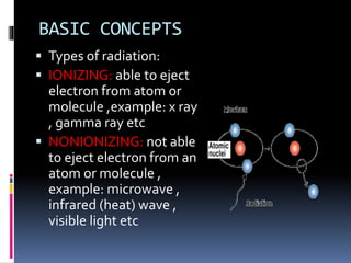
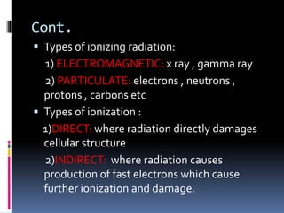
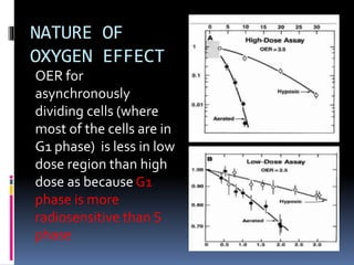
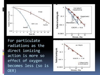
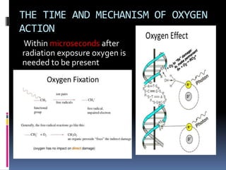
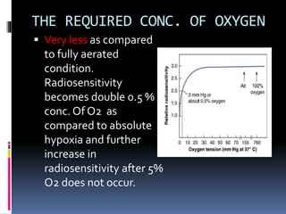
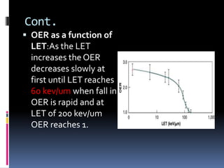
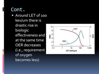
BY DR.SOUMYADIP ROY
MODERATOR: DR.SHYAMA SUDHA
PREM
ADDITIONAL PROFESSOR
RADIATION ONCOLOGY JIPMER
LET RBE OER
OVERVIEW
 BASIC CONCEPTS
 LET
 RBE with their relationship in between
 OER and factors controlling them
 Summary
BASIC CONCEPTS
 Types of radiation:
 IONIZING: able to eject
electron from atom or
molecule ,example: x ray
, gamma ray etc
 NONIONIZING: not able
to eject electron from an
atom or molecule ,
example: microwave ,
infrared (heat) wave ,
visible light etc
Cont.
 Types of ionizing radiation:
1) ELECTROMAGNETIC: x ray , gamma ray
2) PARTICULATE: electrons , neutrons ,
protons , carbons etc
 Types of ionization :
1)DIRECT: where radiation directly damages
cellular structure
2)INDIRECT: where radiation causes
production of fast electrons which cause
further ionization and damage.
Cont.
•Types of radiation
action:
1)Direct action :
Radiation directly
hits the target to
cause damage
2)Indirect action :
production of free
radical which cause
damage.
Cont.
 Types of radiation
as per degree of
ionization:
1) dense: alpha
particle
2) intermediate :
neutron
3) sparse : x ray ,
gamma ray
LET
 Stands for linear energy transfer
 Definition:The LET of charged particles in
medium is the quotient of dE/dl , where dE is
average energy locally imparted to the
medium by a charged particle of specified
energy in traversing a distance of dl. (ICRU ,
1962)
 Unit : kiloelectron volt per micrometer
Cont.
 Methods to calculate
LET:
Cont.
 LET is an average quantity because at
microscopic level , deposition of energy per
unit length of tract varies over a wide range.
 For neutrons biologic properties correlate
better with energy average.
 With increasing LET biologic effectiveness of
a radiation increases upto a level there after it
decreases (discussed later).
Cont.
LET for one
particular type of
radiation
decreases as
energy increases
because with
increasing energy
ionization along
the track becomes
more sparse
RBE
GETTING CELL SURVIVAL CURVE
 A graph is plotted with
surviving fraction in
logarathmic scale in y
axis and dose of
radiation in x axis
RBE:
 Definition:The RBE of some test radiation ®
is defined by the ratio D250/Dr , where D250
and Dr are , respectively, the doses of x rays
and the test radiation required for equal
biologic effect. (National Bureau of Standards
, 1954).
 RBE is not constant for a particular type of
radiation and varies according to the end
point chosen.
Cont.
 RBE for single doses is
large initially and
diminishes gradually
as the dose is
increased because for
neutrons unlike x ray
the initial shoulder is
narrow and the curve
is steeper
Cont.
 For fractionated dose
both of the curves
have several
shoulders but the size
of the shoulders are
more for x ray as the
sublethal damage
repair is more and cell
killing is less So RBE
increases for
neutrons.
Cont.
RBE depends on
1. Radiation quality
2. Radiation dose
3. Fractionation
4. Dose rate
5. Biologic system or end point
Cont.
 RBE as a function of
LET: As LET increases
from 2 kev/um for xrays
to 150 kev/um for alpha
particles survival curve
becomes steeper and
shoulder disappears.
Large shoulder
indicates repair of more
SLD.
Cont.
 RBE increases rapidly
after 10 kev/um and
peaks at 100 kev/um.
After that increase in
LET causes decrease in
RBE. Curve 1,2,3
represents survival
fractions 0.8,0.1 and
0.01 respectively
Cont.
 Optimal LET is 100
kev/um because at this
LET the average
separation of
ionization events just
coincides with the
diameter of DNA (2
nm) and therefore has
highest probability of
causing DSB
OER AND OXYGEN EFFECTS
WHY OXYGEN IS IMPORTANT IN
ONCOLOGY
 Oxygen is important for cell killing effect of
radiation
 Hypoxia has been found to cause tumour
progression and metastsis
 Hypoxia is implicated not only in
radioresistance but also chemoresistance
 Bleomycin , 5FU, cisplatin , methotrexate are
less effective against hypoxic cells (Grau and
Overgaard ,1992).
TUMOUR CONTAINS HYPOXIC
CELLS
Survival curve for mouse
subcutaneous
lymphosarcoma has
two parts. For 1st D0 is
1.1 Gy and for 2nd it is
2.6 Gy , indicating that
after 9 Gy 2nd part of the
curve is due to hypoxic
proportion of cells (1% ).
TYPES OF HYPOXIA IN TUMOURS
1. Acute (Martin Brown ,1980s):Due to
malformed vasculature which
periodically open and close.
1. Chronic (Thomilson and Gray ,1955): Due
to rapid rate of metabolism by tumour
cells oxygen is rapidly used up by initial
layer of cells of 100-180 micron thickness
surrounding capillaries.
OER
 It is the ratio of doses of radiation under
hypoxic to aerated condition needed to produce
same biologic effect.
Oxygen ‘fixes’ the damage produce by indirect
action of ionizing radiation.
 Value: 2.5-3.5 for sparsely ionizing
radiation(X ray , gamma ray) ,1.6 for
intermediate ionizing (neutron) , 1 for densely
ionizing radiation ( alpha particle)
Cont.
Radiation with high
LET has more direct
action of DNA
damage and
indirect action
decreases and so
also the
requirement of O2.
NATURE OF
OXYGEN EFFECT
OER for
asynchronously
dividing cells (where
most of the cells are in
G1 phase) is less in low
dose region than high
dose as because G1
phase is more
radiosensitive than S
phase
For particulate
radiations as the
direct ionizing
action is more so
effect of oxygen
becomes less (so is
OER)
THE TIME AND MECHANISM OF OXYGEN
ACTION
Within microseconds after
radiation exposure oxygen is
needed to be present
THE REQUIRED CONC. OF OXYGEN
 Very less as compared
to fully aerated
condition.
Radiosensitivity
becomes double 0.5 %
conc. Of O2 as
compared to absolute
hypoxia and further
increase in
radiosensitivity after 5%
O2 does not occur.
Cont.
 OER as a function of
LET:As the LET
increases the OER
decreases slowly at
first until LET reaches
60 kev/um when fall in
OER is rapid and at
LET of 200 kev/um
OER reaches 1.
Cont.
 Around LET of 100
kev/um there is
drastic rise in
biologic
effectiveness and
at the same time
OER decreases
(i.e., requirement
of oxygen
becomes less)
SUMMARY
 X ray and gamma rays are sparsely ionizing
radiation because the ionizations are well
separated in time and space
 Alpha particles and neutrons are densely
ionizing radiations
 LET is the energy transferred per unit length
of track.Typical values are 0.2 kev/um for
cobalt-60 gamma ray , 2kev/um for 250kv
xray , 166 kev/um for 2.5 Mev alpha particles
SUMMARY Cont.
 RBE for some test radiation (r) is the ratio
D250/Dr where D250 and Dr are doses of 250
kv X ray and the test radiation respectively to
produce equal biologic effect
 RBE depends on radiation quality (LET) ,
total dose , dose per fraction , dose rate ,
biologic system , end point
 OER is the ratio of doses in hypoxic to
aerated condition to produce same effect
 Small quantity of oxygen is needed for
radiosensitization.
Cont.
 Oxygen makes the free radical mediated
DNA damage permanent

LET RBE OER

  • 1.
    BY DR.SOUMYADIP ROY MODERATOR:DR.SHYAMA SUDHA PREM ADDITIONAL PROFESSOR RADIATION ONCOLOGY JIPMER LET RBE OER
  • 2.
    OVERVIEW  BASIC CONCEPTS LET  RBE with their relationship in between  OER and factors controlling them  Summary
  • 3.
    BASIC CONCEPTS  Typesof radiation:  IONIZING: able to eject electron from atom or molecule ,example: x ray , gamma ray etc  NONIONIZING: not able to eject electron from an atom or molecule , example: microwave , infrared (heat) wave , visible light etc
  • 4.
    Cont.  Types ofionizing radiation: 1) ELECTROMAGNETIC: x ray , gamma ray 2) PARTICULATE: electrons , neutrons , protons , carbons etc  Types of ionization : 1)DIRECT: where radiation directly damages cellular structure 2)INDIRECT: where radiation causes production of fast electrons which cause further ionization and damage.
  • 5.
    Cont. •Types of radiation action: 1)Directaction : Radiation directly hits the target to cause damage 2)Indirect action : production of free radical which cause damage.
  • 6.
    Cont.  Types ofradiation as per degree of ionization: 1) dense: alpha particle 2) intermediate : neutron 3) sparse : x ray , gamma ray
  • 7.
    LET  Stands forlinear energy transfer  Definition:The LET of charged particles in medium is the quotient of dE/dl , where dE is average energy locally imparted to the medium by a charged particle of specified energy in traversing a distance of dl. (ICRU , 1962)  Unit : kiloelectron volt per micrometer
  • 8.
    Cont.  Methods tocalculate LET:
  • 9.
    Cont.  LET isan average quantity because at microscopic level , deposition of energy per unit length of tract varies over a wide range.  For neutrons biologic properties correlate better with energy average.  With increasing LET biologic effectiveness of a radiation increases upto a level there after it decreases (discussed later).
  • 10.
    Cont. LET for one particulartype of radiation decreases as energy increases because with increasing energy ionization along the track becomes more sparse
  • 11.
  • 13.
    GETTING CELL SURVIVALCURVE  A graph is plotted with surviving fraction in logarathmic scale in y axis and dose of radiation in x axis
  • 14.
    RBE:  Definition:The RBEof some test radiation ® is defined by the ratio D250/Dr , where D250 and Dr are , respectively, the doses of x rays and the test radiation required for equal biologic effect. (National Bureau of Standards , 1954).  RBE is not constant for a particular type of radiation and varies according to the end point chosen.
  • 15.
    Cont.  RBE forsingle doses is large initially and diminishes gradually as the dose is increased because for neutrons unlike x ray the initial shoulder is narrow and the curve is steeper
  • 16.
    Cont.  For fractionateddose both of the curves have several shoulders but the size of the shoulders are more for x ray as the sublethal damage repair is more and cell killing is less So RBE increases for neutrons.
  • 17.
    Cont. RBE depends on 1.Radiation quality 2. Radiation dose 3. Fractionation 4. Dose rate 5. Biologic system or end point
  • 18.
    Cont.  RBE asa function of LET: As LET increases from 2 kev/um for xrays to 150 kev/um for alpha particles survival curve becomes steeper and shoulder disappears. Large shoulder indicates repair of more SLD.
  • 19.
    Cont.  RBE increasesrapidly after 10 kev/um and peaks at 100 kev/um. After that increase in LET causes decrease in RBE. Curve 1,2,3 represents survival fractions 0.8,0.1 and 0.01 respectively
  • 20.
    Cont.  Optimal LETis 100 kev/um because at this LET the average separation of ionization events just coincides with the diameter of DNA (2 nm) and therefore has highest probability of causing DSB
  • 21.
  • 22.
    WHY OXYGEN ISIMPORTANT IN ONCOLOGY  Oxygen is important for cell killing effect of radiation  Hypoxia has been found to cause tumour progression and metastsis  Hypoxia is implicated not only in radioresistance but also chemoresistance  Bleomycin , 5FU, cisplatin , methotrexate are less effective against hypoxic cells (Grau and Overgaard ,1992).
  • 23.
    TUMOUR CONTAINS HYPOXIC CELLS Survivalcurve for mouse subcutaneous lymphosarcoma has two parts. For 1st D0 is 1.1 Gy and for 2nd it is 2.6 Gy , indicating that after 9 Gy 2nd part of the curve is due to hypoxic proportion of cells (1% ).
  • 26.
    TYPES OF HYPOXIAIN TUMOURS 1. Acute (Martin Brown ,1980s):Due to malformed vasculature which periodically open and close. 1. Chronic (Thomilson and Gray ,1955): Due to rapid rate of metabolism by tumour cells oxygen is rapidly used up by initial layer of cells of 100-180 micron thickness surrounding capillaries.
  • 27.
    OER  It isthe ratio of doses of radiation under hypoxic to aerated condition needed to produce same biologic effect. Oxygen ‘fixes’ the damage produce by indirect action of ionizing radiation.  Value: 2.5-3.5 for sparsely ionizing radiation(X ray , gamma ray) ,1.6 for intermediate ionizing (neutron) , 1 for densely ionizing radiation ( alpha particle)
  • 28.
    Cont. Radiation with high LEThas more direct action of DNA damage and indirect action decreases and so also the requirement of O2.
  • 29.
    NATURE OF OXYGEN EFFECT OERfor asynchronously dividing cells (where most of the cells are in G1 phase) is less in low dose region than high dose as because G1 phase is more radiosensitive than S phase
  • 30.
    For particulate radiations asthe direct ionizing action is more so effect of oxygen becomes less (so is OER)
  • 31.
    THE TIME ANDMECHANISM OF OXYGEN ACTION Within microseconds after radiation exposure oxygen is needed to be present
  • 32.
    THE REQUIRED CONC.OF OXYGEN  Very less as compared to fully aerated condition. Radiosensitivity becomes double 0.5 % conc. Of O2 as compared to absolute hypoxia and further increase in radiosensitivity after 5% O2 does not occur.
  • 33.
    Cont.  OER asa function of LET:As the LET increases the OER decreases slowly at first until LET reaches 60 kev/um when fall in OER is rapid and at LET of 200 kev/um OER reaches 1.
  • 34.
    Cont.  Around LETof 100 kev/um there is drastic rise in biologic effectiveness and at the same time OER decreases (i.e., requirement of oxygen becomes less)
  • 35.
    SUMMARY  X rayand gamma rays are sparsely ionizing radiation because the ionizations are well separated in time and space  Alpha particles and neutrons are densely ionizing radiations  LET is the energy transferred per unit length of track.Typical values are 0.2 kev/um for cobalt-60 gamma ray , 2kev/um for 250kv xray , 166 kev/um for 2.5 Mev alpha particles
  • 36.
    SUMMARY Cont.  RBEfor some test radiation (r) is the ratio D250/Dr where D250 and Dr are doses of 250 kv X ray and the test radiation respectively to produce equal biologic effect  RBE depends on radiation quality (LET) , total dose , dose per fraction , dose rate , biologic system , end point  OER is the ratio of doses in hypoxic to aerated condition to produce same effect  Small quantity of oxygen is needed for radiosensitization.
  • 37.
    Cont.  Oxygen makesthe free radical mediated DNA damage permanent