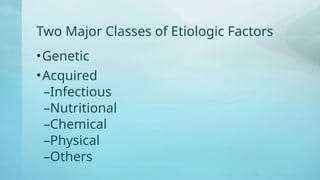
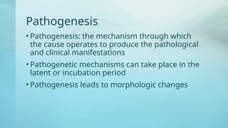
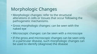
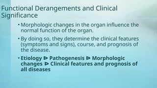
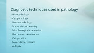
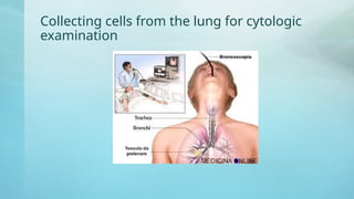
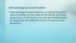
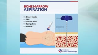
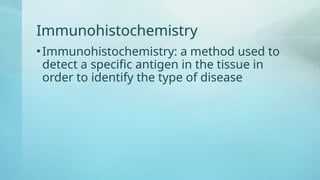
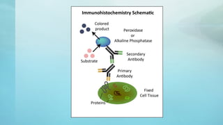
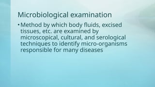
Basic Pathology – The
Diagnostic Process
Ekwendeni College of Health Sciences
Dr. Sue Makin
Learning Objectives
• Define pathology
• Discuss the core aspects of disease in pathology
• Know the diagnostic techniques used in pathology
• Know the various categories of the causes of diseases
• Know the course, outcome, consequences of diseases
Core Aspects of Diseases in Pathology
• Pathology: the study of disease by scientific
methods
• Disease: an abnormal variation in structure or
function of any part of the body
• Four aspects of disease
–Etiology
–Pathogenesis
–Morphologic changes
–Functional derangements and clinical significance
Etiology
• Etiology of a disease: the cause of a disease
• Primary etiology: the cause of a disease
• If the cause of a disease is unknown, the
etiology is idiopathic
• Knowledge of the primary cause is the
backbone on which a diagnosis can be made, a
disease understood, and a treatment can be
developed
Two Major Classes of Etiologic Factors
•Genetic
•Acquired
–Infectious
–Nutritional
–Chemical
–Physical
–Others
Pathogenesis
• Pathogenesis: the mechanism through which
the cause operates to produce the pathological
and clinical manifestations
• Pathogenetic mechanisms can take place in the
latent or incubation period
• Pathogenesis leads to morphologic changes
Morphologic Changes
• Morphologic changes: refer to the structural
alterations in cells or tissues that occur following the
pathogenetic mechanisms.
• Gross morphologic changes: can be seen with the
naked eye
• Microscopic changes: can be seen with a microscope
• If the gross and microscopic changes can be seen only
in a particular disease, such morphologic changes can
be used to identify (diagnose) the disease
Functional Derangements and Clinical
Significance
• Morphologic changes in the organ influence the
normal function of the organ.
• By doing so, they determine the clinical features
(symptoms and signs), course, and prognosis of
the disease.
• Etiology ᐅ Pathogenesis ᐅ Morphologic
changes ᐅ Clinical features and prognosis of
all diseases
Diagnostic techniques used in pathology
• Histopathology
• Cytopathology
• Hematopathology
• Immunohistochemistry
• Microbiological examination
• Biochemical examination
• Cytogenetics
• Molecular techniques
• Autopsy
Histopathological techniques
• The study of tissues under the microscope, looking for
abnormal structure in the tissues
• The tissues for study are obtained by biopsy. Biopsy is a tissue
sample from a living person to identify a disease. Biopsy can be
either incisional or excisional.
• Once the tissue is obtained, it must be immediately fixed by
putting it into 10% formaldehyde (10% formalin) before sending
it to the pathologist
Purpose of Fixation
• 1. To prevent autolysis and bacterial decomposition and
putrefaction
• 2. To coagulate the tissue to prevent loss of easily diffusible
substances
• 3. To strengthen the tissue against the deleterious effects of the
various stages in the preparation of sections and tissue
processing
• 4. To leave the tissues in a condition which helps differential
staining with dyes and other reagents
Cytopathologic Techniques
• Cytopathology: the study of cells from various
body sites to determine the cause or nature of
disease
• Application of cytopathology:
–Screening for the early detection of
asymptomatic cancer
–Diagnosis of symptomatic cancer
–Surveillance of patients treated for cancer
Collecting cells from the lung for cytologic
examination
Hematological examination
• Hematological examination: a method by which
abnormalities of the cells of the blood and their
precursors in the bone marrow are investigated
to diagnose the different kinds of anemia and
leukemia.
Immunohistochemistry
•Immunohistochemistry: a method used to
detect a specific antigen in the tissue in
order to identify the type of disease
Microbiological examination
•Method by which body fluids, excised
tissues, etc. are examined by
microscopical, cultural, and serological
techniques to identify micro-organisms
responsible for many diseases
Biochemical examination
• Method by which the metabolic disturbances of disease are
investigated by assay of various normal and abnormal
compounds in the blood urine, etc.
Clinical genetics (cytogenetics)
• A method in which inherited chromosomal abnormalities in the
germ cells or acquired chromosomal abnormalities in somatic
cells are investigated using the techniques of molecular biology
• Molecular biology: the branch of biology that studies the
molecular basis of biologic activity
Molecular techniques
• Different molecular techniques such as fluorescent in situe
hydbirdization, Southern blot, etc … can be used to detect
genetic diseases
Autopsy
• Autopsy: examination of a dead body to identify the cause of
death. This can be used for forensic or clinical purposes.
Causes of Disease
• Environmental and Genetic
• Environmental
– Physical agents
– Chemicals
– Nutritional deficiencies and excesses
– Infections and infestations
– Immunological factors
– Psychogenic factors
• Genetic factors: hereditary factors that are inherited genetically
from parents
Course of Disease
• Exposure ᐅ Biological Onset ᐅ Clinical Onset ᐅ Permanent
Damage ᐅ Death
• The course of a disease in the absence of any intervention is
called the natural history of the disease.
– Exposure to various risk factors (causative agents)
– Latency: period between exposure and biological onset of disease
– Biological onset of disease: the initiaton of the disease process, however,
without any sign or symptom. Following biologic onset of disease, it may
remain asymptomatic or subclinical (i.e. without any clinical
manifestations), or may lead to overt clinical disease.
– Incubation period: refers to a variable period of time without any obvious
signs or symptoms from the time of exposure
Course of Disease (continued)
– The clinical onset of the disease, when the signs and symptoms of the
disease become apparent.
– Onset of permanent damage
– Death
Outcome and consequences of disease
• Disease may follow any of the following trends:
– Resolution can occur leaving no sequelae (abnormality)
– The disease can settle down, but sequelae(abnormality) are left, or
– The disease may result in death.
Clinical death
• Clinical death: the reversible transition between life and biologic
death. Clinical death is defined as the period of respiratory,
circulatory, and brain arrest during which initiation of
resuscitation can lead to recovery.
• Clinical death begins with either the last agonal inhalation or
the last cardiac contraction.
• Some signs of clinical death
– No pulse or blood pressure detectable
– Pupils are widely dilated(enlarged)
– Recovery can occur with resuscitation (kiss of life)
Biological Death
• Biological death (sure sign of death): an irreversible state of
cellular destruction. It manifests with irreversible cessation of
circulatory and respiratory functions, or irreversible cessation of
all function of the entire brain, including the brain stem.

Lecture 1 - Diag Process.pptx ekwendweni

  • 1.
    Basic Pathology –The Diagnostic Process Ekwendeni College of Health Sciences Dr. Sue Makin
  • 2.
    Learning Objectives • Definepathology • Discuss the core aspects of disease in pathology • Know the diagnostic techniques used in pathology • Know the various categories of the causes of diseases • Know the course, outcome, consequences of diseases
  • 3.
    Core Aspects ofDiseases in Pathology • Pathology: the study of disease by scientific methods • Disease: an abnormal variation in structure or function of any part of the body • Four aspects of disease –Etiology –Pathogenesis –Morphologic changes –Functional derangements and clinical significance
  • 4.
    Etiology • Etiology ofa disease: the cause of a disease • Primary etiology: the cause of a disease • If the cause of a disease is unknown, the etiology is idiopathic • Knowledge of the primary cause is the backbone on which a diagnosis can be made, a disease understood, and a treatment can be developed
  • 5.
    Two Major Classesof Etiologic Factors •Genetic •Acquired –Infectious –Nutritional –Chemical –Physical –Others
  • 6.
    Pathogenesis • Pathogenesis: themechanism through which the cause operates to produce the pathological and clinical manifestations • Pathogenetic mechanisms can take place in the latent or incubation period • Pathogenesis leads to morphologic changes
  • 7.
    Morphologic Changes • Morphologicchanges: refer to the structural alterations in cells or tissues that occur following the pathogenetic mechanisms. • Gross morphologic changes: can be seen with the naked eye • Microscopic changes: can be seen with a microscope • If the gross and microscopic changes can be seen only in a particular disease, such morphologic changes can be used to identify (diagnose) the disease
  • 8.
    Functional Derangements andClinical Significance • Morphologic changes in the organ influence the normal function of the organ. • By doing so, they determine the clinical features (symptoms and signs), course, and prognosis of the disease. • Etiology ᐅ Pathogenesis ᐅ Morphologic changes ᐅ Clinical features and prognosis of all diseases
  • 9.
    Diagnostic techniques usedin pathology • Histopathology • Cytopathology • Hematopathology • Immunohistochemistry • Microbiological examination • Biochemical examination • Cytogenetics • Molecular techniques • Autopsy
  • 10.
    Histopathological techniques • Thestudy of tissues under the microscope, looking for abnormal structure in the tissues • The tissues for study are obtained by biopsy. Biopsy is a tissue sample from a living person to identify a disease. Biopsy can be either incisional or excisional. • Once the tissue is obtained, it must be immediately fixed by putting it into 10% formaldehyde (10% formalin) before sending it to the pathologist
  • 11.
    Purpose of Fixation •1. To prevent autolysis and bacterial decomposition and putrefaction • 2. To coagulate the tissue to prevent loss of easily diffusible substances • 3. To strengthen the tissue against the deleterious effects of the various stages in the preparation of sections and tissue processing • 4. To leave the tissues in a condition which helps differential staining with dyes and other reagents
  • 13.
    Cytopathologic Techniques • Cytopathology:the study of cells from various body sites to determine the cause or nature of disease • Application of cytopathology: –Screening for the early detection of asymptomatic cancer –Diagnosis of symptomatic cancer –Surveillance of patients treated for cancer
  • 14.
    Collecting cells fromthe lung for cytologic examination
  • 15.
    Hematological examination • Hematologicalexamination: a method by which abnormalities of the cells of the blood and their precursors in the bone marrow are investigated to diagnose the different kinds of anemia and leukemia.
  • 17.
    Immunohistochemistry •Immunohistochemistry: a methodused to detect a specific antigen in the tissue in order to identify the type of disease
  • 19.
    Microbiological examination •Method bywhich body fluids, excised tissues, etc. are examined by microscopical, cultural, and serological techniques to identify micro-organisms responsible for many diseases
  • 20.
    Biochemical examination • Methodby which the metabolic disturbances of disease are investigated by assay of various normal and abnormal compounds in the blood urine, etc.
  • 21.
    Clinical genetics (cytogenetics) •A method in which inherited chromosomal abnormalities in the germ cells or acquired chromosomal abnormalities in somatic cells are investigated using the techniques of molecular biology • Molecular biology: the branch of biology that studies the molecular basis of biologic activity
  • 22.
    Molecular techniques • Differentmolecular techniques such as fluorescent in situe hydbirdization, Southern blot, etc … can be used to detect genetic diseases
  • 25.
    Autopsy • Autopsy: examinationof a dead body to identify the cause of death. This can be used for forensic or clinical purposes.
  • 26.
    Causes of Disease •Environmental and Genetic • Environmental – Physical agents – Chemicals – Nutritional deficiencies and excesses – Infections and infestations – Immunological factors – Psychogenic factors • Genetic factors: hereditary factors that are inherited genetically from parents
  • 27.
    Course of Disease •Exposure ᐅ Biological Onset ᐅ Clinical Onset ᐅ Permanent Damage ᐅ Death • The course of a disease in the absence of any intervention is called the natural history of the disease. – Exposure to various risk factors (causative agents) – Latency: period between exposure and biological onset of disease – Biological onset of disease: the initiaton of the disease process, however, without any sign or symptom. Following biologic onset of disease, it may remain asymptomatic or subclinical (i.e. without any clinical manifestations), or may lead to overt clinical disease. – Incubation period: refers to a variable period of time without any obvious signs or symptoms from the time of exposure
  • 28.
    Course of Disease(continued) – The clinical onset of the disease, when the signs and symptoms of the disease become apparent. – Onset of permanent damage – Death
  • 29.
    Outcome and consequencesof disease • Disease may follow any of the following trends: – Resolution can occur leaving no sequelae (abnormality) – The disease can settle down, but sequelae(abnormality) are left, or – The disease may result in death.
  • 30.
    Clinical death • Clinicaldeath: the reversible transition between life and biologic death. Clinical death is defined as the period of respiratory, circulatory, and brain arrest during which initiation of resuscitation can lead to recovery. • Clinical death begins with either the last agonal inhalation or the last cardiac contraction. • Some signs of clinical death – No pulse or blood pressure detectable – Pupils are widely dilated(enlarged) – Recovery can occur with resuscitation (kiss of life)
  • 31.
    Biological Death • Biologicaldeath (sure sign of death): an irreversible state of cellular destruction. It manifests with irreversible cessation of circulatory and respiratory functions, or irreversible cessation of all function of the entire brain, including the brain stem.