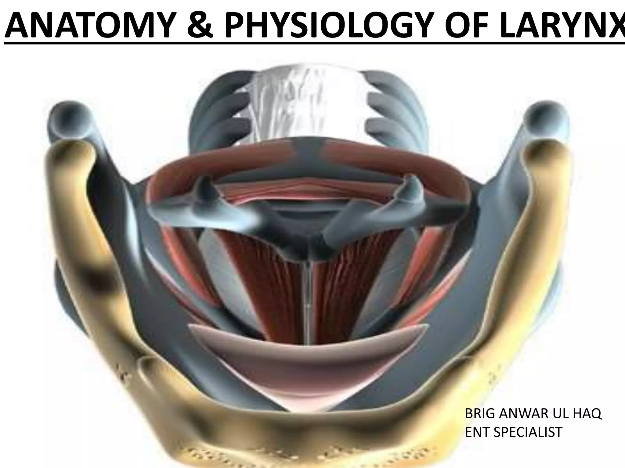
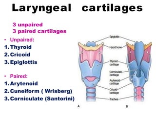
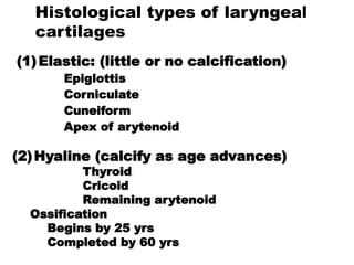
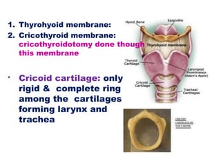
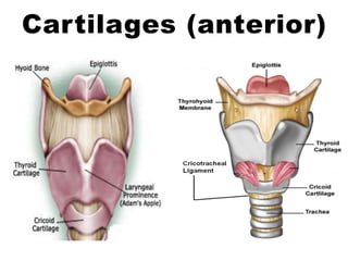
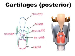
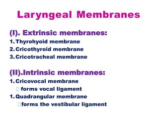
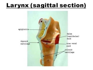
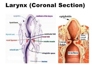
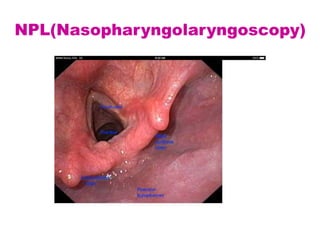
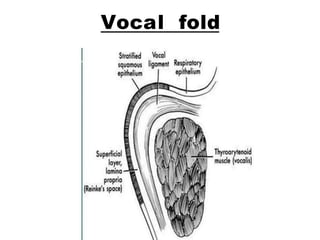
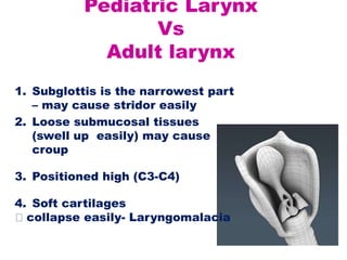
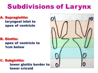
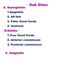
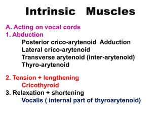
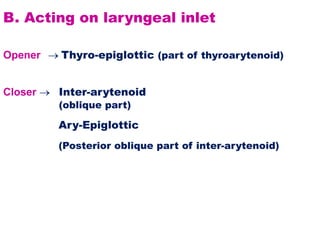
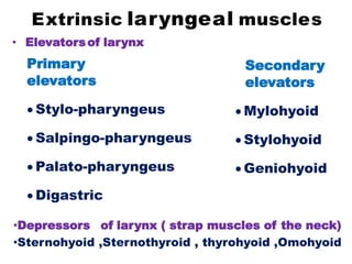
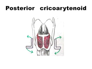
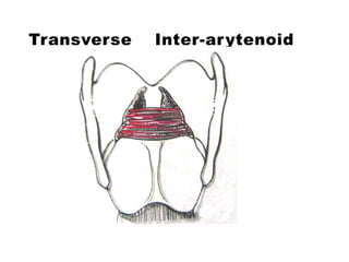
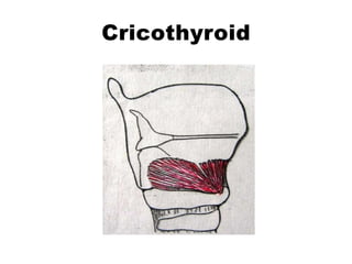
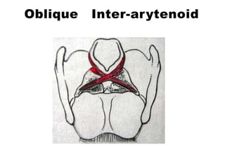
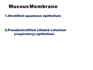
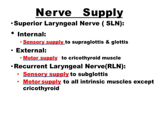
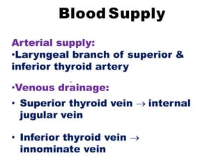
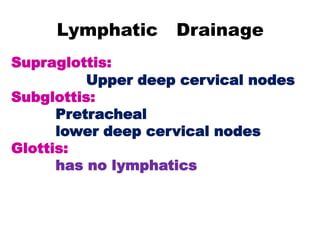
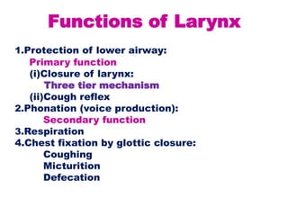
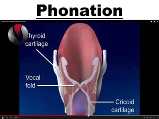
ANATOMY & PHYSIOLOGY OF LARYNX
BRIG ANWAR UL HAQ
ENT SPECIALIST
Laryngeal cartilages
Laryngeal Membranes
Laryngeal muscles function
3 Subdivisions of larynx
Nerve supply of larynx
Functions of larynx
CONTENTS
Anatomy of larynx
Rigid skeleton
cartilages,
muscles
ligaments
membranes
Extension
from hyoid bone above
to cricoid cartilage below
Opposite to C3 to C6
Laryngeal crepitus:
Normally present
Absent in post-cricoid
carcinoma
Laryngeal cartilages
3 unpaired
3 paired cartilages
• Unpaired:
1.Thyroid
2.Cricoid
3.Epiglottis
• Paired:
1.Arytenoid
2.Cuneiform ( Wrisberg)
3.Corniculate (Santorini)
Histological types of laryngeal
cartilages
(1)Elastic: (little or no calcification)
Epiglottis
Corniculate
Cuneiform
Apex of arytenoid
(2)Hyaline (calcify as age advances)
Thyroid
Cricoid
Remaining arytenoid
Ossification
Begins by 25 yrs
Completed by 60 yrs
1. Thyrohyoid membrane:
2. Cricothyroid membrane:
cricothyroidotomy done though
this membrane
• Cricoid cartilage: only
rigid & complete ring
among the cartilages
forming larynx and
trachea
Cartilages (anterior)
Cartilages (posterior)
Laryngeal Membranes
(I). Extrinsic membranes:
1.Thyrohyoid membrane
2.Cricothyroid membrane
3.Cricotracheal membrane
(II).Intrinsic membranes:
1.Cricovocal membrane
forms vocal ligament
1.Quadrangular membrane
forms the vestibular ligament
Larynx (sagittal section)
Larynx (Coronal Section)
NPL(Nasopharyngolaryngoscopy)
Vocal fold
Pediatric Larynx
Vs
Adult larynx
1. Subglottis is the narrowest part
– may cause stridor easily
2. Loose submucosal tissues
(swell up easily) may cause
croup
3. Positioned high (C3-C4)
4. Soft cartilages
collapse easily- Laryngomalacia
Subdivisions of Larynx
A. Supraglottis:
laryngeal inlet to
apex of ventricle
B. Glottis:
apex of ventricle to
1cm below
C. Subglottis:
lower glottis border to
lower cricoid
Sub Sites
A. Supraglottis:
1.Epiglottis
2. AE fold
3. False Vocal Cords
4. Ventricle
B.Glottis:
1.True Vocal Cords
2. Anterior commissure
3. Posterior commissure
C. Subglottis
Intrinsic Muscles
A. Acting on vocal cords
1. Abduction
Posterior crico-arytenoid Adduction
Lateral crico-arytenoid
Transverse arytenoid (inter-arytenoid)
Thyro-arytenoid
2. Tension + lengthening
Cricothyroid
3. Relaxation + shortening
Vocalis ( internal part of thyroarytenoid)
B. Acting on laryngeal inlet
Opener  Thyro-epiglottic (part of thyroarytenoid)
Closer  Inter-arytenoid
(oblique part)
Ary-Epiglottic
(Posterior oblique part of inter-arytenoid)
Extrinsic laryngeal muscles
• Elevatorsof larynx
Primary
elevators
 Stylo-pharyngeus
 Salpingo-pharyngeus
 Palato-pharyngeus
 Digastric
Secondary
elevators
 Mylohyoid
 Stylohyoid
 Geniohyoid
•Depressors of larynx ( strap muscles of the neck)
•Sternohyoid ,Sternothyroid , thyrohyoid ,Omohyoid
Posterior cricoarytenoid
Lateral cricoarytenoid
Transverse Inter-arytenoid
Cricothyroid
Oblique Inter-arytenoid
MucousMembrane
1.Stratified squamous epithelium
2.Pseudostratified ciliated columnar
(respiratory) epithelium
Nerve Supply
•Superior Laryngeal Nerve ( SLN):
• Internal:
• Sensory supply to supraglottis & glottis
• External:
• Motor supply to cricothyroid muscle
•Recurrent Laryngeal Nerve(RLN):
• Sensory supply to subglottis
• Motor supply to all intrinsic muscles except
cricothyroid
Blood Supply
Arterial supply:
•Laryngeal branch of superior &
inferior thyroid artery
•Venous drainage:
• Superior thyroid vein  internal
jugular vein
• Inferior thyroid vein 
innominate vein
Lymphatic Drainage
Supraglottis:
Upper deep cervical nodes
Subglottis:
Pretracheal
lower deep cervical nodes
Glottis:
has no lymphatics
Functions of Larynx
1.Protection of lower airway:
Primary function
(i)Closure of larynx:
Three tier mechanism
(ii)Cough reflex
2.Phonation (voice production):
Secondary function
3.Respiration
4.Chest fixation by glottic closure:
Coughing
Micturition
Defecation
DUGLITATION
SWALLOWING
Phonation
OPERA SINGING
THANK YOU

Larynx anatomy and physiology