Download to read offline

![is normalized as follows: The mean of pixel intensity is
1
computed by G =
n (x, y) ∈Ω
G(x, y) and the standard deviation by
1
σG =
n (x, y) ∈Ω
(G(x, y) – G)2
, and the pixels are normalized by
Gσ(x, y) =
G(x, y) – G
.
σG
After similar normalization of the nuclear intensity R, a cor-
relation value between R and G is computed via Corr (R, G) =
(x, y) ∈Ω
Rσ(x, y)Gσ(x, y). Thus, the degree of co-localization between
NF-κB labeling and the nuclear stain is quantified, irrespective
of the geometry and the number of cells.
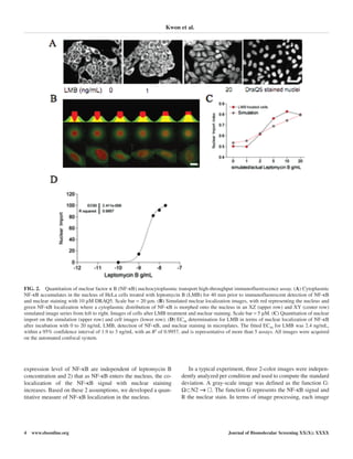
Validation of the image analysis algorithm
A simulation of NF-κB entry into the nucleus was used to
validate the image analysis algorithm. NF-κB distribution in
the cell was represented by a subtraction of 2 evolving
Gaussian shapes, and the nuclear stain was represented by a
nonevolving thresholded Gaussian (Fig. 2B). This permitted
the volume of the shape(s) to remain constant and thus
respected assumption 1 that leptomycin B treatment would not
alter intensities of NF-κB or the nucleus. The simulation is
depicted in a vertical and XY image in Figure 2B and C. The
green intensity was linearly evolved to fill the center and thus
tended to increase the number of pixels that had a high value of
G and R, satisfying assumption 2.
Figure 2B shows the evolution between the 2 situations:
where the NF-κB simulation (green) shape was entirely absent
from the nucleus and where it morphed linearly from an NF-
κB–free empty nucleus to a filled nucleus. The identical simu-
lation is shown in Figure 2B but in 2 dimensions and with
additive white Gaussian noise added to mimic the predicted
real experimental situation. These simulations were conceived
to represent the repression of nuclear export of NF-κB through
an increasing exposure of cells to leptomycin B. The measure-
ment algorithm extracted a linearly increasing coefficient for
nuclear entry, as was expected (Fig. 2C). It was then applied to
images of HeLa cells that were treated with increasing concen-
trations of leptomycin B for 40 min before fixation, indirect
immunofluorescent detection of the NF-κB distribution, and
nuclear staining with DRAQ5 (Fig. 2A). The nuclear localiza-
tion coefficient across a logarithmic gradient of leptomycin B
was fitted to a variable slope model and gave an EC50 for lep-
tomycin B of 2.4 ng/mL (4.4 nM), within the 95% confidence
limits of 1 ng/mL and 3 ng/mL.
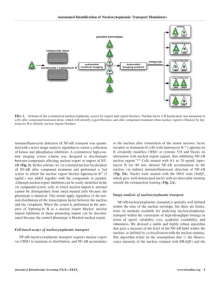
Nucleocytoplasmic import/export screen
A symmetrical screen was devised as a suitable method to
allow the discrimination between compounds affecting nuclear
import or nuclear export (Fig. 3A). It comprised 2 parallel
screens: In the 1st screen, compounds were screened for their
effects on endogenous NF-κB localization in wild-type cells,
allowing the identification of compounds that caused nuclear
localization of NF-κB through an inhibition of export. In the
2nd screen, cells were treated with compounds and leptomycin
B, and in this case, inhibitors of nuclear import could be dis-
tinguished from compounds that had a null phenotype in the 1st
screen (Fig. 3A).
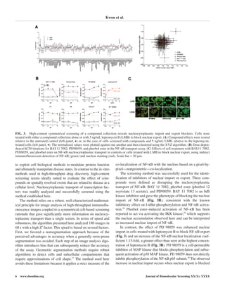
A collection of kinase and phosphatase inhibitors covering
most of the kinase families in the genome were used in this
screen, and the collection also contained leptomycin B as an
internal control. Leptomycin B was identified as a pure export
blocker in the screens, and the collected screen data are pre-
sented in Figure 3A. Molecules acting on nuclear import or
export were identified using cluster analyses of the effect of a
compound on both screens, shown in the XY plot in Figure 3A.
This resolved an import blocker, export blocker, and import
enhancer compound well outside the distribution of the control.
Three compounds were defined as disrupting the nucleocyto-
plasmic transport of NF-κB: BAY 11 7082, phorbol ester (phor-
bol 12 myristate 13 acetate), and PD98059. BAY 11 7082 is an
IκB-α kinase inhibitor and gave the phenotype of blocking the
nuclear import of NF-κB (Fig. 3B). In contrast, the effect of PD
98059 was to enhance nuclear import and was demonstrated only
in cells treated with leptomycin B to block NF-κB export (Fig. 3),
whereas PD 98059 increased the NF-κB nuclear localization
coefficient 1.15-fold (Fig. 3B) over the highest concentration of
leptomycin B. We determined the concentration dependence
(AC50) of the selected compounds on nucleocytoplasmic trans-
port. BAY 11 7082 had an AC50 of 5 µM for the inhibition of
nuclear import when measured in the presence of leptomycin B.
In contrast, phorbol ester promoted nuclear localization of NF-
κB 1.3-fold at 10 nM irrespective of the addition of leptomycin
B, and it is classified as a nuclear export inhibitor. The AC50 for
PD98059 was 1 µM, with a strong nuclear localization pheno-
type observed when NF-κB export was blocked, implying that its
target (p38 mitogen-activated protein [MAP] kinase) may regu-
late the rate of nuclear import (Fig. 3B).
DISCUSSION
A method is presented and validated for the identification of
small-molecule disruptors of nuclear import or export. This
method may have applications in the chemical genomic identi-
fication of molecules involved in nucleocytoplasmic transport
and their evaluation as therapeutic drugs.
Chemical biology—the application of high-throughput and
high-content methods for identifying small molecules that are suit-
able to assign function to the genome and its protein complement—
is emerging as a tool for both cell biology and drug discovery.23,24
In this context, image-based, high-content methods are gaining
interest for their combination of image-based analyses, automa-
tion, and compound library screening, and they provide a means
Automated Identification of Nucleocytoplasmic Transport Modulators
Journal of Biomolecular Screening XX(X); XXXX www.sbsonline.org 5](https://image.slidesharecdn.com/kwonetal2007jbs-141219151239-conversion-gate01/85/Kwon-et-al-2007-jbs-5-320.jpg)

The document discusses a high-content screening method to identify compounds that affect nucleocytoplasmic import and export of transcription factors, particularly NF-κB. The study confirmed efficacy and selectivity of the nuclear export inhibitor leptomycin B and established a quantitative imaging approach for assessing NF-κB localization in cells. Additionally, it revealed the involvement of MAP kinases in NF-κB nuclear import and provided results on specific inhibitors that affect this transport mechanism.


