1. Abstract
Mitochondria are vital organelles, which serves as a major energy sink
for many organisms and interface for protein transport and protein regulation
with the cytoplasm, as well as have their own set of translated proteins.
Tetrahymena thermophilia is a good eukaryotic model organism to use to better
understand processes such as protein translation and apoptotic-pathways in
mitochondria. A gene in the Tetrahymena genome, called voltage dependent
anion selective channel 4 (VDAC4) may have important roles in such
mitochondrial processes. VDAC4 encodes for a porin trans-membrane protein,
whose orthologs, such as VDAC1 and Tom40, are involved in communication
among organelles and trans-membrane transport. Evidence gathered from
bioinformatics analysis and protein localization studies support VDAC4
involvement in mitochondrial membrane processes. To better understand the
dynamics of this protein in the mitochondria and their interactions with other
organelles, we formed a YFP protein fusion construct to determine protein
localization, along with co-localizing and staining. The potential insights
possible from these results can be valuable in understanding cell life and death
cycles, as well as protein folding and trafficking.
Title
Studies of Mitochondrial-Associated VDAC4 in Tetrahymena Thermophilia
Authors
George C. Rizk and Douglas L. Chalker
Features
TTHERM_00117590
Voltage Dependent Anion Selective Channel 4 (VDAC4)
Experiment type: Fluorophore tag
Introduction
The purpose of this study is to investigate the localization and function
of VDAC4 in Tetrahymena thermophila. Tetrahymena is an organism of
constant movement, propelled by longitudinal rows of cilia along the length of
its body. High densities of mitochondria tend to congregate near such sites of
high-energy consumption to continue fueling the cilia. The cilia are attached to
the cell’s body at basal bodies, which take on the 9+2 microtubule formation
and serve as an interface between the cilia and the cell. The axonemal shaft of
2. the cilia receives nutrients and other cellular materials through intraflagellar
transportation across the basal body. In other organisms, VDACs are often
found associated with mitochondrial and basilar trans-membrane processes.
VDAC homologs are involved in ionic transport, mitochondria-mediated
apoptotic pathways, cell signaling, and inter-organelle communication. Such
homologs are often beta-barrel porins with several beta strands spanning the
outer mitochondrial membrane, whose conformations are voltage-dependent.
Previous research of VDACs such as “VDAC, a multi-functional mitochondrial
protein regulating cell life and death” (Shoshan-Barmatz et al. 2010) and
“Biophysical properties of porin pores from mitochondrial outer membrane of
eukaryotic cells” (Benz 1990) provide much foundational knowledge into their
mechanisms and properties. Such insight was possible by the well-rehearsed
but sophisticated method of channel purification and reconstitution in a planar
bilayer. This study aims to understand VDAC4 through bioinformatic,
genomic, and localization analyses as a novel protein and in the context of
known homologs.
Methods
1. PCR Amplification of Gene
Bioinformatic analysis involved downloading the gene’s sequence from
Tetrahymena Genome Database (ciliate.org), and designing specific oligonucleotide
primers to span its 1765bp region using the Primer 3 program. I then amplified the
gene from Tetrahymena genomic DNA using PCR. Reaction composed of 5x buffer,
upstream and downstream primers, dNTPs, NEB Phusion polymerase, water,
genomic DNA, and 3 different amounts of MgCl2 (0, 1, and 1.5 ul). Confirmed gene’s
size with electrophoresis gel.
2. Topoisomerase cloning reaction (pENTR plasmid insertion)
PCR products were initially cloned into the pENTR plasmid vector using TOPO
cloning kit (Life Technologies) in a 6 ul reaction containing X ng PCR product, 1 ul
salt solution and 1 ul Confirmed presence of insert and plasmid with gel.
3. E coli. Transformation and Electroporation
Added the TOPO cloning reaction to chemically competent E. coli and spread the
transformation culture on selective plates of kanamycin antibiotic to select for
transformed E. coli. Transformed E coli. Cells with the ligation mix from LR-
recombination by electroporating TOP10 electrocompetent cells with pICY+gene
insertion DNA.
4. pENTR plasmid DNA Isolation
Extracted, concentrated, and lysed the transformed E. coli cells to release plasmid
DNA. First centrifuged cells and added 250ul Resuspension solution. Added 250
Lysis solution followed by 350ul of Neutralization solution. pENTR plasmid + gene
3. insertion was washed in a mini-column with 500ul Wash solution. then eluted with
50ul of Elution buffer. Stored for use in restriction enzyme analysis.
5. Restriction enzyme analysis
Digested aliquots of the pENTR plasmid DNA with restriction enzymes BsrGI, BamHI
+ NotI, and HincII. Plasmid DNA (0.4-1 ug) was digested in enzyme reactions
containing the 1x buffer (as specified by the manufacturer) and 3-6 units of the
appropriate enzyme. Reactions were incubated at 37 degrees for 45min before
sample fragment sizes were confirmed by gel electrophoresis.
6. LR-recombination reaction (pICY plasmid insertion)
To create a YFP fusion, 0.75 ul of LR Clonase II enzyme was mixed with 400 ng of the
pICY-gtw vector and 44 ng of pENTR-GCR in a 3.75 ul reaction which would remove
the coding sequence from the pENTR plasmid and replace the gateway cassette in
the pICY-gtw. Treated the LR recombination reaction with 0.5ul of proteinase K,
added 2ul water, and transferred 1ul of mixture to electrocompetent E. Coli cells.
Electroporated cells and resuspended in 600ul S.O.C. medium. Spread cells on LB
plates containing ampicillin for selection of transformants. Confirmed insertion into
pICY with another restriction enzyme digest using BsrGI and BamHI +NotI, and
checked for presence of insertion by running gel electrophoresis of the digest
against blank pICY plasmids.
7. Alkaline lysis plasmid prep of pICY-induced E coli
Extracted, concentrated, and lysed the pICY-transformed E. coli cells to release
plasmid DNA. Spun down 20ml of pICY cells, resuspended in 2.5 ml of buffer 1,
followed by 2.5 ml buffer 2, and then 2.5ml buffer 3, mixing in between additions.
Centrifuged at 12000 RPM for 7min and added 8ml Isopropanol. Spun another 7min,
removed supernatant, and washed with 70% ethanol. After final spin, resuspended
in 400ul water and added 5ul of 5mg/ml RNAse A. Performed phenol/chloroform
extraction of DNA by adding 45ul 3M NaOAC and 400ul phenol/chloroform,
centrifuging, and extracting aqueous layer. Completed two wash cycles with ethanol
and combined with PEG solution to create a prepared DNA sample.
8. Electroporation of Tetrahymena cells to uptake pICY
Tetrahymena cells (45 mls) 8.5-9.5 hours into conjugation were harvested by
centrifugation for 3minutes at 1500x g. The cell pellet was resuspended in 30 mls of
10 mM Hepes. After cells were left in Hepes for 5 minutes, they were harvested by
centrifugation as above and the cell pellet was resuspended in 500-600 ul 10 mM
Hepes. The cells (150-200 ul) were mixed with 12 ug of pICY plasmid DNA, and
electroporated at 2.45 kVolts, 125 ohms resistance, and 50 uF capacitance for 6.5
msec. Added cells to Neff’s medium containing 30mls of 1x spp + 1x penicillin + 1x
fungizone + 200ug/ml paramomycin drug to select for pICY transformants, since
pICY contains allele that confers resistance to paramomycin. Plated cells in multi-
well plates and incubated for a couple nights.
9. YFP localization & microscopy
4. After observing successful transformants, isolated cells from medium by centrifuge,
and prepared microscope slides of transformed cells using methyl-cellulose for
immobilization. Viewed the fluorescent localizations of the YFP-fused protein under
microscope and imaged the results for documentation. Tagged a duplicate set of the
same transformants with a 0.1 ug/ml Mitotracker Red to stain mitochondria, viewed
localization with microscope, and imaged the results.
Results
The gene of study was predicted to express a eukaryotic porin protein
(VDAC4) containing a Porin3 superfamily conserved functional domain, as
shown in Figure 1. The domain has an E-value of 4.2e-12 and is found in
eukaryotic mitochondrial porin homologues as well as Tom40 proteins. The
291 AA domain spans most of the protein, suggesting that the predicted
protein’s primary function is akin to that of eukaryotic mitochondrial porins
and Tom40 proteins. According to the Simple Modular Architecture Research
Tool and De Pinto et al, mitochondrial porins’ molecular function is voltage-
dependent, anion-selective, channel (VDAC) activity found in the outer
membrane of the mitochondria. In biological processes they act as regulatory or
diffusion pores for transmembrane transport of small hydrophilic molecules.
The porins are beta-barrel proteins with 12-16 beta strands spanning the outer
mitochondrial membrane and have open conformations at low membrane
potentials and closed conformations at 30-40mV potentials. Tom40 is a
mitochondrial, outer membrane, integral protein found in the TOM complex,
which imports protein precursors into the mitochondrion. Tom40 is composed
of subunits Tom5, Tom6, and Tom7. The predicted protein was expected to
have similar structure and VDAC functions. VDAC porins form vital interfaces
between mitochondrial and cellular metabolisms, and are involved in apoptotic
pathways, potentially insightful for cancer-related research.
The predicted protein’s expression profile indicates high levels of
expression of this gene (~55,000 units) in growing cells during periods L-l, L-
m and L-h. The protein’s expression also peaks, though lower in magnitude, in
conjugating cells (~37,000 units) during periods C4 and C6 (conjugation of
cells B2086 and CU428 collected 4hrs and 6hrs after mixing). Given the
dominant presence of the Porin3 superfamily domain in this gene and the
documented localization data of such eukaryotic porins, I used mitochondrial
porins and Tom40 proteins as functional and structural homologues for
comparison to predict that my porin protein’s gene-YFP localization would
occur in the outer membrane of the Tetrahymena mitochondrion.
5. After initial PCR amplification of the gene, gel electrophoresis (Figure
3.) confirmed the gene size at 1.8kb as expected from bioinformatics data in
Figure 1. The band intensity was compared to the intensity of the 1kb ladder
bands to estimate DNA concentration. Successful gene insertion into pENTR
plasmid was confirmed in Figure 4, where bands at 1.8kb and 3kb indicate
presence of gene insertion and pENTR plasmid respectively. Gel bands confirm
correct ligation and digest of insertion and plasmid, as shown by the expected
restriction enzyme digest patterns. Figure 9 displays the schematic of the
correctly ligated pENTR vector. Correct transfer of VDAC4 from pENTR to
pICY vector was confirmed (Figure 5.) with bands at 1.8kb and 20kb for
presence of gene insertion and pICY plasmid respectively. Gel bands again
confirmed the correct ligation and digest of insertion and plasmid. Faint
intensities of bands in lanes 3-5 and 7-9 indicate low concentration, but correct
sizes. Figure 8 displays the schematic of the correctly ligated pICY vector.
Since RT primers spanned ~250bp around the spliced intron region,
bands in the 1:3 cDNA dilution (Figure 6) confirm correct mRNA to cDNA
synthesis and lack of intron. Gene expression similar at all stages except 6
hours. Higher intensity bands than 1:10 cDNA dilution confirms greater DNA
concentration. gDNA lane confirms the correct and expected product size of
intron + RT-spanning region (886bp + 250bp) of ~1100bp. Figure 7 confirms
similar data at the 1:10 dilution, where lower intensity bands than 1:3 cDNA
dilution confirm lower DNA concentration.
VDAC4 appeared to localize in the membrane of the mitochondria as
expected. It also appeared to localize at the basal bodies of the cilia of
Tetrahymena, suggesting further studies to delineate whether the protein co-
localizes in both regions. A subsequent mitochondrial stain on the YFP-fused
transformants to determine possible co-localization of the protein indeed
highlighted the mitochondria. YFP fluorescence proved bright enough to
distinguish that VDAC also localized outside of the mitochondria and
potentially in the ciliary basal bodies. In Figure 11, mitochondria appear as
small, cloudy, pea-pod shaped objects. The localization appears clustered near
the outer membrane of the cell and aligned parallel with the longitudinal,
ciliary rows of cell. The localization seems uniformly spread and ordered
throughout all mitochondria of the cell, not favoring certain regions over
others. Other images, as depicted in Figure 12, displayed extra-mitochondrial
localization. In this image, mitochondria appear as cloudy clusters spread
throughout the cell beneath its outer membrane. In addition smaller, bright
spots or points now appear along the length of the cell, parallel with the ciliary
rows and with the mitochondrial patterns. The point-like localization is very
6. distinct and also of brighter fluorescence than in the mitochondria. A
mitochondrial stain in Figure 13 confirms that the dotted localization is not
confined to mitochondria alone, since comparison of Figures 12 and 13 reveals
that the points localize outside the regions of mitochondrial staining.
Discussion
VDAC4’s peaked expression during growth is consistent with the
expected high demand for porins during cellular growth to transport nutrients
across the mitochondrial membrane to power organelle processes. Extra porins
may be embedded in the outer membrane to compensate for the elevated
nutritional need. Peaked expression during early conjugation also seems
consistent with the need to transport negatively-charged DNA molecules into
the mitochondrion during transfer of genetic material, but since mitochondrial
DNA only composes a small portion of the genomic DNA, this may account for
the lower magnitude of expression observed.
The point-like localization along the ciliary rows but outside the
mitochondria suggests that VDAC4 may also localize to the ciliary basal
bodies, which are located at the base of the cilia and thus would follow the
ciliary patterns. Cellular materials need to cross the basal body to be
transported into the cilia. As a porin, it would make sense for VDAC4 to
localize at the basal body interface to facilitate intraflagellar transport, and this
is consistent with the trans-membrane transport functions in its homologs.
Related studies (Majumder and Fisk, 2013) have found that VDAC3
(mitochondrial porin in humans) depletion causes inappropriate ciliogenesis,
which suggests that it’s involved in regulation of ciliary growth and apoptosis.
If VDAC4 has similar functions, this would support its apparent localization
and proximity to the ciliary basal bodies.
The localization of VDAC4 to the mitochondria near the cilia and
surface of the cell is consistent with the prediction that VDAC4 is involved
with ionic and molecular transport functions, especially across the outer
mitochondrial membrane and towards the cilia to fuel cellular movement and
transport into or out of the cytosol. In VDAC4 homologs, mitochondria-
mediated, caspase-associated, apoptosis pathways are found at the outer
membrane of mitochondria and are dependent on transport of cytokines across
the membrane. This supports the predicted role of VDAC4 in apoptotic
pathways. Inter-organelle communication, another function of VDAC4
homologs, would also rely on a trans-membrane channel to allow the passage
of hydrophilic messenger molecules, further supporting the advantageous
location of VDAC4 for such functions.
7. There remains much room for further research and discovery of
VDAC4’s functions and roles. It has not yet been confirmed to be a porin
protein, it may yet be protein lacking a channel but embedded in the membrane.
The characteristics of the pore, if there is one, have yet to be determined in
shape, size, and biophysical properties. Future experiments may involve the
purification of VDAC4 using agents such as Triton X-100 and run SDS
electrophoresis to determine size. Then reconstitution of the channel in a planar
lipid bilayer would allow for controlled measures of ion and current flow,
voltage-dependent conformations, channel conductance, selectivity, and pore
characteristics based on the molecules that it is permeable to. Patch-clamp
experiments could study single channels and detect more electrophysiological
features of the pore. Nuclear magnetic resonance could be used to determine
the 3D structure of the pore to then propose potential gating mechanisms of the
channel.
Figures
TTHERM_00117590
625 1250 1875 2500 nts
1765 bp
Predicted
Protein
309 AA Porin3 Super family domain (17-308aa)
Conserved Domains
Porin3 Super family domain
Primers
Intron coding region
Exons/UTR
Figure 1. Ttherm_00117590 Predicted Gene, Protein, and Conserved Domains: Ttherm_00117590 is a 1765
bp gene containing one predicted and confirmed intron. The gene coding region is depicted in blue and the
gene’s loca on in the genome is between 1016.1 kb to 1017.9 kb. Conserved domain (in red) on the 309 AA
predicted protein (green strip) includes a Porin3 Super Family domain (ID: cl03224) that is predicted to span
the protein in the region 17-308 AA. Oligonucleo de primers were chosen to amplify from 6 bp upstream of
the ATG start site to the last base before the gene’s TGA stop codon. rtPCR primers were chosen to amplify a
886 bp sec on star ng 720 bp into the gene.
8. TTHERM_00117590
500 1500
Figure 2. Oligonucleo de-sequenced regions: Total of 1489 base pairs sequenced from oligonucleo de
primers, 858 bp from the forward reac on and 631 bp from the reverse reac on. Mismatches in
forward reac on, in order, at bp posi ons: 821, 845, 856, 858. Mismatch in reverse reac on at bp
posi on 1771. rtPCR-sequenced region: Total of 575 base pairs sequenced, en rely from the reverse
rtPCR primer reac on. Base pair mismatches, in order from le to right, at bp posi ons: 469 and 499.
2000 bp1000
= Oligo-sequenced regions
* * **
+821 +845 +856 +858
*
+1771
* = Base pair mismatches
A to T A to T T to C T to A T to G
= rtPCR sequenced region
* *
A to C T to A
+469 +499
Gene Map
9. Figure 4. Confirming gene inser on into pENTR plasmid: Each lane contains a possible pENTR clone digested with
one of the three indicated enzymes or combina ons. Lane 5 contains a 1kbp size ladder to es mate band sizes,
the size of selected ladder bands are indicated on the le .
pENTR plasmid digest
BamH1+Not1(1)
BamH1(2)
BamH1(3)
BamH1(4)
Ladder(1kb)
BsrG1(1)
BsrG1(2)
BsrG1(3)
HincII(1)
HincII(2)
HincII(3)
HincII(4)
5000bp
1500
500
2000
10.
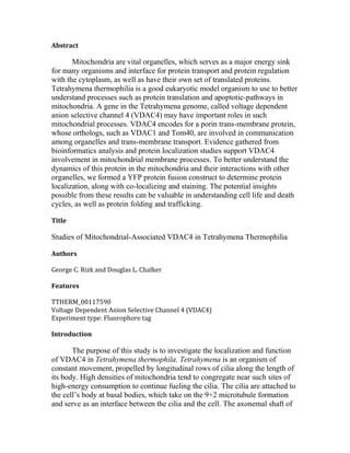
11.
12. Figure 11. Intracellular localiza on of VDAC4: Image on the le represents YFP
fluorescence of VDAC4 in a live cell during growth. Contrasted with bright-field image
on the right.
13. Figure 12. Intracellular localiza on of VDAC4: Image represents YFP fluorescence of
VDAC4 in live cells during growth.
Figure 13. Mitotracker Red stain: Image represents mitochondrial staining of live cell
during growth.
14. References
1. De Pinto, V., A. Messina, and Et. al. "New functions of an old protein: the
eukaryotic porin or voltage dependent anion selective channel (VDAC)." Mar.
2003. Digital file.
2. "Gene Model Identifier TTHERM_00117590." Tetrahymena Genome
Database Wiki. N.p., n.d. Web. 17 Feb. 2015.
<http://ciliate.org/index.php/feature/details/TTHERM_00117590>.
3. "Gene Model Identifier Contig10439.0.g72." Oxytricha Genome Database
Wiki. N.p., n.d. Web. 17 Feb. 2015.
http://oxy.ciliate.org/index.php/feature/details/Contig10439.0.g72
4. "TTHERM_00117590." Tetrahymena Functional Genomics Database. N.p.,
n.d. Web. 17 Feb. 2015.
<http://tfgd.ihb.ac.cn/search/detail/gene/TTHERM_00117590>.
5. "Domains within Tetrahymena thermophila proteins." Simple Modular
Architecture Research Tool. N.p.,n.d. Web. 17 Feb. 2015. <http://smart.embl-
heidelberg.de/smart/job_status.pl?jobid
=128252110215489011424201884LUidfDllBH>.
6. Majumder, Shubra, and Harold Fisk. "VDAC3 and Mps1 negatively regulate
ciliogenesis." Feb. 2013. Digital file.