kavita jaidiya pathology.pptx
•Download as PPTX, PDF•
0 likes•7 views
The document summarizes autophagy and electron microscopy. It defines autophagy as the degradation of cells through lysosomes and discusses its importance in survival, cancer, and disease. Three forms of autophagy are identified: macroautophagy, microautophagy, and chaperone-mediated autophagy. Electron microscopes use electron beams for high magnification imaging of cells and materials. Transmission electron microscopes image thin samples while scanning electron microscopes image surface structure of bulk samples. Sample preparation for electron microscopy involves cleaning, coating, and thin film deposition depending on the analysis needs.
Report
Share
Report
Share

Recommended
Techniques in Cell Biology
1. The document discusses various cytological techniques used in the study and manipulation of cells including microtomy, microscopy, staining, and centrifugation.
2. Microtomy involves sectioning tissues into thin slices using a microtome for microscopic observation while staining allows cellular structures to be visualized through selective uptake of dyes.
3. Centrifugation separates particles according to properties like density, shape and size through application of centrifugal force, and is used to isolate cellular components through differential pelleting or density gradient centrifugation.
lecture3andlecture4techniquesincellbiology-210201105723.pdf
1. The document discusses various cytological techniques used in the study and manipulation of cells including microtomy, microscopy, staining, and centrifugation.
2. Microtomy involves sectioning tissues into thin slices using a microtome for microscopic examination after fixation, dehydration, embedding, and staining.
3. Centrifugation separates particles in a solution based on properties like density, shape, and size through application of centrifugal force. Types include differential pelleting and density gradient centrifugation.
Microbial Staining
This is a rundown of some staining techniques used in microbiology, including simple staining, negative staining, gram staining, acid-fast staining, endospore staining, and flagellar staining.
Staining
The document describes several microscopy experiments involving microscopy, staining techniques, and aseptic techniques.
The microscopy experiment involves observing samples under different magnifications of a compound microscope and identifying structures. Staining techniques are used to make microorganisms visible, including simple staining using methylene blue dye and Gram staining to differentiate bacteria.
Aseptic techniques aim to minimize contamination and prepare pure cultures, including cleaning work areas, sterilizing equipment in flame, and isolation techniques like streak plating or pour plating that allow single colonies to grow separated on nutrient agar.
Isolation of microbes , its all types, handling, advantages and dis advantage...
This document discusses various methods for isolating microbes. It begins by defining microbes and their importance industrially, environmentally, economically, and commercially. There are two main strategies for isolation - from the environment or from sites with microbes of interest. Common classical plating methods include streak plate, spread plate, and pour plate methods. Streak plating involves spreading microbes on media with an inoculating needle to thin them out into separate colonies. Newer single-cell isolation methods like laser tweezers use an infrared laser to optically trap and manipulate individual cells. Whichever method is used, aseptic technique is critical to prevent contamination.
Histopathological technique
Histopathology techniques are used to demonstrate minute structural alterations in tissues caused by disease. Key techniques include fixing tissues in formalin to preserve structure, processing tissues through dehydration, clearing and infiltration steps, embedding in paraffin wax, sectioning with a microtome, staining, and mounting slides. Histopathology allows diagnosis of diseases through microscopic examination of tissue structures and any pathological changes present. It is a crucial technique when other testing may not be possible or provides definitive confirmation of diseases.
Culture techniq and type of animal cell culture
A primary culture refers to the initial culture of cells directly taken from an organism before the first subculture. A cell line refers to the propagation of cells after the first subculture. Primary cultures contain a variety of differentiated cell types and require higher cell quantities due to lower survival rates. Tissues are disaggregated into single cells using mechanical or enzymatic techniques for primary culture. Organ cultures involve culturing whole organs or tissues to preserve their structure and function in vitro. Various techniques like plasma clot, raft, and grid methods are used to culture different organ explants.
diagnosticmicrobiology-140523042250-phpapp01.pdf
This document provides an overview of diagnostic microbiology. It discusses the goals of clinical microbiology laboratories in testing specimens to identify microorganisms causing illness and providing antimicrobial susceptibility results. It also describes various laboratory procedures used, including microscopy, culture-based techniques, immunological and molecular assays. Specimen collection, processing, staining methods, and interpretation of culture results are discussed in detail.
Diagnostic microbiology.
The document discusses diagnostic microbiology and the role of the clinical microbiology laboratory. The key responsibilities of the laboratory include testing specimens to identify microorganisms causing illness, providing antimicrobial susceptibility results, and advising physicians. Important techniques used in diagnosis include microscopy, culture, antigen detection methods like ELISA, and molecular methods like PCR. Proper specimen collection, transport, and processing are essential for accurate diagnostic testing.
Histopathological techniques -sectioning, STAINING, EMBEDDING, fixaton, micro...
Histopathology examines minute tissue alterations from disease. Samples come from cadavers, autopsies, animal tissues, or biopsies. Histopathological examination is useful for establishing disease pathogenesis and diagnosing diseases that are difficult to diagnose by other means. It typically begins with surgery or biopsy to collect tissue samples, which are then fixed, processed, and examined microscopically. Common fixatives include formaldehyde and glutaraldehyde, which cross-link proteins to preserve tissue morphology and prevent autolysis.
Plant tissue culture ⅱ isolation of protoplast
Protoplast isolation involves removing the cell wall from plant cells to leave only the plasma membrane and intracellular components. The isolated protoplasts can be cultured and genetically manipulated. There are two main methods for isolating protoplasts - mechanical and enzymatic. The enzymatic method uses enzymes like cellulase and pectinase to break down the cell wall. Isolated protoplasts are purified through centrifugation and cultured in an osmotic medium to prevent bursting due to the lack of a cell wall. Protoplasts are useful for studies in cell fusion and genetic transformation.
Plant histology
This document provides an overview of histology and tissue preparation techniques. It discusses the following key points:
- Histology is the study of tissues and how cells are organized into tissues and organs. The four main types of tissues are epithelial, connective, nervous, and muscular tissue.
- Tissue samples are obtained through biopsies and prepared through a process including fixation, dehydration, clearing, embedding in paraffin wax or resin, sectioning with a microtome, and staining. Special staining techniques can identify structures like glycogen or calcium.
- Histochemistry uses chemical reactions to identify certain molecules in tissues, while immunohistochemistry uses labeled antibodies to identify antigens within tissues under a microscope.
Pure culture techniques
The document discusses various techniques for obtaining pure cultures of microorganisms, including streak plating, spread plating, serial dilution, and single cell isolation. Streak plating involves streaking bacteria across nutrient agar plates using a loop to separate individual colonies. Serial dilution uses successive dilutions of a mixed culture in liquid media to isolate a predominant microorganism. Single cell isolation picks a single cell from a culture using a micromanipulator or capillary pipette and allows it to multiply, producing a pure culture. These pure culture techniques are important for laboratory study of individual microbial species.
Bacteriology&immunology lab2
Culture K and J were identified through examination of their appearance on culture media, gram staining reactions, and catalase tests. Gram staining revealed that culture K was gram-positive, staining purple, while culture J was gram-negative, staining pink. Gram-positive bacteria have a thick peptidoglycan layer in their cell walls, staining purple with gram staining. Gram-negative bacteria have a thinner peptidoglycan layer and an outer membrane, staining pink. Identification of the bacteria was achieved through analysis of cell structure and staining characteristics.
10.analytical biochemistry
This document discusses analytical biochemistry techniques for isolating and sequencing proteins. It covers cell disruption methods like sonication, centrifugation types including differential and density gradient, and spectrophotometry. Cell disruption is needed to extract proteins from cells. Centrifugation separates particles based on size, shape and density. Differential centrifugation separates with increasing g-force while density gradient centrifugation uses a medium with increasing density. Spectrophotometry analyzes light absorption of substances using Lambert's and Beer's laws.
P1 Tissue engineering abhishek mishra
Tissue engineering is defined as an interdisciplinary field that applies engineering and life science principles to develop biological substitutes that restore or improve tissue function. It involves understanding tissue growth principles to produce functional replacement tissue for clinical use. Examples include artificial skin, bone, pancreas, and bone marrow. Tissue engineering utilizes living cells as engineering materials, such as fibroblasts for skin repair. Cells are extracted from tissues and embedded in scaffolds to support tissue formation.
bacterial staining.pptx
This document discusses various staining techniques used to visualize microorganisms under a microscope. It begins by explaining why staining is necessary since microorganisms cannot be seen with the naked eye. It then covers different types of staining including simple staining, Gram staining, acid-fast staining, negative staining, and specialized staining techniques for flagella, capsules, and spores. Each staining method is described in 1-2 sentences and includes the basic procedure and results.
Isolation of protoplast in plant tissue culture.
The document discusses the isolation of protoplasts from plant cells. There are two main methods for isolating protoplasts - mechanical and enzymatic. The enzymatic method uses enzymes to digest the cell wall and is more widely used as it works for a variety of plant tissues and causes less damage to the cells. Key steps in the enzymatic method include incubating plant tissue in enzyme solutions, filtering to separate protoplasts from debris, and centrifugation to purify the protoplasts. Isolated protoplasts can be used for cell fusion between unrelated plant species and for genetic modification of plants. Factors like plant species, age of donor tissue, and pre-treatment of tissue affect the viability
Pg departnment of biochemistry sim (1)
This document summarizes methods for isolating and culturing plant protoplasts. Protoplasts are plant cells that have had their cell walls removed. The document describes two methods for isolating protoplasts - mechanical and enzymatic. It also discusses purifying the protoplasts, assessing their viability, culturing them in liquid media to regenerate cell walls, and the potential applications of protoplast culture and regeneration including plant breeding techniques.
Gen. histology (introduction) 1
This document provides an overview of histology, the study of tissues. It discusses that tissues are made up of cells and an extracellular matrix that interact closely. The key steps in preparing tissues for microscopic examination are fixation, dehydration, clearing, embedding, sectioning, and staining. Fixation preserves tissues using chemicals like formaldehyde. Sections are cut very thin using a microtome and mounted on slides for staining with dyes like hematoxylin and eosin or periodic acid-Schiff to visualize different tissue components under the microscope. The interactions between cells and extracellular matrix allow tissues to form and carry out specialized functions in organs.
Primary cell culture anjana.pptx
There are three main types of cell culture: primary, secondary, and continuous. Primary cultures are derived directly from tissue and maintain characteristics of the original tissue but are heterogeneous. Secondary cultures are derived from primary cultures after the first subculture. Continuous cell lines can be subcultured indefinitely and have increased growth rates but lose tissue-specific properties. Various enzymes are commonly used to disaggregate tissues for primary culture, including trypsin and collagenase. Mechanical and explant methods are also used to establish primary cultures.
Plant Cell Structures
This document discusses various structures found in plant and animal cells. It provides descriptions of 20 different cell structures found in animal cells such as the nuclear pore, chromatin, nucleolus, nuclear envelope, and centrioles. It also describes cell structures found in plant cells like the cell wall, chloroplasts, vacuoles, and mitochondria. The functions of various organelles are explained, including the Golgi body which processes proteins and lipids, and peroxisomes which contain enzymes to break down hydrogen peroxide. The document provides a detailed overview of the key components of both animal and plant cells.
pureculturetechnic-160204060702.pdf
Pure cultures contain only a single type of microorganism and are important for accurate identification, testing, and experimentation. They are obtained through techniques like streak plating that isolate individual cells on nutrient-rich solid media to promote separate colony growth. Purity is confirmed when all colonies appear identical under microscopy and in biochemical tests. Pure cultures are maintained through refrigeration, inclusion in paraffin, or preservation through cryopreservation, lyophilization, or freezing to halt metabolism while retaining viability for long-term storage.
pureculturetechnic-160204060702.pdf
Pure cultures contain only a single type of microorganism and are important for accurate identification, testing, and experimentation. They are obtained through techniques like streak plating that isolate individual cells on nutrient-rich agar plates. Once isolated, various methods are used to prove purity and maintain cultures, like ensuring uniform colony morphology and biochemical reactions. Pure cultures are then preserved long-term through refrigeration, paraffin coating, cryopreservation in liquid nitrogen, or lyophilization to stop metabolic activity and allow viability for years.
pureculturetechnic-160204060702.pdf
Pure cultures contain only a single type of microorganism and are important for accurate identification, testing, and experimentation. They are obtained through techniques like streak plating that isolate individual cells on nutrient-rich solid media to promote separate colony growth. Purity is confirmed when all colonies appear identical under microscopy and in biochemical tests. Pure cultures are preserved long-term through cryopreservation, lyophilization, or storage in paraffin or refrigeration to maintain viability without contamination.
attachment general microbiology course .pptx
This document provides an overview of the General Microbiology course offered at Wallaga University's Department of Biology in 2015/2022. It covers key topics including the definition and branches of microbiology, methods of culturing and identifying microorganisms, bacterial cell structure and growth conditions, microbial genetics, and an introduction to mutations. The course instructor is listed as kumsa.adm12@gmail.com and will take place in Room B9R20.
tissue culture hybridization
Tissue culture is a technique where small pieces of plant or animal tissue are cultured in a sterile medium outside of the organism. It was first developed in 1885 and has since been used extensively in medicine, agriculture, and research. It allows for the rapid duplication of plant materials while eliminating diseases and maintaining genetic traits. However, it requires specialized facilities and equipment and reduces genetic diversity.
Single cell culture
Single cell culture involves isolating single cells from plant tissue and culturing them on a nutrient medium. There are mechanical and chemical methods for isolation. Cells can be cultured using various techniques like microchamber, microdroplet, or nurse culture techniques. The paper raft nurse culture places isolated cells on nutrient-soaked paper placed on actively growing callus tissue. Single cell culture is important for fundamental studies, mutation analysis, and industrial applications like crop improvement and production of medicinal compounds.
Haemorrhagic_Septicemia in Ruminant ,Gal ghotus
Pasteruella multocida and Mannheimia haemolytica are bacteria that can cause pasteurellosis in cattle and other animals. P. multocida causes haemorrhagic septicemia in cattle, fowl cholera in fowl, and atropic rhinitis in pigs. M. haemolytica causes shipping fever in cattle. Haemorrhagic septicemia is an acute disease in cattle characterized by fever, respiratory distress, and diarrhea. The bacteria enter through ingestion or inhalation and spread systemically, causing hemorrhages on membranes. Johne's disease is a chronic wasting disease of ruminants caused by Mycobacterium paratuberculosis.
ADDISON(1).pptx
Canine hypoadrenocorticism, also known as Addison's disease, is a disorder caused by a lack of hormones produced by the adrenal glands. It results in electrolyte and blood pressure issues and can be fatal if not treated. Diagnosis involves ruling out other causes through tests of electrolyte and cortisol levels before and after administration of ACTH. Treatment focuses on rapid fluid therapy and steroid hormone replacement to correct electrolyte imbalances and restore blood volume and pressure. Long term management relies on mineralocorticoid and glucocorticoid supplementation through oral or injectable medications.
More Related Content
Similar to kavita jaidiya pathology.pptx
Diagnostic microbiology.
The document discusses diagnostic microbiology and the role of the clinical microbiology laboratory. The key responsibilities of the laboratory include testing specimens to identify microorganisms causing illness, providing antimicrobial susceptibility results, and advising physicians. Important techniques used in diagnosis include microscopy, culture, antigen detection methods like ELISA, and molecular methods like PCR. Proper specimen collection, transport, and processing are essential for accurate diagnostic testing.
Histopathological techniques -sectioning, STAINING, EMBEDDING, fixaton, micro...
Histopathology examines minute tissue alterations from disease. Samples come from cadavers, autopsies, animal tissues, or biopsies. Histopathological examination is useful for establishing disease pathogenesis and diagnosing diseases that are difficult to diagnose by other means. It typically begins with surgery or biopsy to collect tissue samples, which are then fixed, processed, and examined microscopically. Common fixatives include formaldehyde and glutaraldehyde, which cross-link proteins to preserve tissue morphology and prevent autolysis.
Plant tissue culture ⅱ isolation of protoplast
Protoplast isolation involves removing the cell wall from plant cells to leave only the plasma membrane and intracellular components. The isolated protoplasts can be cultured and genetically manipulated. There are two main methods for isolating protoplasts - mechanical and enzymatic. The enzymatic method uses enzymes like cellulase and pectinase to break down the cell wall. Isolated protoplasts are purified through centrifugation and cultured in an osmotic medium to prevent bursting due to the lack of a cell wall. Protoplasts are useful for studies in cell fusion and genetic transformation.
Plant histology
This document provides an overview of histology and tissue preparation techniques. It discusses the following key points:
- Histology is the study of tissues and how cells are organized into tissues and organs. The four main types of tissues are epithelial, connective, nervous, and muscular tissue.
- Tissue samples are obtained through biopsies and prepared through a process including fixation, dehydration, clearing, embedding in paraffin wax or resin, sectioning with a microtome, and staining. Special staining techniques can identify structures like glycogen or calcium.
- Histochemistry uses chemical reactions to identify certain molecules in tissues, while immunohistochemistry uses labeled antibodies to identify antigens within tissues under a microscope.
Pure culture techniques
The document discusses various techniques for obtaining pure cultures of microorganisms, including streak plating, spread plating, serial dilution, and single cell isolation. Streak plating involves streaking bacteria across nutrient agar plates using a loop to separate individual colonies. Serial dilution uses successive dilutions of a mixed culture in liquid media to isolate a predominant microorganism. Single cell isolation picks a single cell from a culture using a micromanipulator or capillary pipette and allows it to multiply, producing a pure culture. These pure culture techniques are important for laboratory study of individual microbial species.
Bacteriology&immunology lab2
Culture K and J were identified through examination of their appearance on culture media, gram staining reactions, and catalase tests. Gram staining revealed that culture K was gram-positive, staining purple, while culture J was gram-negative, staining pink. Gram-positive bacteria have a thick peptidoglycan layer in their cell walls, staining purple with gram staining. Gram-negative bacteria have a thinner peptidoglycan layer and an outer membrane, staining pink. Identification of the bacteria was achieved through analysis of cell structure and staining characteristics.
10.analytical biochemistry
This document discusses analytical biochemistry techniques for isolating and sequencing proteins. It covers cell disruption methods like sonication, centrifugation types including differential and density gradient, and spectrophotometry. Cell disruption is needed to extract proteins from cells. Centrifugation separates particles based on size, shape and density. Differential centrifugation separates with increasing g-force while density gradient centrifugation uses a medium with increasing density. Spectrophotometry analyzes light absorption of substances using Lambert's and Beer's laws.
P1 Tissue engineering abhishek mishra
Tissue engineering is defined as an interdisciplinary field that applies engineering and life science principles to develop biological substitutes that restore or improve tissue function. It involves understanding tissue growth principles to produce functional replacement tissue for clinical use. Examples include artificial skin, bone, pancreas, and bone marrow. Tissue engineering utilizes living cells as engineering materials, such as fibroblasts for skin repair. Cells are extracted from tissues and embedded in scaffolds to support tissue formation.
bacterial staining.pptx
This document discusses various staining techniques used to visualize microorganisms under a microscope. It begins by explaining why staining is necessary since microorganisms cannot be seen with the naked eye. It then covers different types of staining including simple staining, Gram staining, acid-fast staining, negative staining, and specialized staining techniques for flagella, capsules, and spores. Each staining method is described in 1-2 sentences and includes the basic procedure and results.
Isolation of protoplast in plant tissue culture.
The document discusses the isolation of protoplasts from plant cells. There are two main methods for isolating protoplasts - mechanical and enzymatic. The enzymatic method uses enzymes to digest the cell wall and is more widely used as it works for a variety of plant tissues and causes less damage to the cells. Key steps in the enzymatic method include incubating plant tissue in enzyme solutions, filtering to separate protoplasts from debris, and centrifugation to purify the protoplasts. Isolated protoplasts can be used for cell fusion between unrelated plant species and for genetic modification of plants. Factors like plant species, age of donor tissue, and pre-treatment of tissue affect the viability
Pg departnment of biochemistry sim (1)
This document summarizes methods for isolating and culturing plant protoplasts. Protoplasts are plant cells that have had their cell walls removed. The document describes two methods for isolating protoplasts - mechanical and enzymatic. It also discusses purifying the protoplasts, assessing their viability, culturing them in liquid media to regenerate cell walls, and the potential applications of protoplast culture and regeneration including plant breeding techniques.
Gen. histology (introduction) 1
This document provides an overview of histology, the study of tissues. It discusses that tissues are made up of cells and an extracellular matrix that interact closely. The key steps in preparing tissues for microscopic examination are fixation, dehydration, clearing, embedding, sectioning, and staining. Fixation preserves tissues using chemicals like formaldehyde. Sections are cut very thin using a microtome and mounted on slides for staining with dyes like hematoxylin and eosin or periodic acid-Schiff to visualize different tissue components under the microscope. The interactions between cells and extracellular matrix allow tissues to form and carry out specialized functions in organs.
Primary cell culture anjana.pptx
There are three main types of cell culture: primary, secondary, and continuous. Primary cultures are derived directly from tissue and maintain characteristics of the original tissue but are heterogeneous. Secondary cultures are derived from primary cultures after the first subculture. Continuous cell lines can be subcultured indefinitely and have increased growth rates but lose tissue-specific properties. Various enzymes are commonly used to disaggregate tissues for primary culture, including trypsin and collagenase. Mechanical and explant methods are also used to establish primary cultures.
Plant Cell Structures
This document discusses various structures found in plant and animal cells. It provides descriptions of 20 different cell structures found in animal cells such as the nuclear pore, chromatin, nucleolus, nuclear envelope, and centrioles. It also describes cell structures found in plant cells like the cell wall, chloroplasts, vacuoles, and mitochondria. The functions of various organelles are explained, including the Golgi body which processes proteins and lipids, and peroxisomes which contain enzymes to break down hydrogen peroxide. The document provides a detailed overview of the key components of both animal and plant cells.
pureculturetechnic-160204060702.pdf
Pure cultures contain only a single type of microorganism and are important for accurate identification, testing, and experimentation. They are obtained through techniques like streak plating that isolate individual cells on nutrient-rich solid media to promote separate colony growth. Purity is confirmed when all colonies appear identical under microscopy and in biochemical tests. Pure cultures are maintained through refrigeration, inclusion in paraffin, or preservation through cryopreservation, lyophilization, or freezing to halt metabolism while retaining viability for long-term storage.
pureculturetechnic-160204060702.pdf
Pure cultures contain only a single type of microorganism and are important for accurate identification, testing, and experimentation. They are obtained through techniques like streak plating that isolate individual cells on nutrient-rich agar plates. Once isolated, various methods are used to prove purity and maintain cultures, like ensuring uniform colony morphology and biochemical reactions. Pure cultures are then preserved long-term through refrigeration, paraffin coating, cryopreservation in liquid nitrogen, or lyophilization to stop metabolic activity and allow viability for years.
pureculturetechnic-160204060702.pdf
Pure cultures contain only a single type of microorganism and are important for accurate identification, testing, and experimentation. They are obtained through techniques like streak plating that isolate individual cells on nutrient-rich solid media to promote separate colony growth. Purity is confirmed when all colonies appear identical under microscopy and in biochemical tests. Pure cultures are preserved long-term through cryopreservation, lyophilization, or storage in paraffin or refrigeration to maintain viability without contamination.
attachment general microbiology course .pptx
This document provides an overview of the General Microbiology course offered at Wallaga University's Department of Biology in 2015/2022. It covers key topics including the definition and branches of microbiology, methods of culturing and identifying microorganisms, bacterial cell structure and growth conditions, microbial genetics, and an introduction to mutations. The course instructor is listed as kumsa.adm12@gmail.com and will take place in Room B9R20.
tissue culture hybridization
Tissue culture is a technique where small pieces of plant or animal tissue are cultured in a sterile medium outside of the organism. It was first developed in 1885 and has since been used extensively in medicine, agriculture, and research. It allows for the rapid duplication of plant materials while eliminating diseases and maintaining genetic traits. However, it requires specialized facilities and equipment and reduces genetic diversity.
Single cell culture
Single cell culture involves isolating single cells from plant tissue and culturing them on a nutrient medium. There are mechanical and chemical methods for isolation. Cells can be cultured using various techniques like microchamber, microdroplet, or nurse culture techniques. The paper raft nurse culture places isolated cells on nutrient-soaked paper placed on actively growing callus tissue. Single cell culture is important for fundamental studies, mutation analysis, and industrial applications like crop improvement and production of medicinal compounds.
Similar to kavita jaidiya pathology.pptx (20)
Histopathological techniques -sectioning, STAINING, EMBEDDING, fixaton, micro...
Histopathological techniques -sectioning, STAINING, EMBEDDING, fixaton, micro...
More from KavitaJaidiya
Haemorrhagic_Septicemia in Ruminant ,Gal ghotus
Pasteruella multocida and Mannheimia haemolytica are bacteria that can cause pasteurellosis in cattle and other animals. P. multocida causes haemorrhagic septicemia in cattle, fowl cholera in fowl, and atropic rhinitis in pigs. M. haemolytica causes shipping fever in cattle. Haemorrhagic septicemia is an acute disease in cattle characterized by fever, respiratory distress, and diarrhea. The bacteria enter through ingestion or inhalation and spread systemically, causing hemorrhages on membranes. Johne's disease is a chronic wasting disease of ruminants caused by Mycobacterium paratuberculosis.
ADDISON(1).pptx
Canine hypoadrenocorticism, also known as Addison's disease, is a disorder caused by a lack of hormones produced by the adrenal glands. It results in electrolyte and blood pressure issues and can be fatal if not treated. Diagnosis involves ruling out other causes through tests of electrolyte and cortisol levels before and after administration of ACTH. Treatment focuses on rapid fluid therapy and steroid hormone replacement to correct electrolyte imbalances and restore blood volume and pressure. Long term management relies on mineralocorticoid and glucocorticoid supplementation through oral or injectable medications.
TRIPS.pptx
This document discusses the Trade-Related Aspects of Intellectual Property Rights (TRIPS) agreement. It notes that TRIPS sets minimum standards for intellectual property protection as part of the World Trade Organization agreements. It was signed by 153 parties and subjects intellectual property rights to enforcement mechanisms, national treatment principles, and the WTO dispute settlement process. The document outlines some of the key provisions of TRIPS regarding availability and scope of intellectual property rights, enforcement, procedures, transitional agreements, and dispute resolution.
1525488865092_presented_Credit_seminar_canine_hypothyroidism[1].pptx
Hypothyroidism is a common endocrine disease in dogs that results from a deficiency of thyroid hormones. It is primarily caused by lymphocytic thyroiditis or idiopathic thyroid atrophy in 95% of cases. Clinical signs vary but commonly include dermatological abnormalities like hair loss and skin infections, obesity, lethargy, mental dullness, and cold intolerance. Untreated hypothyroidism can also impact the nervous, cardiovascular, ocular, and reproductive systems. Diagnosis is based on abnormal thyroid hormone levels and is typically treated with lifelong thyroid hormone supplementation.
bio 597 theileriosis(2).pptx
East Coast Fever is caused by the parasite Theileria parva, which is transmitted between cattle via ticks. It causes high mortality in cattle and poses a major economic burden for farmers in sub-Saharan Africa. The study examined treating cattle naturally infected with ECF early through diagnosis and chemotherapy. It found a high recovery rate with this approach and that it can help cattle develop immunity against future infections, providing an alternative to vaccination.
_canine_hypothyroidism.pptx
Hypothyroidism is a common endocrinopathy in dogs resulting from a deficiency of thyroid hormones. It can be primary, secondary, or tertiary in origin. Primary hypothyroidism accounts for over 95% of cases and is usually caused by lymphocytic thyroiditis or idiopathic thyroid atrophy. Clinical signs vary but commonly include dermatological abnormalities such as hair loss and skin infections, obesity, lethargy, neurologic issues like peripheral neuropathy, and cardiovascular effects like bradycardia. Untreated hypothyroidism can impact multiple body systems and result in long-term health problems.
medicine.pptx
The document provides information on hepatic (liver) diseases in canines and felines. It discusses the structure and functions of the liver. It describes infectious causes of hepatitis including viral, parasitic, and bacterial sources. Non-infectious causes such as drugs/toxins, autoimmunity, and endocrine disorders are also outlined. Clinical signs, diagnosis including liver enzymes and imaging, and treatment approaches including medications, fluid therapy, and nutrition are summarized.
History of Microbiology CHITRA.pptx
This document provides an overview of microbiology and principles of laboratory diagnosis of infectious diseases. It discusses the history of microbiology from the discovery era to the modern era. Key figures who contributed to early discoveries include Van Leeuwenhoek, Pasteur, Koch, Fleming and others. The document also summarizes different sample types used for diagnosis, specimen collection and transport, staining techniques including Gram staining and acid-fast staining, and isolation of bacteria through culture. The overall principles of laboratory diagnosis involve direct examination, culture, antigen detection and antibody detection.
malassia pachydermatitis.pptx
Malassezia dermatitis is a common skin infection in dogs caused by the yeast Malassezia pachydermatis. It develops due to disorders that alter the skin environment or immune system. Clinically, it presents as erythematous, alopecic, and scaly skin with pruritus. Diagnosis is based on cytology and culture. Treatment involves systemic antifungals like ketoconazole for 3-4 weeks along with topical shampoos. Prognosis is good with prolonged therapy and follow up to prevent recurrence.
More from KavitaJaidiya (9)
1525488865092_presented_Credit_seminar_canine_hypothyroidism[1].pptx
1525488865092_presented_Credit_seminar_canine_hypothyroidism[1].pptx
Recently uploaded
Cell Therapy Expansion and Challenges in Autoimmune Disease
There is increasing confidence that cell therapies will soon play a role in the treatment of autoimmune disorders, but the extent of this impact remains to be seen. Early readouts on autologous CAR-Ts in lupus are encouraging, but manufacturing and cost limitations are likely to restrict access to highly refractory patients. Allogeneic CAR-Ts have the potential to broaden access to earlier lines of treatment due to their inherent cost benefits, however they will need to demonstrate comparable or improved efficacy to established modalities.
In addition to infrastructure and capacity constraints, CAR-Ts face a very different risk-benefit dynamic in autoimmune compared to oncology, highlighting the need for tolerable therapies with low adverse event risk. CAR-NK and Treg-based therapies are also being developed in certain autoimmune disorders and may demonstrate favorable safety profiles. Several novel non-cell therapies such as bispecific antibodies, nanobodies, and RNAi drugs, may also offer future alternative competitive solutions with variable value propositions.
Widespread adoption of cell therapies will not only require strong efficacy and safety data, but also adapted pricing and access strategies. At oncology-based price points, CAR-Ts are unlikely to achieve broad market access in autoimmune disorders, with eligible patient populations that are potentially orders of magnitude greater than the number of currently addressable cancer patients. Developers have made strides towards reducing cell therapy COGS while improving manufacturing efficiency, but payors will inevitably restrict access until more sustainable pricing is achieved.
Despite these headwinds, industry leaders and investors remain confident that cell therapies are poised to address significant unmet need in patients suffering from autoimmune disorders. However, the extent of this impact on the treatment landscape remains to be seen, as the industry rapidly approaches an inflection point.
TEST BANK For An Introduction to Brain and Behavior, 7th Edition by Bryan Kol...
TEST BANK For An Introduction to Brain and Behavior, 7th Edition by Bryan Kol...rightmanforbloodline
TEST BANK For An Introduction to Brain and Behavior, 7th Edition by Bryan Kolb, Ian Q. Whishaw, Verified Chapters 1 - 16, Complete Newest Versio
TEST BANK For An Introduction to Brain and Behavior, 7th Edition by Bryan Kolb, Ian Q. Whishaw, Verified Chapters 1 - 16, Complete Newest Version
TEST BANK For An Introduction to Brain and Behavior, 7th Edition by Bryan Kolb, Ian Q. Whishaw, Verified Chapters 1 - 16, Complete Newest VersionIntegrating Ayurveda into Parkinson’s Management: A Holistic Approach
Explore the benefits of combining Ayurveda with conventional Parkinson's treatments. Learn how a holistic approach can manage symptoms, enhance well-being, and balance body energies. Discover the steps to safely integrate Ayurvedic practices into your Parkinson’s care plan, including expert guidance on diet, herbal remedies, and lifestyle modifications.
8 Surprising Reasons To Meditate 40 Minutes A Day That Can Change Your Life.pptx
8 Surprising Reasons To Meditate 40 Minutes A Day That Can Change Your Life.pptxHolistified Wellness
We’re talking about Vedic Meditation, a form of meditation that has been around for at least 5,000 years. Back then, the people who lived in the Indus Valley, now known as India and Pakistan, practised meditation as a fundamental part of daily life. This knowledge that has given us yoga and Ayurveda, was known as Veda, hence the name Vedic. And though there are some written records, the practice has been passed down verbally from generation to generation.REGULATION FOR COMBINATION PRODUCTS AND MEDICAL DEVICES.pptx
It includes regulation of combination products and medical devices. FDA and industry liaisons.
TEST BANK For Community Health Nursing A Canadian Perspective, 5th Edition by...
TEST BANK For Community Health Nursing A Canadian Perspective, 5th Edition by Stamler, Verified Chapters 1 - 33, Complete Newest Version Community Health Nursing A Canadian Perspective, 5th Edition by Stamler, Verified Chapters 1 - 33, Complete Newest Version Community Health Nursing A Canadian Perspective, 5th Edition by Stamler Community Health Nursing A Canadian Perspective, 5th Edition TEST BANK by Stamler Test Bank For Community Health Nursing A Canadian Perspective, 5th Edition Pdf Chapters Download Test Bank For Community Health Nursing A Canadian Perspective, 5th Edition Pdf Download Stuvia Test Bank For Community Health Nursing A Canadian Perspective, 5th Edition Study Guide Test Bank For Community Health Nursing A Canadian Perspective, 5th Edition Ebook Download Stuvia Test Bank For Community Health Nursing A Canadian Perspective, 5th Edition Questions and Answers Quizlet Test Bank For Community Health Nursing A Canadian Perspective, 5th Edition Studocu Test Bank For Community Health Nursing A Canadian Perspective, 5th Edition Quizlet Test Bank For Community Health Nursing A Canadian Perspective, 5th Edition Stuvia Community Health Nursing A Canadian Perspective, 5th Edition Pdf Chapters Download Community Health Nursing A Canadian Perspective, 5th Edition Pdf Download Course Hero Community Health Nursing A Canadian Perspective, 5th Edition Answers Quizlet Community Health Nursing A Canadian Perspective, 5th Edition Ebook Download Course hero Community Health Nursing A Canadian Perspective, 5th Edition Questions and Answers Community Health Nursing A Canadian Perspective, 5th Edition Studocu Community Health Nursing A Canadian Perspective, 5th Edition Quizlet Community Health Nursing A Canadian Perspective, 5th Edition Stuvia Community Health Nursing A Canadian Perspective, 5th Edition Test Bank Pdf Chapters Download Community Health Nursing A Canadian Perspective, 5th Edition Test Bank Pdf Download Stuvia Community Health Nursing A Canadian Perspective, 5th Edition Test Bank Study Guide Questions and Answers Community Health Nursing A Canadian Perspective, 5th Edition Test Bank Ebook Download Stuvia Community Health Nursing A Canadian Perspective, 5th Edition Test Bank Questions Quizlet Community Health Nursing A Canadian Perspective, 5th Edition Test Bank Studocu Community Health Nursing A Canadian Perspective, 5th Edition Test Bank Quizlet Community Health Nursing A Canadian Perspective, 5th Edition Test Bank Stuvia
Top 10 Best Ayurvedic Kidney Stone Syrups in India
we have mentioned the best 10 ayurvedic Kidney Stone Syrups. You can check all these products and choose the best one for yourself.
Artificial Intelligence Symposium (THAIS)
Artificial Intelligence Symposium (THAIS). Parc Taulí. Sabadell
Ketone bodies and metabolism-biochemistry
This slide consists of all the topics of ketone . This can be used for exam purpose for writing about Diabetic keto acidosis etc . Thank you
Histololgy of Female Reproductive System.pptx
Dive into an in-depth exploration of the histological structure of female reproductive system with this comprehensive lecture. Presented by Dr. Ayesha Irfan, Assistant Professor of Anatomy, this presentation covers the Gross anatomy and functional histology of the female reproductive organs. Ideal for students, educators, and anyone interested in medical science, this lecture provides clear explanations, detailed diagrams, and valuable insights into female reproductive system. Enhance your knowledge and understanding of this essential aspect of human biology.
Does Over-Masturbation Contribute to Chronic Prostatitis.pptx
In some case, your chronic prostatitis may be related to over-masturbation. Generally, natural medicine Diuretic and Anti-inflammatory Pill can help mee get a cure.
Osteoporosis - Definition , Evaluation and Management .pdf
Osteoporosis is an increasing cause of morbidity among the elderly.
In this document , a brief outline of osteoporosis is given , including the risk factors of osteoporosis fractures , the indications for testing bone mineral density and the management of osteoporosis
Recently uploaded (20)
Cell Therapy Expansion and Challenges in Autoimmune Disease
Cell Therapy Expansion and Challenges in Autoimmune Disease
TEST BANK For An Introduction to Brain and Behavior, 7th Edition by Bryan Kol...
TEST BANK For An Introduction to Brain and Behavior, 7th Edition by Bryan Kol...
Integrating Ayurveda into Parkinson’s Management: A Holistic Approach
Integrating Ayurveda into Parkinson’s Management: A Holistic Approach
8 Surprising Reasons To Meditate 40 Minutes A Day That Can Change Your Life.pptx
8 Surprising Reasons To Meditate 40 Minutes A Day That Can Change Your Life.pptx
REGULATION FOR COMBINATION PRODUCTS AND MEDICAL DEVICES.pptx
REGULATION FOR COMBINATION PRODUCTS AND MEDICAL DEVICES.pptx
TEST BANK For Community Health Nursing A Canadian Perspective, 5th Edition by...
TEST BANK For Community Health Nursing A Canadian Perspective, 5th Edition by...
Top 10 Best Ayurvedic Kidney Stone Syrups in India
Top 10 Best Ayurvedic Kidney Stone Syrups in India
Tests for analysis of different pharmaceutical.pptx
Tests for analysis of different pharmaceutical.pptx
Does Over-Masturbation Contribute to Chronic Prostatitis.pptx
Does Over-Masturbation Contribute to Chronic Prostatitis.pptx
Osteoporosis - Definition , Evaluation and Management .pdf
Osteoporosis - Definition , Evaluation and Management .pdf
kavita jaidiya pathology.pptx
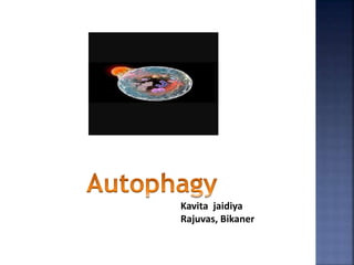
- 2. Autophagy is natural, conserverd, degradation of the cell that removes unnecessary or dysfunctional components through a lysosome dependent regulated mechanism. Autopahgy
- 3. The concept emerged during 1960 s till recently mechanism was not known. 1990- Y. Ohsumni demostrated mechanism in yeast. History
- 4. Mechanism
- 5. Three forms of autophagy have been identified- 1) Macro autophagy 2) Micro autophagy 3) Chaperone mediated autophagy Classification
- 7. Autophagy functions as a survival mechanism under various stress conditions, maintaining the integrity of cells by recyclingessectial metabolites and clearing debris. Autophagy cann both promote cancer growth and act as a defence against cancer. Many pathogens are degraded by autophagy, these include mycobacteria, shigella spp. Dysregulation of autophagy occurs in many disease states including cancers, inflammatory bowel disease and neurodegenerative disorders. Autophagy plays a role in host defense against certain microbes. Importance of Autophagy
- 8. Etosis is a cell death mechanism defined by the release of extracellular traps, which can foster inflammation and exert microbicidal activity. Etosis
- 9. Electrone microscope are used to investigate the ultrastructure of wide range of biological and inorganic specimens including microorganisms cells, large molecules, biopsy samples , metals and crystals. Electroen microscope uses a beam of electrons an image of the specimen. It is capable of much higher magnification and has a greater resolving power than a light microscope.
- 11. There are two types of electrone microscope- 1) Transmission electron microscope ( TEM) 2) Scanning electron microscope ( SEM) For SEM samples can be classified into three categories- 1) Sample for fracture study 2) Sample for Microstructural study 3) Powder samples of morphology i.e size, shapes of powder particles
- 12. For Fractography- Fracture sample is carefully cut according to the size that specimen holder can accommodate. Sample is cleaned using some solvent eg. Acetone, carbon tetrachloride, alchohal, oil, grease , dust, oil or any other extraneous material. For Microscopical study- Sample of suitable size is taken. It is gound and polished than etched using suitable etchant. Then cleaned etched sample is ready for observation in the SEM.
- 13. For particle size and shape determination of powder sample- The powder must be distributed evenly over a clean substrate. A small quantity of powder is taken in test tube filled with sovent or distilled water. Then it is agitated in ultrasonic cleaner to disperse the particles, with the help of dropper one or more drops of the solvent is dropped on clean glass slides. When the slides is dried a self adhesive conducting tapes is pressed over the glass slide, so that all particles comes out of slides, when tape is stripped out. The tape is then put on a mount and coated with gold or carbon before examination in SEM.
- 14. For TEM- Thin film of material are prepared by physical vapour deposition technique. The material is kept in a tungsten or Molybdenum basket and clean substrate is place above or below the basket. The basket is then heated by passing current through it. When the material melts it evaporates and get deposits on substrate. A clean glass slide coated with uniform layer of soap or Nacl can be used as substrate. When thin flim is form on slide , it is striped out by dipping the slide in distilled water . The soap and Nacl dissolve in distilled water and thin film floats on water surface then can be loaded in TEM specimen hloder and ready for examination.