Download to read offline

![5
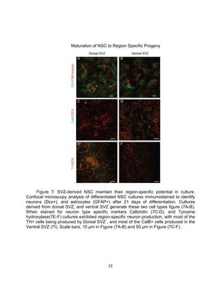
Western Blot Analysis
Tissue was micro-dissected from the dorsal wedge area, ventral SVZ, and corpus
callosum of wildtype mice. Tissue extracts were prepared by lysing the tissue with
radioimmunoprecipitation assay (RIPA) buffer (50 mM Tris [pH 7.5], 150 mM NaCl, 1
mMEDTA, 1% Triton X-100, 0.1% SDS, 1% sodium deoxycholate) containing freshly
added phosphatase inhibitor cocktails I and II (P-2850 and P-5726; Sigma), Complete
protease inhibitor cocktail (Roche), and 1 mM phenylmethylsulfonyl fluoride (PMSF;
Sigma) on ice for 30 min. Insoluble debris was removed by centrifugation at 16,000 g for
20 min at 4°C.
Lysate concentrations were determined with the bicinchoninic acid (BCA) kit
(Pierce, Rockford, IL) and compared to BSA standards in RIPA buffer, and the samples
were equalized with RIPA buffer. 30µg aliquots of the lysates were separated on a 4 to
10% Tris-glycine SDS-PAGE with Precision Plus protein standards (Bio-Rad, Hercules,
CA) and transferred to nitrocellulose membranes (all reagents from Invitrogen,
Carlsbad, CA). Membranes were incubated with 2 to 5µg/ml anti-Gli antibodies in 5%
(wt/vol) milk in Tris-buffered saline–0.05% Tween 20 followed by horseradish
peroxidase(HRP)-conjugated secondary antibodies. Endogenous Gli bands were
visualized with a rabbit monoclonal antibody against Gli3, gli1 antibody (1:500, Cell
Signaling), and tubulin loading controls (1:10,000 dilution of 1A2 MAb; Sigma T9028)
were visualized with ECL reagent(GE HealthCare).](https://image.slidesharecdn.com/20573ee9-450d-433c-be25-2dad64fe755d-160627222637/85/JustinDEvans-8-320.jpg)

![18
Experimental Procedures
Animals
The following mice lines were used and genotyped as previously described: CD1
WT, Gli3lox (Huang et al Dev Dyn, 2008.) and Ai14lox (Jackson Labs). Mice of either
sex were used for the analysis. Experiments were performed in accordance to protocols
approved by Institutional Animal Care and Use Committee at Vanderbilt University.
Cell Culture
SVZ cells from postnatal day 1 (P1) (Timed pregnant CD1 wild-type females, The
Harlan Laboratory or Gli3 conditional mice, in house) were placed in DMEM/F-12 (DF)
containing 100 units/ml penicillin, 100 μg/ml streptomycin, and 50 µg/ml Gentamicin.
Extracted tissues were triturated in 0.25% trypsin (15 min, 37°C, pH 7.3) and placed
overnight in 24 well plastic tissue-culture dishes in N5 medium [DF containing N2
supplements, 35 μg/ml bovine pituitary extract, 5% FBS (Sigma), and 20 ng/ml EGF and
basic FGF]. Unattached cells were collected, gently triturated, and replated onto
uncoated plastic dishes, and grown to confluency in N5 media was added every other
day. To induce differentiation, the cells were plated on glass 8 well chamber slides
coated with 0.1 mg/ml poly-D-Lysine (Sigma) and 1 μg/ml laminin at densities of ≈5 ×
104
cells per cm2. The cells were proliferated to 90–100% confluency and were induced
to differentiate by removing growth factors from the culture media. Neurons were
allowed to differentiate in N6 media [DF containing N2 supplements, 35 μg/ml bovine](https://image.slidesharecdn.com/20573ee9-450d-433c-be25-2dad64fe755d-160627222637/85/JustinDEvans-21-320.jpg)
![19
pituitary extract, and 2.5% FBS].The media were changed every other day. For
immunostaining, cultures were fixed with 4% paraformaldehyde.
Immunostaining
Immunostainings were carried out on paraformaldehyde-fixed cells according to
standard procedures. Cells were blocked with 1% goat serum plus 0.10% Trition X-
100(Sigma) in PBS for 30 min at 25 C, before primary antibody incubation at 4 C
overnight. Primary antibodies used were mouse anti-GFAP (1:1000, Chemicon),
chicken anti-GFAP (1:1000, Abcam), rabbit anti-doublecortin (1:1000, Cell Signaling),
Guinea pig anti-doublecortin (1:1000, Cell Signaling), rabbit anti-tyrosine hydroxylase
(1:500, Pel-Freez Biologicals), monoclonal anti-calbindin D28k(1:500, Sigma), rabbi
anti-calretinin(1:500, Swant), rat anti-Ki67(1:1000, Novocastra), mouse anti-βIII
tubulin(1:1000,Millipore), and rabbi anti-calbindin D28k (1:1000, Swant). The secondary
antibodies used were conjugated to AlexaFluor dyes (1:1000, Invitrogen/Molecular
Probes) and nuclei were counterstained with DAPI (Sigma).
Microscopic Analysis and Quantification
Fluorescent staining in the eight well chamber slide (Millipore) was visualized
using a LSM 710 Meta Inverted confocal microscope. Fluorescent staining in the BD 96
well imaging plate was visualized using the CellAvista (Dynamic Devices) and analyzed
the data using a MATLAB script developed by Stephen Hummel (Quaranta lab, MS in
prep.). Data were quantified and analyzed using GraphPad Prism 5.](https://image.slidesharecdn.com/20573ee9-450d-433c-be25-2dad64fe755d-160627222637/85/JustinDEvans-22-320.jpg)


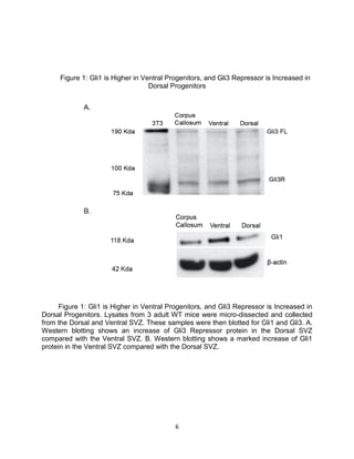
This thesis examines the role of the transcription factor Gli3 in regulating neural stem cell proliferation and neuronal fate specification in the subventricular zone (SVZ). The author finds that: 1) Expression of the Gli3 repressor form is higher in dorsal SVZ neural stem cells, while expression of the activator form Gli1 is higher in ventral SVZ stem cells. 2) Conditional ablation of Gli3 in dorsal SVZ stem cells results in their progeny adopting aberrant positions deeper in the olfactory bulb compared to controls. 3) Loss of Gli3 also decreases expression of the dopamine marker TH in dorsal SVZ-derived neurons, making their fate resemble that of ventral


