Integrin V can form heterodimers with several subunits to mediate cell-cell and cell-extracellular matrix interactions. During zebrafish gastrulation, V is expressed maternally and zygotically. Here, we used a morpholino-mediated V knockdown strategy to study V function. Although V morphants displayed vascular defects, they also exhibited left-right body asymmetry defects affecting multiple visceral organs. This was preceded by mislocalization of dorsal forerunner cells (DFCs) and malformation of the Kupffer’s vesicle (KV) laterality organ
![3449RESEARCH ARTICLE
INTRODUCTION
Integrins mediate cell-cell and cell-extracellular matrix (ECM)
interactions. The eighteen and eight integrin subunits in
mammals can assemble into 24 different heterodimers (Hynes,
2002). Specific integrins recognize a restricted range of ECM
ligands (Plow et al., 2000; Wendel et al., 1998), and integrin
affinity for these ligands is controlled by ‘inside-out’ signaling
pathways that converge upon the integrin cytoplasmic and
transmembrane domains to activate the extracellular domains.
Ligand binding to integrin triggers ‘outside-in’ signals that mediate
anchorage-dependent events, including cell migration, proliferation,
differentiation and survival (Hynes, 2002; Luo et al., 2007).
In mammals, the V integrin subunit can associate with any of
five subunits (1, 3, 5, 6 or 8) and V integrins typically
recognize Arg-Gly-Asp-containing ligands, such as fibronectin,
vitronectin, osteopontin and latency-associated peptide-TGF1
(Hynes, 2002; Takada et al., 2007). Postnatally, V integrins have
been implicated in cellular responses to injury, immunity,
angiogenesis and aspects of tumor progression (Nemeth et al.,
2007; Takada et al., 2007). During vertebrate development, V
integrins exhibit a wide distribution of overlapping expression
domains in mammalian, avian and zebrafish embryos (Ablooglu et
al., 2007; Delannet et al., 1994; Neugebauer et al., 1991; Testaz et
al., 1999; Yamada et al., 1995). The developmental importance of
V is illustrated by the phenotype of V (itgav – Zebrafish
Information Network) knockout mice, which demonstrate improper
formation of embryonic cerebral blood vessels and defective axon
and glia interactions in the postnatal central nervous system (Bader
et al., 1998; McCarty et al., 2005; McCarty et al., 2002). However,
as V is maternally deposited in mice (Sutherland et al., 1993) and
up to 20% of V-null mice survive to birth (Bader et al., 1998), the
opportunity to uncover potential roles for V in very early mouse
development has been limited.
Here, we used antisense morpholino oligonucleotides (MOs) to
transiently knockdown integrin V in zebrafish (Eisen and Smith,
2008; Nasevicius and Ekker, 2000). We provide the first evidence
that depletion of V, along with depletion of one of its potential
subunit partners, 1b, leads to defective dorsal forerunner cell
(DFC) migration during gastrulation. Recent reports have shown
that DFC migration is important for the formation of Kupffer’s
vesicle (KV), a ciliated organ involved in left-right body axis
specification in zebrafish (Amack et al., 2007; Amack and Yost,
2004; Essner et al., 2005; Essner et al., 2002; Oishi et al., 2006;
Schneider et al., 2008). Indeed, we find that KV is abnormally
formed in V and 1b morphants and that both exhibit body
asymmetry defects later in development.
MATERIALS AND METHODS
Zebrafish maintenance and stocks
Wild-type Danio rerio and Tg[sox17:gGFP] embryos were raised at
28.5°C. Embryos from natural matings were kept in 1-phenyl-2-thiourea
(PTU; 0.003%) to inhibit pigmentation and staged according to Kimmel et
al. (Kimmel et al., 1995). Zebrafish were housed in the UCSD animal
facility and experiments were performed in accordance with the guidelines
of UCSD Institutional Animal Care and Use Committee.
Antisense depletion of integrins V and b
MOs used (see Figs S1 and S5 in the supplementary material) were: V1,
5Ј-AGTGTTTGCCCATGTTTTGAGTCTC-3Ј; V2, 5Ј-AGTAGATGG-
AGATCGCGCTGTTTGT-3Ј; VEI10, 5Ј-GTCAGTGCAAATCATT-
ACTCACCCA-3Ј; V1miss (mutated residues in lower case), 5Ј-
AcTcTTTcCCgATcTTTTcAGTgTC-3Ј; standard control MO, 5Ј-
CCTCTTACCTCAGTTACAATTTATA-3Ј; 1b1, 5Ј-GGAGCAGCCTTA-
CGTCCATCTTAAC-3Ј; 1bEI10, 5Ј-GCCAGTTTGAGTGAATAAC-
TCACCT-3Ј. All MOs were obtained from Gene Tools (Philomath, OR,
USA). MOs were injected at the 1- to 4-cell-stage blastulae except where
Development 137, 3449-3458 (2010) doi:10.1242/dev.045310
© 2010. Published by The Company of Biologists Ltd
Department of Medicine, University of California San Diego, La Jolla, CA 92093-
0726, USA.
*Author for correspondence (aablooglu@ucsd.edu)
Accepted 6 August 2010
SUMMARY
Integrin V can form heterodimers with several subunits to mediate cell-cell and cell-extracellular matrix interactions. During
zebrafish gastrulation, V is expressed maternally and zygotically. Here, we used a morpholino-mediated V knockdown strategy
to study V function. Although V morphants displayed vascular defects, they also exhibited left-right body asymmetry defects
affecting multiple visceral organs. This was preceded by mislocalization of dorsal forerunner cells (DFCs) and malformation of the
Kupffer’s vesicle (KV) laterality organ. These defects were rescued with morpholino-resistant V mRNA. Like V, integrin 1b was
expressed in DFCs, and 1b knockdown largely recapitulated the laterality phenotype of V morphants. When tracked in real-
time, individual DFCs of both morphants showed defects in DFC migration, preventing them from organizing into a KV of normal
shape and size. Thus, we propose that V1b mediates cellular interactions that are necessary for DFC clustering and movements
necessary for Kupffer’s vesicle formation, uncovering an early contribution of integrins to the regulation of vertebrate laterality.
KEY WORDS: Alpha V, Beta 1b, Dorsal forerunner cells, Gastrulation, Integrin, Zebrafish
Integrin V is necessary for gastrulation movements that
regulate vertebrate body asymmetry
Ararat J. Ablooglu*, Eugene Tkachenko, Jian Kang and Sanford J. Shattil
DEVELOPMENT
Development ePress online publication date 15 September 2010](https://image.slidesharecdn.com/56283763-e379-4cab-b423-1e58b9adf57e-160718113855/85/Ablooglu-AJ-2010-Development-1-320.jpg)


![3452
wild-type embryos (Fig. 2I-L), their heart tube location was
randomized compared with controls (DFCV1
, 48.2±5.9% left,
n223; DFCcontrol
, 87.0±4.5% left, n251) (Fig. 2M; see Table S3
in the supplementary material). However, when V1 was delivered
into the yolk cell at an even later stage [dome stage to 30% epiboly
(30% E)], when DFC connections with yolk cells are considered to
be closed (D’Amico and Cooper, 1997; Essner et al., 2005), these
yolkV1
morphants displayed normal heart tube asymmetry (Fig.
2M; see Table S3 in the supplementary material). Thus, selective
knockdown of V supported its presence in DFCs by
phenocopying the heart laterality defect.
Given the prominent role of V integrins in mammalian cell
migration (Hynes, 2002), we asked whether V might be required
for DFC migration. DFCs were identified in gastrulating embryos
at 80% E by utilizing cas (Kikuchi et al., 2001), sox17 (Alexander
and Stainier, 1999) or ntl (Amack and Yost, 2004) as markers (Fig.
3; see Fig. S4 in the supplementary material). Over 76% of
uninjected embryos or embryos injected with V1miss showed
ovoid DFC clustering forming 5-6 tiers of cells from the margin
(Fig. 3A,B). By contrast, when embryos were injected with V1,
V2 or VEI10, 53-69% of V morphant DFCs were confined to
a linear domain that had occasional gaps (Fig. 3C-E). This mutant
DFC phenotype was independent of the markers used to identify
the cells (see Fig. S4 in the supplementary material) and it could
be rescued by co-injection of mV mRNA. For example, although
only 17.6±6.3% (n69) of V1 morphants showed an ovoidal wild-
type DFC clustering pattern, 56.3±1.1% (n23) of morphants co-
injected with mV mRNA demonstrated the wild-type pattern (see
Table S4 in the supplementary material). Rescue by mV mRNA
was also observed in V2 morphants (Fig. 3F). Finally, DFC-
selective, DFCV1
morphants also exhibited DFC phenotypes
similar to that of the V1, V2 or VEI10 morphants (see Table S3
in the supplementary material). Thus, the loss of V function
during gastrulation appears to impair DFC migration but not
specification.
Integrin 1b is the likely partner for V in dorsal
forerunner cells
Of the several subunits that can pair with V (Bouvard et al.,
2001), zebrafish express 5, 6, 8 and multiple forms of 1 (1a,
1b, 1b.1, 1b.2) and 3 (3a, 3b) (Ablooglu et al., 2007; Julich
et al., 2005; Julich et al., 2009; Mould et al., 2006; Thisse et al.,
2001). Based on the reported spatial and temporal expression
patterns of these subunits and on overlapping expression patterns
with V during gastrulation, 1b (itgb1b – Zebrafish Information
Network) and 5 (itgb5 - Zebrafish Information Network) appeared
to be the only potential partners for V in DFCs. Consequently, we
examined their localization patterns at gastrulation to establish the
identity of the potential V partner in DFCs. Both integrins
showed distinct localization patterns at mid-gastrulation stages
(Fig. 4A,B). For example, although 1b transcripts were mainly
present in the embryonic axis at 80% E (Fig. 4A), 5 transcripts
were only present in the marginal cells and there was a gap in its
expression field (Fig. 4B). When embryos were examined at 80%
E for cas expression in DFCs and for integrin subunit expression
by double wholemount in situ hybridization (WISH), the gap in the
5 expression field in the marginal cells overlapped the embryonic
axis and only 1b was present in DFCs (Fig. 4C,D). Consequently,
the binding partner for V in DFCs might be 1b.
To study 1b function, a translation-blocking MO (1b1) and a
splice-inhibiting MO (1bE10) were employed (see Fig. S5 in the
supplementary material). After injection of either MO at the 1-4
cell stage, DFC markers were still expressed but there was a DFC
mutant phenotype similar to that observed in V morphants (Fig.
4E-H). Later in development at 32 hours post-fertilization (hpf),
1b1 morphants had pericardial edema and showed shorter and
undulated midlines associated with U-shaped somites (Fig. 4I,J).
When 0.7 ng 1b1 or 5 ng 1bEI10 were delivered at the 1-4 cell
stage, these morphants showed reduced left-sided location of the
liver (see Table S2 in the supplementary material). At these MO
doses, considerable numbers of 1b1 morphants exhibited an
absence of liver primordium and some 1bEI10 morphants had
situs inversus totalis. When both 1b1 and 1bEI10 were co-
injected, no liver primordium was evident (see Table S2 in the
supplementary material). These results suggested that pleiotropic
phenotypes might be caused by midline defects (Biemar et al.,
2001) or lack of endoderm (Alexander and Stainier, 1999; Kikuchi
et al., 2000; Komada and Soriano, 1999). As 1b is deposited
maternally (Ablooglu et al., 2007; Mould et al., 2006) and its
zygotic expression is maintained exclusively in the developing
midline during gastrulation (Fig. 4A) (Julich et al., 2005), knocking
down of maternal 1b with 1b1 MO could contribute pleiotropic
defects. Consequently, we used a lower dose of 1b1 (0.5 ng) to
minimize possible pleiotropic defects. Under these conditions,
1b1 alone did not significantly alter the asymmetric spaw
expression pattern (Fig. 4L; see Table S2 in the supplementary
material). However, when 0.5 ng 1b1 and 5 ng 1bEI10 were
each co-injected, the severity of randomized spaw expression
RESEARCH ARTICLE Development 137 (20)
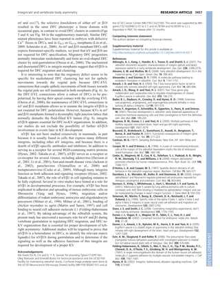
Fig. 3. Migratory DFCs are not properly formed in V morphants.
(A-E)Dorsal views of MO-injected embryos were slightly tilted to
visualize cas expression (arrows, black) in DFCs at 80% E. Gaps in DFC
field are indicated with bracketed arrows. (F)Bar graph showing scores
from DFC phenotypes. Phenotypic classification of DFCs were as
follows: Wild-type (WT), ovoid DFC cluster; mutant, a linear array of
DFCs with occasional gaps; none, no visible DFCs. Data expressed are
similar to those in Fig. 1E. Scale bars: 20m. See also Table S4 in the
supplementary material.
DEVELOPMENT](https://image.slidesharecdn.com/56283763-e379-4cab-b423-1e58b9adf57e-160718113855/85/Ablooglu-AJ-2010-Development-4-320.jpg)


![Pells et al [2015] PLoS ONE 10[7] e0131102](https://cdn.slidesharecdn.com/ss_thumbnails/f79bb09e-8eb1-41e7-8042-28c0aa4a48c6-150720142907-lva1-app6891-thumbnail.jpg?width=640&height=640&fit=bounds)


