1. Filter
Identification of a Transcription Factor Required for the Apoptotic Response to
Synapsis Checkpoint Activation in C. elegans!
Cody Wong and Needhi Bhalla!
Department of Molecular, Cell, and Developmental Biology, University of California, Santa Cruz!
References:
Bhalla, N. and Dernburg, A.. “A Conserved Checkpoint Monitors Meiotic Chromosome Synapsis in Caenorhabditis Elegans.” Science 310, no.
5754 (December 9, 2005): 1683–86. doi:10.1126/science.1117468.
Conradt, B. and Xue D. Programmed cell death (October 06, 2005), WormBook, ed. The C. elegans Research Community, WormBook, doi/
10.1895/wormbook.1.32.1, http://www.wormbook.org.
Gartner, A., Milstein, S.,, Ahmed,, S., Hodgkin,. J., and Hengartner, M.. “A Conserved Checkpoint Pathway Mediates DNA Damage–Induced
Apoptosis and Cell Cycle Arrest in C. elegans.” Molecular Cell 5, no. 3 (March 2000): 435–43. doi:10.1016/S1097-2765(00)80438-4.
Lu, Nan, Xiaomeng Yu, Xiangwei He, and Zheng Zhou. “Detecting Apoptotic Cells and Monitoring Their Clearance in the Nematode Caenorhabditis
elegans.” Methods in Molecular Biology (Clifton, N.J.) 559 (2009): 357–70. doi:10.1007/978-1-60327-017-5_25.
Schumacher, B., Hanazawa, M., Lee, M.-H., Nayak, S., Volkmann, K., Hofmann, R., … Gartner, A. (2005). Translational Repression of C. elegans
p53 by GLD-1 Regulates DNA Damage-Induced Apoptosis. Cell, 120(3), 357–368. http://doi.org/10.1016/j.cell.2004.12.009
Shaham, S., ed., WormBook: Methods in Cell Biology (January 02, 2006), WormBook, ed. The C. elegans Research Community, WormBook, doi/
10.1895/wormbook.1.49.1, http://www.wormbook.org.
Ye, A., Ragle, M., Conradt, B., and Bhalla, N. “Differential Regulation of Germline Apoptosis in Response to Meiotic Checkpoint Activation.”
Genetics 198, no. 3 (November 1, 2014): 995–1000. doi:10.1534/genetics.114.170241.
Why C. elegans?
*
**
B.!
Background
Visualizing Apoptotic Nuclei using Plim-7ced-1::gfp
.
The Bhalla Lab studies
chromosome dynamics during
meiotic prophase to better
understand proper
chromosome segregation.
The C.elegans germline allows
us to visualize different stages
of meiotic prophase. Other
reasons are easy-to-propagate,
transparent, and eukaryotic.
Image from
WormClassroom.org, “C.
elegans germline”
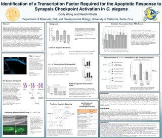
Identification of
Candidate
Transcription Factors
Filter Number of
Genes
Germline-expressed
Genes
4699
Germline-expressed
Genes w/ GLD-1 binding
site
1084
Germline-expressed
Transcription Factors w/
GLD-1 binding site
55
Germline-expressed and
Oogenesis-enriched
Transcription Factors w/
GLD-1 binding site
18
To quantify checkpoint-
activated apoptosis, I used
a CED-1::GFP reporter.
CED-1 is a transmembrane
protein expressed on
phagocytic cells that engulf
apoptotic cells (Lu et al.,
2009). Therefore, I looked
for cells that were
fluorescently outlined
(completely encircled by
CED-1) .
Figure 15 from Shaham,
S., 2006
Figure 3, Image B from Bhalla and Dernburg,
2005
To avoid aneuploidy, cells maintain vigilance
over the progression of meiosis through the
use of checkpoints. The synapsis checkpoint
activates apoptosis when a developing
oocyte exhibits unsynapsed chromosomes
during pachytene. How do these nuclei
activate apoptosis?
The pro-apoptotic gene, egl-1, was found to be transcriptionally
upregulated in response to synapsis checkpoint activation (Ye et
al., 2014). Therefore, I looked for transcription factors that could
promote transcription of pro-apoptotic genes in response to
activation of the synapsis checkpoint.
Figure 3, Images A and C from Ye et al., 2014
All cells that undergo apoptosis follow this core pathway.
Adapted from Figure 2 from Conradt and Xue, 2005
Core Cell Apoptotic Machinery
egl-1 is Transcriptionally Upregulated
A GLD-1-regulated Transcription
Factor
Schumacher et al. investigated GLD-1’s, a RNA-binding
repressor, involvement in germline apoptosis. Dr. Bhalla
noticed a significant increase in apoptosis in gld-1 mutants.
We thought that this could suggest that GLD-1 was
suppressing a pro-apoptotic synapsis checkpoint protein,
specifically, a transcription factor of interest.
Figure 3, Image A from Schumacher et al., 2005
Characterization of nhr-84’s involvement in the Synapsis Checkpoint
0
5
10
15
20
wild−type
(Physiological
Apoptosis)
syp−1
(Physiological
Apoptosis +
DNA−Damage
Checkpoint +
Synapsis
Checkpoint)
syp−1;spo−11
(Physiological
Apoptosis +
Synapsis
Checkpoint)
NumberofApoptoticNucleiperGonadArm
No RNAi
nhr−84 RNAi
nhr−84 RNAi in Meiotic Checkpoint Background
The Synapsis Checkpoint
Predicted Apoptotic Signaling
Conclusion:
Previously, it was unknown how the synapsis checkpoint activates the apoptotic pathway. The results of the candidate
transcription factor RNAi screen revealed that nhr-84 could be involved in linking synapsis checkpoint activation with apoptosis.
Even though nhr-84 RNAi in a syp-1;spo-11 background reduced apoptosis to wild-type levels, it was still possible that nhr-84
could be involved in physiological apoptosis rather than synapsis checkpoint-induced apoptosis. Therefore, I performed nhr-84
RNAi in wild-type and syp-1 backgrounds. Since nhr-84 RNAi in a wild-type background did not significantly reduce apoptosis
levels, nhr-84 was not involved in promoting physiological apoptosis. Whether nhr-84 is involved in synapsis checkpoint-induced
apoptosis is contingent upon there only being two apoptotic pathways in syp-1;spo-11 mutants. Nevertheless, it is highly
probable that it is involved in synapsis checkpoint-induced apoptosis. Having identified a transcription factor that acts as the
synapsis checkpoint’s manipulator of the apoptotic pathway, this essential protein can be investigated to see what happens in
cases of aneuploidy.
Future Work:
-set-16, die-1, rcor-1, ztf-22, and dmd-9 RNAi in Synapsis Checkpoint Background
-if any of those return a positive result, that gene would need to be silenced in syp-1 and wild-type backgrounds
-A negative result could mean that the transcription factor is not involved in activating the apoptosis pathway in response to
synapsis checkpoint activation. It could also mean that the gene is RNAi-resistant. To test this, antibody staining for the
respective protein would be conducted to see if RNAi effectively silenced the gene.
-It is unknown whether NHR-84 promotes transcription of egl-1 in response to synapsis checkpoint activation. This would be
tested through qPCR.
-to determine if NHR-84 is GLD-1-regulated, I would have to do antibody staining to see if NHR-84 expression is increased in a
gld-1 mutant. For biochemical analysis, I would do a pull-down assay to see if GLD-1 co-precipitates nhr-84 mRNA
Here, we see the different mutants
experimented with and what meiotic
checkpoints they activate.
I tested to see if nhr-84 was involved in DNA-Damage
checkpoint-induced apoptosis, Synapsis Checkpoint-
induced Apoptosis, and Physiological Apoptosis.
Candidate Transcription Factor RNAi Screen
There are three main meiotic prophase
events that ensure proper chromosome
segregation: the pairing of homologous
chromosomes, the loading of the
synaptonemal complex between
homologous chromosomes (synapsis),
and DNA exchange (recombination).
There are currently two identified
checkpoints that monitor these
processes: the synapsis checkpoint and
the DNA-damage checkpoint. A
nucleus activates the synapsis
checkpoint if the X chromosomes or all
of the chromosomes are unable to
synapse (Bhalla and Dernburg, 2005).
Alternatively, a nucleus activates the
DNA-damage checkpoint if
chromosomes are unable to recombine
(Gartner et al., 2000).
Here is Dr. Bhalla’s theory on how meiotic checkpoints
activate apoptosis. The DNA-damage checkpoint
activates apoptosis through CEP-1 and CED-13. The
synapsis checkpoint activates apoptosis through an
unidentified transcription factor. The transcription factor
of interest is encircled.
I used multiple filters to narrow down the
number of possible transcription factors.
Here, I present the number of genes that
fulfilled each filter. I ended up with 18
potential candidates.
0
5
10
W
ild−typeC
ontrol
egl−1
R
N
Ai
aha−1
R
N
Ai
m
xl−1
R
N
Ai
tbx−36
R
N
Ai
nhr−71
R
N
Ai
nhr−84
R
N
Ai
sm
a−9
R
N
Ai
m
ex−5
R
N
Ai
om
a−2
R
N
Ai
pzf−1
R
N
Ai
bed−2
R
N
Ai
pal−1
R
N
Ai
m
ex−6
R
N
Ai
ceh−20
R
N
Ai
NumberofApoptoticNucleiperGonadArm
RNAi Candidate Screen in Synapsis Checkpoint Background
Abstract
Meiosis is a specialized form of cell division that produces haploid gametes, such as
eggs and sperm. During meiosis, programmed cell-death, or apoptosis, removes
defective cells that could compromise the accuracy of gametic inheritance. Improper
chromosome segregation can result in aneuploidy or cancer. To ensure proper
chromosome segregation, the synaptonemal complex must get loaded between
homologues (synapsis) during meiotic prophase I. In C. elegans, germline cells in
meiotic prophase that display unsynapsed chromosomes have been shown to undergo
cell apoptosis. To better understand how asynapsis triggers cellular apoptosis, I have
been looking for a pro-apoptotic transcription factor that is activated by a cellular
meiotic checkpoint that activates if chromosomes are unsynapsed, termed the
synapsis checkpoint. My goal was to find this transcription factor that responds to the
synapsis checkpoint. By cross-referencing published databases, I have narrowed down
4699 germline-expressed genes identified by Wang et al. to 18 possible transcription
factors using the conditions of the synapsis checkpoint and predicting that this
transcription factor is GLD-1 regulated. I performed RNAi on each transcription factor
with the goal of identifying the link between the synapsis checkpoint and the cell
apoptosis pathway. Here, we present the identification of nhr-84 and characterize its
involvement in the apoptotic response to synapsis checkpoint activation.
I performed a RNAi screen of 18 candidate
transcription factors in a syp-1;spo-11 background. In
these mutants, nuclei contain unsynapsed
chromosomes, resulting in activation of the synapsis
checkpoint (Bhalla and Dernburg, 2005). Here is a
bar graph of the RNAi data. The RNAi experiments
are labeled on the x-axis. syp-1;spo-11 mutants are
labeled as controls. Silencing most of the
transcription factors did not decrease the number of
apoptotic cells. Silencing of nhr-84 decreased
apoptotic levels to around wild-type levels, suggesting
that it is involved in either physiological apoptosis or
synapsis checkpoint-induced apoptosis.
Why C. elegans?