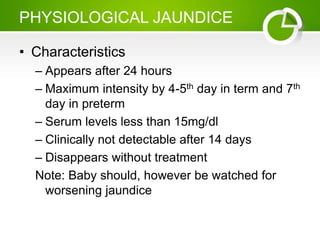
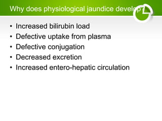
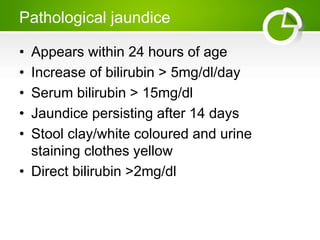
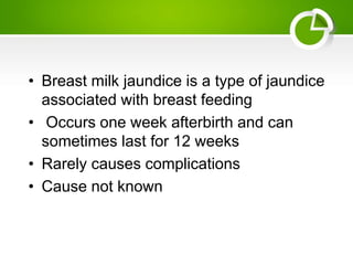
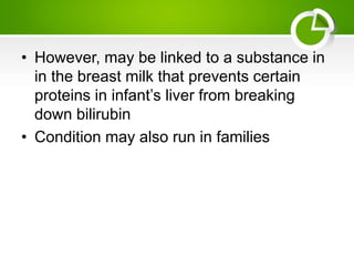
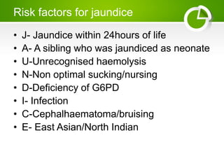
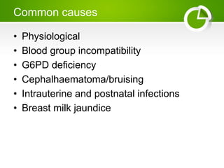
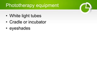
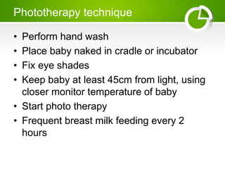
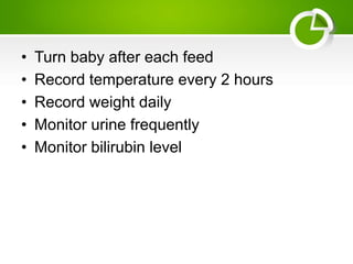
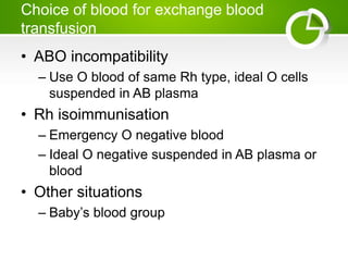
JAUNDICE
DR MUG,OMBA
objectives
• By the end of the lesson, learners must be
able to;
– Define neonatal jaundice
– Outline causes of jaundice
– Outline management of physiological jaundice
– Relate neonatal jaundice to clinical practice
definition
• Visible form of bilirubinemia
– Newborn skin > 5mg/dl
• Occurs in 60% of term and 80% of preterm
neonates
• However, significant jaundice occurs in 6%
of term babies
Clinical assessment of jaundice
• Area of body bilirubin levels in mg/dl
face 4-8
upper trunk 5-12
lower trunk 8-16
and thighs
arms and lower 11-18
limbs
palms and soles >15
PHYSIOLOGICAL JAUNDICE
• Characteristics
– Appears after 24 hours
– Maximum intensity by 4-5th day in term and 7th
day in preterm
– Serum levels less than 15mg/dl
– Clinically not detectable after 14 days
– Disappears without treatment
Note: Baby should, however be watched for
worsening jaundice
Why does physiological jaundice develop?
• Increased bilirubin load
• Defective uptake from plasma
• Defective conjugation
• Decreased excretion
• Increased entero-hepatic circulation
Pathological jaundice
• Appears within 24 hours of age
• Increase of bilirubin > 5mg/dl/day
• Serum bilirubin > 15mg/dl
• Jaundice persisting after 14 days
• Stool clay/white coloured and urine
staining clothes yellow
• Direct bilirubin >2mg/dl
Causes of jaundice
• Appearing within 24 hours of age
– Haemolytic disease of new born Rh, AOB
– Infections; TORCH, malaria, bacterial
– G6PD deficiency
TORCH syndrome refers to infection of
developing foetus or newborn. TORCH stands
for Toxoplasmosis, Other agents, Rubella,
Cytomegalo viruses, and Herpes simplex
Appearing between 24-72 hours of life
• Physiological
• Sepsis
• Polycythemia
• Concealed haemorrhage
• Intraventricular haemorrhage
• Increased entero-hepatic circulation
After 72 hours of age
• Sepsis
• Cephalhaematoma
• Neonatal hepatitis
• Extra hepatic biliary atresia
• Breast milk jaundice
• Metabolic disorders
• Breast milk jaundice is a type of jaundice
associated with breast feeding
• Occurs one week afterbirth and can
sometimes last for 12 weeks
• Rarely causes complications
• Cause not known
• However, may be linked to a substance in
in the breast milk that prevents certain
proteins in infant’s liver from breaking
down bilirubin
• Condition may also run in families
Risk factors for jaundice
• J- Jaundice within 24hours of life
• A- A sibling who was jaundiced as neonate
• U-Unrecognised haemolysis
• N-Non optimal sucking/nursing
• D-Deficiency of G6PD
• I- Infection
• C-Cephalhaematoma/bruising
• E- East Asian/North Indian
Common causes
• Physiological
• Blood group incompatibility
• G6PD deficiency
• Cephalhaematoma/bruising
• Intrauterine and postnatal infections
• Breast milk jaundice
Approaches to jaundiced baby
• Ascertain birth weight, gestation and post
natal age
• Assess clinical condition (well or ill)
• Decide whether jaundice physiological or
pathological
• Look for evidence of kernicterus* in deeply
jaundiced newborn
• *Lethargy and poor feeding, convulsions
investigations
• Maternal and perinatal history
• Physical examination
• Laboratory tests (must in all)
– Total and direct bilurubin
– Blood group and Rh for mother and baby
– Haematocrit, retic count and peripheral smear
– Sepsis screen
• Liver and thyroid function
• TORCH titers, liver scan when conjugated
hyperbilirubinemia
management
• Rationale: reduce level of bilurubin and
prevent bilurubin toxicity
• Prevention of hyperbilirubinemia; early
feeds, adequate hydration
• Reduction of bilirubin levels ; photo
therapy, exchange transfusion, drugs
Phototherapy equipment
• White light tubes
• Cradle or incubator
• eyeshades
Phototherapy technique
• Perform hand wash
• Place baby naked in cradle or incubator
• Fix eye shades
• Keep baby at least 45cm from light, using
closer monitor temperature of baby
• Start photo therapy
• Frequent breast milk feeding every 2
hours
• Turn baby after each feed
• Record temperature every 2 hours
• Record weight daily
• Monitor urine frequently
• Monitor bilirubin level
Side effects of phototherapy
• Increased sensible water loss
• Loose stools
• Skin rash
• Bronze baby syndrome
• Hyperthermia
• Upsets maternal baby interaction
• May result in hypocalcaemia
Choice of blood for exchange blood
transfusion
• ABO incompatibility
– Use O blood of same Rh type, ideal O cells
suspended in AB plasma
• Rh isoimmunisation
– Emergency O negative blood
– Ideal O negative suspended in AB plasma or
blood
• Other situations
– Baby’s blood group
Pronged indirect jaundice
• Causes
– Crigler Najja’s syndrome
– Breast milk jaundice
– Hypothyroidism
– Pyloric stenosis
– Ongoing haemolysis, malaria
Conjugated hyperbilirubinemia
• Suspect
– High coloured urine
– White or clay coloured stool
Caution : always refer to hospital for
investigations so that biliary atresia or
metabolic disorders can be diagnosed and
managed early
Causes
• Idiopathic neonatal hepatitis
• Infections- hepatitis B, TORCH. sepsis
• Biliary atresia, choledechal cyst
• Metabolic- Galactosemia, tyrosinemia,
hypothyroidism
• Total parenteral nutrition

JAUNDICE.pptx

  • 1.
  • 2.
    objectives • By theend of the lesson, learners must be able to; – Define neonatal jaundice – Outline causes of jaundice – Outline management of physiological jaundice – Relate neonatal jaundice to clinical practice
  • 3.
    definition • Visible formof bilirubinemia – Newborn skin > 5mg/dl • Occurs in 60% of term and 80% of preterm neonates • However, significant jaundice occurs in 6% of term babies
  • 4.
    Clinical assessment ofjaundice • Area of body bilirubin levels in mg/dl face 4-8 upper trunk 5-12 lower trunk 8-16 and thighs arms and lower 11-18 limbs palms and soles >15
  • 5.
    PHYSIOLOGICAL JAUNDICE • Characteristics –Appears after 24 hours – Maximum intensity by 4-5th day in term and 7th day in preterm – Serum levels less than 15mg/dl – Clinically not detectable after 14 days – Disappears without treatment Note: Baby should, however be watched for worsening jaundice
  • 6.
    Why does physiologicaljaundice develop? • Increased bilirubin load • Defective uptake from plasma • Defective conjugation • Decreased excretion • Increased entero-hepatic circulation
  • 7.
    Pathological jaundice • Appearswithin 24 hours of age • Increase of bilirubin > 5mg/dl/day • Serum bilirubin > 15mg/dl • Jaundice persisting after 14 days • Stool clay/white coloured and urine staining clothes yellow • Direct bilirubin >2mg/dl
  • 8.
    Causes of jaundice •Appearing within 24 hours of age – Haemolytic disease of new born Rh, AOB – Infections; TORCH, malaria, bacterial – G6PD deficiency TORCH syndrome refers to infection of developing foetus or newborn. TORCH stands for Toxoplasmosis, Other agents, Rubella, Cytomegalo viruses, and Herpes simplex
  • 9.
    Appearing between 24-72hours of life • Physiological • Sepsis • Polycythemia • Concealed haemorrhage • Intraventricular haemorrhage • Increased entero-hepatic circulation
  • 10.
    After 72 hoursof age • Sepsis • Cephalhaematoma • Neonatal hepatitis • Extra hepatic biliary atresia • Breast milk jaundice • Metabolic disorders
  • 11.
    • Breast milkjaundice is a type of jaundice associated with breast feeding • Occurs one week afterbirth and can sometimes last for 12 weeks • Rarely causes complications • Cause not known
  • 12.
    • However, maybe linked to a substance in in the breast milk that prevents certain proteins in infant’s liver from breaking down bilirubin • Condition may also run in families
  • 13.
    Risk factors forjaundice • J- Jaundice within 24hours of life • A- A sibling who was jaundiced as neonate • U-Unrecognised haemolysis • N-Non optimal sucking/nursing • D-Deficiency of G6PD • I- Infection • C-Cephalhaematoma/bruising • E- East Asian/North Indian
  • 14.
    Common causes • Physiological •Blood group incompatibility • G6PD deficiency • Cephalhaematoma/bruising • Intrauterine and postnatal infections • Breast milk jaundice
  • 15.
    Approaches to jaundicedbaby • Ascertain birth weight, gestation and post natal age • Assess clinical condition (well or ill) • Decide whether jaundice physiological or pathological • Look for evidence of kernicterus* in deeply jaundiced newborn • *Lethargy and poor feeding, convulsions
  • 16.
    investigations • Maternal andperinatal history • Physical examination • Laboratory tests (must in all) – Total and direct bilurubin – Blood group and Rh for mother and baby – Haematocrit, retic count and peripheral smear – Sepsis screen
  • 17.
    • Liver andthyroid function • TORCH titers, liver scan when conjugated hyperbilirubinemia
  • 18.
    management • Rationale: reducelevel of bilurubin and prevent bilurubin toxicity • Prevention of hyperbilirubinemia; early feeds, adequate hydration • Reduction of bilirubin levels ; photo therapy, exchange transfusion, drugs
  • 19.
    Phototherapy equipment • Whitelight tubes • Cradle or incubator • eyeshades
  • 20.
    Phototherapy technique • Performhand wash • Place baby naked in cradle or incubator • Fix eye shades • Keep baby at least 45cm from light, using closer monitor temperature of baby • Start photo therapy • Frequent breast milk feeding every 2 hours
  • 21.
    • Turn babyafter each feed • Record temperature every 2 hours • Record weight daily • Monitor urine frequently • Monitor bilirubin level
  • 22.
    Side effects ofphototherapy • Increased sensible water loss • Loose stools • Skin rash • Bronze baby syndrome • Hyperthermia • Upsets maternal baby interaction • May result in hypocalcaemia
  • 23.
    Choice of bloodfor exchange blood transfusion • ABO incompatibility – Use O blood of same Rh type, ideal O cells suspended in AB plasma • Rh isoimmunisation – Emergency O negative blood – Ideal O negative suspended in AB plasma or blood • Other situations – Baby’s blood group
  • 24.
    Pronged indirect jaundice •Causes – Crigler Najja’s syndrome – Breast milk jaundice – Hypothyroidism – Pyloric stenosis – Ongoing haemolysis, malaria
  • 25.
    Conjugated hyperbilirubinemia • Suspect –High coloured urine – White or clay coloured stool Caution : always refer to hospital for investigations so that biliary atresia or metabolic disorders can be diagnosed and managed early
  • 26.
    Causes • Idiopathic neonatalhepatitis • Infections- hepatitis B, TORCH. sepsis • Biliary atresia, choledechal cyst • Metabolic- Galactosemia, tyrosinemia, hypothyroidism • Total parenteral nutrition